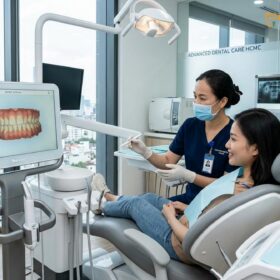
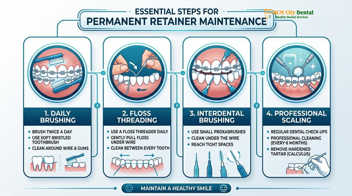
Permanent retainer plaque removal requires a combination of specialized at-home interdental cleaning tools and routine clinical scaling. Because the bonded wire creates a physical barrier, daily use of floss threaders and biannual professional cleanings are essential to help prevent calculus formation, gingival inflammation, and periodontal disease.
Clinical Summary:
Removing plaque from a fixed lingual retainer demands meticulous oral hygiene and professional intervention. The bonded wire naturally traps food particles and bacterial biofilm, which rapidly calcifies into tartar if left undisturbed. Effective management relies on specialized tools like a floss threader and interdental brushes for daily maintenance. Clinically, a professional scale and polish is generally required every six months to safely remove hardened calculus without damaging the orthodontic composite or the wire itself. Neglecting this area can lead to localized periodontitis, gingival recession, and potential retainer failure.
Key Takeaways:
- Fixed lingual retainers act as a physical trap for bacterial biofilm and salivary minerals.
- Daily interdental cleaning using specialized floss threaders is critical for preventing calculus.
- Hardened tartar cannot be removed at home and requires ultrasonic clinical scaling.
- Biannual dental visits help ensure the structural integrity of the bonding composite remains intact.
- Ignoring plaque accumulation can contribute to gingival recession, bone loss, and wire detachment.
The Clinical Anatomy of a Permanent Retainer and Plaque Accumulation
A permanent retainer consists of a custom-fitted wire bonded to the lingual surfaces of anterior teeth, creating microscopic ledges where plaque and salivary minerals rapidly accumulate.
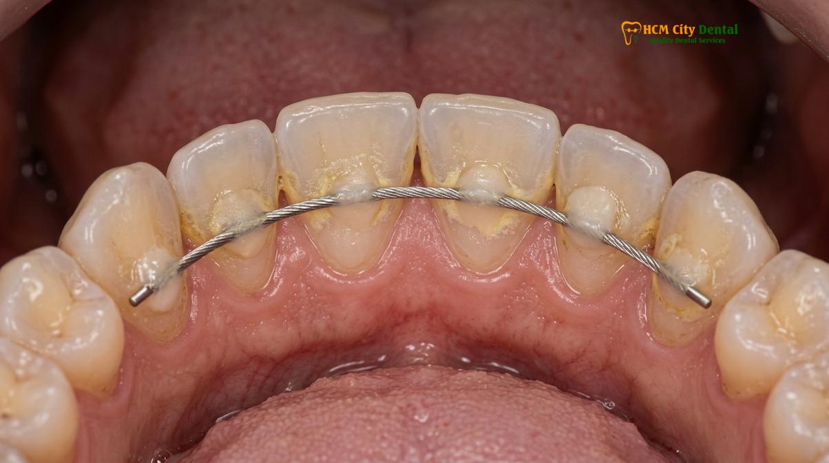
To understand the complexities of permanent retainer plaque removal, one must first examine the clinical anatomy of the retention system and the biological environment of the oral cavity. A fixed retainer, typically placed after active Orthodontics treatment, involves a multi-stranded stainless steel or titanium wire bonded to the lingual (tongue-facing) surfaces of the anterior teeth, most commonly the lower six incisors and canines. The wire is secured using small pads of orthodontic composite resin. While highly effective at maintaining tooth position, this architectural addition creates numerous artificial undercuts, ledges, and stagnation areas that are inaccessible to a standard toothbrush bristle[1].
The Role of Salivary Glands in Calculus Formation
The location of the lower fixed retainer is particularly problematic from a periodontal perspective. The lingual surfaces of the mandibular incisors sit directly opposite the submandibular and sublingual salivary glands. These glands secrete saliva through Wharton’s duct, pooling mineral-rich fluid directly onto the backs of the lower front teeth. Saliva is supersaturated with calcium and phosphate ions, which are essential for enamel remineralization. However, when bacterial plaque biofilm is allowed to stagnate around the retainer wire and composite pads, these salivary minerals precipitate into the biofilm matrix.
This biochemical process transforms soft, easily removable plaque into hard, calcified dental calculus (tartar) rapidly. Once the biofilm calcifies, it forms a rough, porous surface that attracts even more bacterial colonization, creating a vicious cycle of rapid plaque buildup bonded wire complications. The resulting calculus cannot be brushed or flossed away; it requires professional mechanical removal.
The Biofilm Maturation Process
Plaque is not merely food debris; it is a highly organized, complex microbial ecosystem. Initially, a pellicle layer of salivary proteins coats the tooth and the retainer. Pioneer bacteria, primarily aerobic Gram-positive cocci, adhere to this pellicle. If not mechanically disrupted by brushing and flossing, the biofilm matures. The bacterial population shifts toward anaerobic Gram-negative rods and spirochetes, which are highly pathogenic and responsible for initiating gingival inflammation[2]. The physical barrier of the permanent wire protects these maturing colonies from the natural cleansing action of the tongue and salivary flow, making proactive, targeted cleaning absolutely critical for long-term oral health.
Daily At-Home Protocols to Clean Permanent Wire
Effective daily cleaning requires threading floss underneath the wire and using interdental brushes to disrupt biofilm before it hardens into calculus.
Maintaining periodontal health with a fixed retainer requires a paradigm shift in daily oral hygiene. Standard flossing techniques are rendered ineffective because the continuous wire prevents the floss from passing down through the contact points between the teeth. Therefore, patients must adopt specialized techniques to clean permanent wire structures effectively. The goal of at-home care is the daily mechanical disruption of the biofilm before mineralization occurs.
The Floss Threader Technique
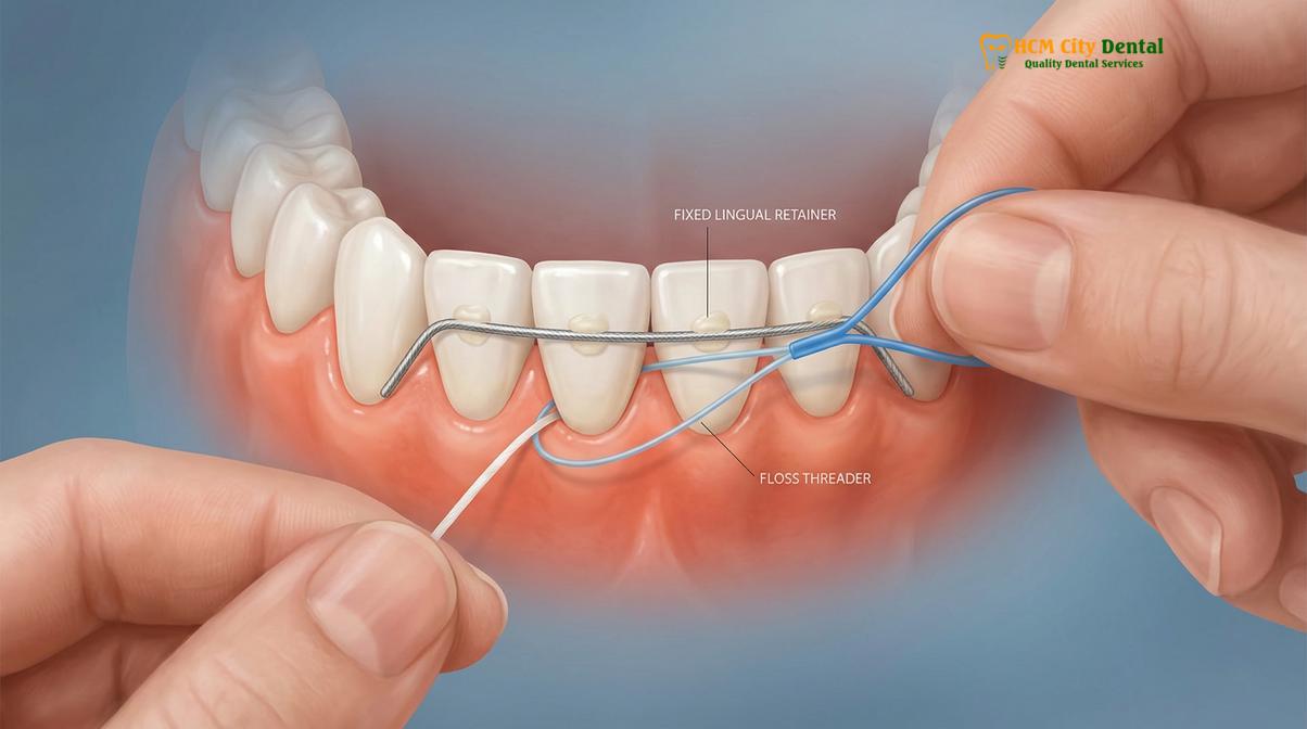
The gold standard for cleaning under a fixed retainer is the use of a floss threader or specialized orthodontic floss. A floss threader acts much like a needle carrying thread. The stiff, flexible loop of the threader is passed between the teeth, underneath the bonded wire, and close to the gumline. Once threaded through, the patient pulls the attached floss under the wire. The floss is then adapted into a “C-shape” around the side of the tooth and moved gently up and down to scrape the root surface and the interproximal enamel[3].
Alternatively, a superfloss threader combines a stiffened end, a spongy middle section, and regular floss into a single strand. The stiff end is threaded under the wire, while the spongy section is highly effective at sweeping away larger amounts of plaque and debris from the wide embrasure spaces and the wire itself. This process must be repeated for every interproximal space spanned by the retainer.
“The mechanical friction provided by string floss against the tooth surface and the gingival sulcus is irreplaceable. While it requires patience and dexterity, threading floss beneath the retainer wire is one of the most effective at-home measures to help prevent localized periodontal disease in orthodontic patients.”
Interdental Brushes and Adjunctive Tools
In addition to flossing, interdental brushes (often called proxy brushes) are invaluable tools. These small, cone-shaped or cylindrical brushes are designed to slide between the teeth, below the contact point and the retainer wire. They are particularly effective at cleaning the lateral surfaces of the composite bonding pads, which are notorious for harboring plaque. When selecting an interdental brush, it is crucial to choose the correct size; the brush should fit snugly but pass through without requiring excessive force, which could traumatize the gingival papilla or bend the retainer wire.
Water flossers (oral irrigators) serve as an excellent adjunctive tool. By utilizing a pulsating stream of water, they create hydrodynamic shear forces that flush out loose food debris and disrupt loosely attached biofilm. However, clinical studies consistently show that water irrigation alone cannot detach the sticky, mature biofilm matrix from the tooth surface. Therefore, a water flosser should be used in conjunction with, rather than as a replacement for, mechanical flossing and interdental brushing[4].
The Role of Professional Scale and Polish in Retainer Maintenance
A professional scale and polish utilizes ultrasonic instrumentation to safely shatter hardened calculus around the retainer wire without compromising the composite bonding resin.
Despite the most meticulous at-home care, microscopic amounts of plaque will inevitably be missed and subsequently calcify, particularly in the high-risk mandibular anterior region. Once calculus forms, it bonds tenaciously to the enamel and the orthodontic composite. Attempting to remove this hardened tartar at home is dangerous and ineffective. Therefore, a professional scale and polish is a mandatory component of long-term retainer maintenance.
Ultrasonic Scaling Mechanics
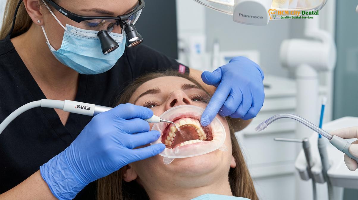
During a clinical cleaning, the dental hygienist or dentist utilizes an ultrasonic scaler. This sophisticated instrument features a specialized metal tip that vibrates at high frequencies. The rapid vibration physically shatters the brittle calculus deposits. Simultaneously, the instrument emits a continuous stream of water, which serves two critical purposes: it cools the tip to prevent thermal damage to the tooth pulp, and it creates a phenomenon known as cavitation. Cavitation produces microscopic bubbles that implode, releasing energy that disrupts bacterial cell walls and flushes debris from the periodontal pocket[5].
The clinician must carefully navigate the ultrasonic tip around the delicate multi-stranded wire and the composite pads. Excessive pressure or incorrect angulation can inadvertently debond the composite or distort the wire, leading to orthodontic relapse. Understanding how long to wear retainers after braces also involves committing to these regular professional maintenance sessions.
Hand Instrumentation and Composite Polishing
Following ultrasonic scaling, the clinician uses fine hand instruments, such as curettes and scalers, to meticulously remove any residual calculus from the root surfaces and the intricate margins of the bonding pads. This tactile phase ensures that the subgingival environment is completely free of irritants.
The final step is the polishing phase. Using a slow-speed handpiece, a rubber cup, and a specialized prophylaxis paste, the clinician polishes the enamel and the composite resin. Polishing removes extrinsic stains and, more importantly, smooths the surfaces. A highly polished composite pad is significantly less retentive to future plaque accumulation compared to a rough, unpolished surface.
| Cleaning Modality | Primary Tools Used | Target Biofilm State | Frequency | Clinical Objective |
|---|---|---|---|---|
| At-Home Maintenance | Floss threaders, interdental brushes, water flossers | Soft, uncalcified plaque biofilm and food debris | Daily (1-2 times) | Mechanical disruption to prevent mineralization |
| Professional Scaling | Ultrasonic scalers, hand curettes, prophy paste | Hardened calculus (tartar) and mature biofilm | Every 6 months (or as prescribed) | Complete removal of calcified deposits and surface smoothing |
Managing Severe Plaque Buildup Bonded Wire Complications
Chronic plaque retention around a fixed retainer can trigger localized gingivitis, periodontal attachment loss, and eventual failure of the orthodontic bonding.
When permanent retainer plaque removal is neglected, the consequences extend beyond simple aesthetic concerns. The chronic presence of bacterial biofilm and calculus initiates a cascade of inflammatory responses that can compromise both periodontal health and the structural integrity of the orthodontic retention system.
Calculus Bridge Formation and Periodontal Implications
In severe cases of neglect, calculus does not merely form isolated nodules; it coalesces into a solid mass known as a calculus bridge. This bridge can completely encase the retainer wire, the composite pads, and the interproximal spaces, effectively splinting the teeth together with tartar. The rough surface of the calculus harbors billions of pathogenic bacteria directly against the gingival margin.
The immune system responds to this bacterial invasion by triggering inflammation—gingivitis. The gums become erythematous (red), edematous (swollen), and bleed easily upon probing or brushing. If the calculus is not professionally removed, the chronic inflammation progresses to periodontitis. The inflammatory mediators begin to destroy the periodontal ligament and the alveolar bone supporting the teeth. Over time, this leads to gingival recession, exposing the sensitive root surfaces, and increasing tooth mobility. This aligns with the guidelines from the Vietnam Odonto-Stomatology Association (VOSA) regarding post-orthodontic periodontal maintenance[6].
Clinical Case Review: Calculus-Induced Wire Detachment
A 24-year-old patient presented to HCMC Dental Clinic in Ho Chi Minh City complaining of a “loose tooth” and bleeding gums. Clinical examination revealed a massive calculus bridge encasing the mandibular 3-to-3 fixed retainer. The severe tartar buildup had wedged between the enamel and the composite resin on tooth #41, causing the bonding to fail. The calculus itself was acting as a temporary splint, masking the detached wire. Treatment required comprehensive ultrasonic scaling under local anesthesia to remove the calculus, followed by periodontal therapy to resolve the gingivitis, and finally, the complete replacement of the compromised permanent retainer.
Wire Detachment and Orthodontic Relapse
Beyond periodontal destruction, severe plaque buildup directly threatens the retainer itself. As calculus accumulates, it can physically wedge itself between the tooth surface and the composite bonding pad. The expansive force of the growing calculus, combined with the acidic byproducts of the plaque bacteria that weaken the enamel-composite bond, can cause the pad to detach. Once a segment of the wire debonds, the affected tooth is no longer stabilized and is highly susceptible to orthodontic relapse, shifting back toward its pre-treatment position. In such cases, patients may also need to consider an Essix retainer replacement to maintain alignment while the fixed retainer is repaired.
“The longevity of a fixed retainer is inextricably linked to the periodontal health of the surrounding tissues. A retainer cannot function effectively if the foundational support—the enamel bond and the alveolar bone—is compromised by chronic calculus accumulation.”
When to See a Doctor for Retainer Issues
Immediate clinical evaluation is necessary if you experience bleeding gums, notice a loose wire segment, or see visible hardened tartar that cannot be brushed away.
While diligent home care is the first line of defense, certain clinical signs indicate that professional intervention is urgently required. Patients should not wait for their scheduled biannual cleaning if they experience symptoms of periodontal distress or mechanical failure of the retainer. Early intervention is critical to helping prevent irreversible bone loss or unwanted tooth movement.
You should schedule an appointment for a clinical evaluation if you notice any of the following warning signs:
- Persistent Gingival Bleeding: Gums that bleed consistently during brushing or flossing indicate active inflammation and the presence of subgingival calculus.
- Visible Calculus Deposits: If you can see hard, yellow, or brown deposits forming around the wire or between the teeth that cannot be removed with a toothbrush.
- Tactile Changes: If you feel a sharp edge, a loose segment of wire, or if one of the composite pads feels rough or missing when you run your tongue over it.
- Tooth Mobility or Shifting: Any sensation that a tooth is moving or if you notice a visible change in your dental alignment, which may indicate a failed bond.
- Halitosis: Persistent bad breath or a bad taste in the mouth, which is often a byproduct of anaerobic bacteria thriving in the plaque around the retainer.
Important Clinical Warning: Do Not Scrape Tartar at Home
Never attempt to remove hardened calculus from your permanent retainer using metal picks, tweezers, or DIY dental kits. At-home scraping can easily scratch the enamel, debond the composite resin, bend the orthodontic wire, and cause severe lacerations to the gingival tissue. Calculus removal requires calibrated ultrasonic instrumentation performed by a licensed dental professional.
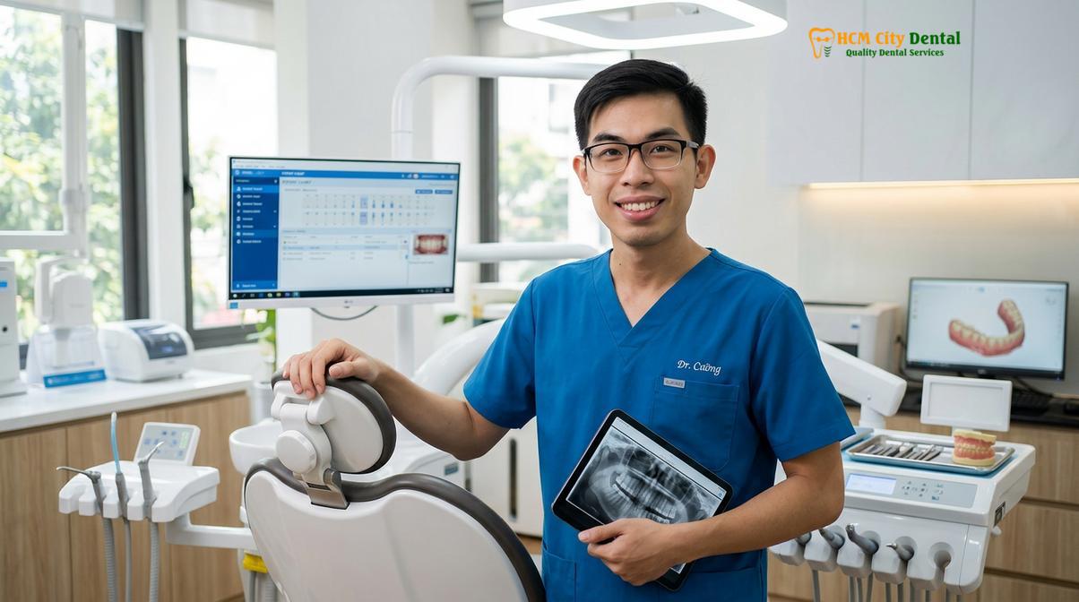
Dr. Nguyen Van Cuong, a leading specialist at HCMC Dental Clinic, emphasizes that proactive periodontal care is inseparable from orthodontic success. He routinely advises patients that while fixed retainers provide excellent stability, they require a strict daily hygiene protocol to prevent localized gingivitis. Regular clinical monitoring ensures that both the bonding composite and the surrounding gum tissues remain in optimal health.
If you are struggling with permanent retainer plaque removal or suspect that your bonded wire needs professional attention, do not delay treatment. Contact HCMC Dental Clinic in Ho Chi Minh City today to schedule a comprehensive periodontal evaluation and professional scaling to protect your orthodontic investment.
References
- Journal of Clinical Orthodontics. Plaque accumulation and periodontal health adjacent to fixed lingual retainers.
- American Journal of Orthodontics and Dentofacial Orthopedics. Long-term effects of orthodontic retention protocols on gingival health.
- Journal of Periodontology. The role of submandibular salivary flow in calculus formation on mandibular incisors.
- European Journal of Orthodontics. Efficacy of interdental cleaning devices in patients with bonded retainers.
- International Journal of Dental Hygiene. Ultrasonic scaling effects on orthodontic composite resins.
- Vietnam Odonto-Stomatology Association (VOSA). National clinical guidelines on post-orthodontic periodontal maintenance.