Teeth grinding in sleep, clinically known as sleep bruxism, is an involuntary movement disorder characterized by clenching and gnashing the jaws during rest. It often leads to enamel wear, jaw pain, and disrupted sleep, requiring professional dental intervention to protect oral structures and alleviate muscular tension.
Clinical Summary:
Sleep bruxism is a complex, multifactorial condition that occurs unconsciously during the sleep cycle, subjecting the teeth and temporomandibular joints (TMJ) to extreme, destructive forces. Unlike conscious daytime clenching, nocturnal grinding is deeply intertwined with central nervous system micro-arousals, airway resistance, and psychological stress. Because patients are asleep, the condition often goes unnoticed until significant dental damage occurs or a bed partner reports the disruptive noise. According to Dr. Nguyen Van Cuong, early clinical diagnosis is critical to preventing irreversible enamel loss, tooth fractures, and chronic myofascial pain. The gold standard for conservative management involves the fabrication of custom occlusal splints (night guards) to dissipate bite forces, alongside complementary therapies addressing the root neurological or respiratory triggers. Comprehensive care requires a multidisciplinary approach, bridging restorative dentistry and sleep medicine to ensure long-term oral health and systemic well-being.
Key Takeaways:
- Sleep bruxism is an involuntary sleep-related movement disorder, not merely a bad habit.
- The jaw muscles can exert up to 250 pounds of force during nocturnal grinding episodes.
- Unmanaged bruxism leads to severe enamel attrition, cracked teeth, and chronic TMJ dysfunction.
- Custom-fitted night guards are the primary defense, protecting teeth from structural damage.
- Underlying issues like obstructive sleep apnea and chronic stress must be evaluated for comprehensive treatment.
To protect crown restorations from chipping, using a custom nightguard for zirconia crowns is highly recommended. Similarly, cosmetic veneers are highly vulnerable to grinding forces, making a bruxism nightguard for veneers a vital preventative measure.
What Is Sleep Bruxism?
Sleep bruxism is a sleep-related movement disorder involving the involuntary clenching and grinding of teeth, distinct from awake bruxism and often linked to micro-arousals during the sleep cycle.
To understand the clinical severity of teeth grinding in sleep, it is essential to differentiate it from normal chewing functions and daytime clenching habits. Sleep bruxism is officially classified by sleep medicine authorities as a sleep-related movement disorder. It is characterized by rhythmic masticatory muscle activity (RMMA) that occurs without the patient’s conscious awareness, typically during the transition between different stages of sleep. While normal chewing generates approximately 20 to 40 pounds of force per square inch, the involuntary contractions during a bruxism episode can generate up to 250 pounds of force on the molars[1]. This immense pressure is sustained over prolonged periods, leading to rapid and devastating consequences for the oral cavity.
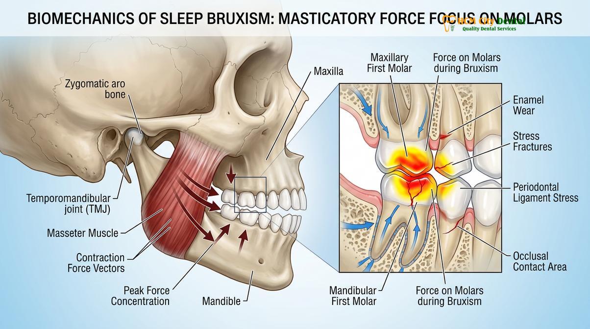
The condition is distinct from awake bruxism, which is often a subconscious reaction to stress, deep concentration, or emotional tension during the day. Awake bruxism primarily involves static clenching of the jaw without the lateral grinding motions. In contrast, nocturnal bruxism involves both forceful clenching and aggressive lateral gnashing, which grinds the upper and lower teeth against each other. This friction acts like sandpaper on the dentition, progressively stripping away the protective outer layer of enamel and exposing the softer, sensitive dentin underneath.
From a physiological standpoint, sleep bruxism is not an isolated dental issue but a complex neurological event. Clinical studies indicate that episodes of grinding are frequently preceded by a sequence of physiological changes known as micro-arousals. During a micro-arousal, the brain briefly shifts from deep sleep to a lighter stage of sleep. This shift is accompanied by a sudden spike in autonomic nervous system activity: the heart rate increases, blood pressure rises, breathing becomes faster, and finally, the masseter and temporalis muscles of the jaw contract violently[2]. Because the patient does not fully wake up, they remain entirely unaware of the event, waking only with the secondary symptoms of the trauma.
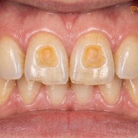
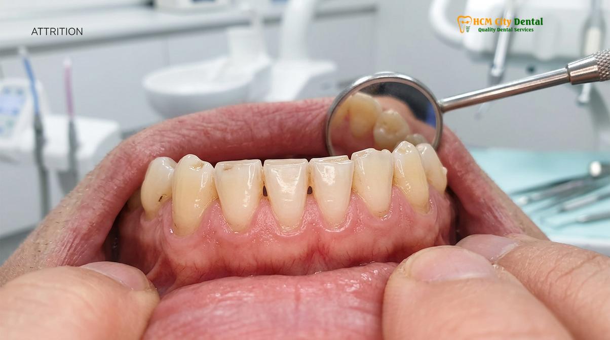
The structural damage caused by this condition is profound. Dentists frequently observe advanced attrition, where the natural cusps and valleys of the teeth are worn completely flat. Over time, the vertical dimension of the bite collapses, leading to a prematurely aged facial appearance. Furthermore, the constant flexing of the teeth under extreme pressure can cause abfraction lesions—V-shaped notches at the gumline where the enamel has literally snapped off due to stress. The periodontal ligaments, which act as shock absorbers holding the teeth in the bone, become inflamed and widened, leading to tooth mobility. Without intervention, the cumulative trauma of sleep bruxism compromises the entire stomatognathic system.
The Neurobiology of Sleep Bruxism
Sleep bruxism originates in the central nervous system, where autonomic cardiac and respiratory fluctuations precede rhythmic masticatory muscle activity.
For decades, the dental and medical communities largely attributed teeth grinding in sleep to morphological factors, such as malocclusion or anatomical discrepancies in the jaw. However, contemporary sleep medicine has fundamentally shifted this paradigm. Current clinical consensus establishes that sleep bruxism is primarily a central nervous system (CNS) phenomenon. The genesis of this involuntary movement lies deep within the brain’s complex regulatory systems, specifically involving the autonomic nervous system and the basal ganglia, which are responsible for motor control.
The neurobiological cascade leading to a bruxism episode is intricate. It begins with a shift in the autonomic nervous system from a state of parasympathetic dominance (rest and digest) to sympathetic dominance (fight or flight). This shift is characterized by a sudden, albeit brief, surge in autonomic cardiac activity. Heart rate variability alters, and respiratory patterns may become irregular. These physiological changes occur entirely beneath the threshold of conscious awareness but are highly detectable via polysomnographic monitoring. The basal ganglia, which modulate voluntary and involuntary movements, receive these autonomic signals and subsequently misfire, sending aberrant motor commands to the trigeminal motor nucleus. This nucleus controls the muscles of mastication—primarily the masseter, temporalis, and medial pterygoid muscles.
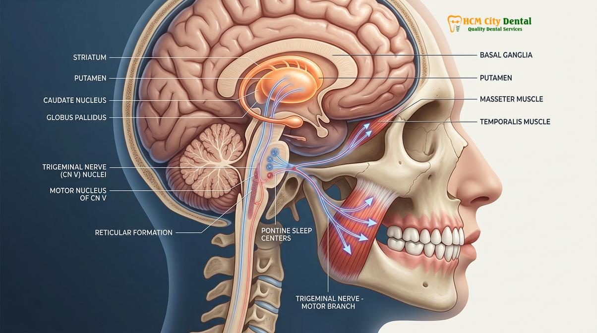
Furthermore, neurotransmitter imbalances play a critical role in the pathophysiology of this condition. Research indicates that the dopaminergic and serotonergic systems are heavily implicated in the regulation of jaw movements during sleep. Alterations in dopamine receptor sensitivity or availability in the striatum can disrupt the smooth execution of motor pathways, leading to the rhythmic or sustained contractions characteristic of parasomnia teeth grinding. This neurochemical link also explains why certain pharmacological agents, particularly selective serotonin reuptake inhibitors (SSRIs) used for depression and anxiety, can inadvertently induce or exacerbate sleep bruxism as a secondary side effect. The intricate dance of neurotransmitters dictates the tone of the masticatory muscles, and any disruption can lead to pathological grinding forces [1].
Psychological factors, particularly chronic stress and anxiety, act as potent modulators of this neurobiological framework. Stress does not directly cause the mechanical grinding, but it significantly amplifies the underlying central nervous system excitability. Elevated levels of cortisol and adrenaline keep the brain in a state of hyper-vigilance, lowering the threshold required for motor activation during sleep. Consequently, patients experiencing high psychosocial stress exhibit a markedly higher frequency and intensity of bruxism episodes. Understanding this central origin is crucial for effective comprehensive temporomandibular joint therapy, as treatments must address both the mechanical damage and, when possible, the neurological triggers.
Why You Grind Without Knowing
The exact etiology of nocturnal grinding is multifactorial, combining central nervous system arousals, psychological stress, airway resistance, and certain medications that trigger involuntary jaw muscle contractions.
The mystery of why individuals engage in grinding teeth at night has been the subject of extensive medical research. Historically, the dental profession believed that bruxism was caused primarily by malocclusion—a misaligned bite that the body was subconsciously trying to “grind away” to find a comfortable resting position. However, modern clinical evidence has shifted the paradigm. We now understand that sleep bruxism is centrally mediated, meaning it originates in the brain rather than the mouth. The etiology is highly multifactorial, requiring a comprehensive evaluation of the patient’s overall health, lifestyle, and psychological state.
One of the most significant discoveries in recent years is the strong correlation between sleep bruxism and sleep-disordered breathing, particularly Obstructive Sleep Apnea (OSA). When a patient with OSA falls asleep, the soft tissues at the back of the throat collapse, obstructing the airway and causing oxygen levels in the blood to drop. The brain senses this dangerous hypoxia and triggers a survival response—a micro-arousal. As part of this arousal, the brain commands the jaw muscles to contract and thrust the lower jaw forward in a desperate attempt to open the airway[3]. This forward thrusting and clenching manifest clinically as severe teeth grinding. In these cases, bruxism is actually a compensatory mechanism to keep the patient breathing.
“We can no longer view nocturnal bruxism purely as a dental problem. When a patient presents with severely worn dentition, our first clinical responsibility is to evaluate their airway. Treating the teeth without addressing underlying sleep apnea is like treating the smoke while ignoring the fire.”
Psychological factors also play a massive role in the pathogenesis of the condition. Chronic stress, anxiety, and emotional tension elevate the body’s baseline levels of cortisol and adrenaline. This heightened state of sympathetic nervous system arousal prevents the brain from entering deep, restorative sleep stages. Instead, the brain hovers in lighter sleep stages where micro-arousals—and consequently, bruxism episodes—are far more likely to occur. Patients often report a direct correlation between highly stressful periods in their personal or professional lives and an increase in morning jaw pain and dental sensitivity.
Furthermore, exogenous substances and medications are potent triggers for involuntary jaw movements. The consumption of central nervous system stimulants, such as caffeine and nicotine, particularly in the evening, disrupts sleep architecture and increases muscle hyperactivity. Alcohol, while initially acting as a depressant that helps initiate sleep, causes significant sleep fragmentation and airway relaxation during the second half of the night, leading to a sharp increase in grinding episodes. Additionally, certain prescription medications, most notably Selective Serotonin Reuptake Inhibitors (SSRIs) used to treat depression and anxiety, have a well-documented side effect of inducing secondary bruxism by altering dopamine pathways in the brain.
How Micro-Arousals Trigger Involuntary Jaw Activity
Sleep micro-arousals are brief, unconscious awakenings accompanied by a sudden increase in heart rate and brain activity, directly triggering jaw muscle contractions.
To fully comprehend the mechanics of teeth grinding in sleep, one must examine the phenomenon of sleep micro-arousals. These are transient, subtle shifts in the electroencephalogram (EEG) that last anywhere from three to fifteen seconds. Unlike full awakenings, the individual remains entirely unaware of these events and has no memory of them upon waking. However, from a physiological standpoint, a micro-arousal represents a massive, sudden storm of neurological and cardiovascular activity. They are a natural part of the sleep architecture, occurring multiple times per hour in healthy individuals to help the body shift positions or respond to minor environmental stimuli. Yet, in patients with sleep bruxism, these arousals are the primary catalyst for destructive jaw activity.
The temporal sequence of a bruxism episode triggered by a micro-arousal is highly predictable and has been extensively mapped in clinical sleep laboratories. The sequence initiates approximately four to eight seconds before any actual jaw movement occurs. The first measurable event is a sharp increase in sympathetic cardiac activity, manifesting as a sudden spike in heart rate. Following this cardiovascular surge, the brain’s EEG patterns shift from slow, synchronized waves to faster, desynchronized frequencies, indicating cortical arousal. Finally, this wave of neurological excitation reaches the brainstem, specifically the trigeminal motor nucleus, resulting in rhythmic masticatory muscle activity (RMMA) or sustained nocturnal clenching [2].
“The cascade of physiological events during a micro-arousal demonstrates that sleep bruxism is a secondary response to central nervous system shifts, rather than a localized dental issue. It is a motor manifestation of a brain attempting to transition between sleep states.”
This precise sequence highlights that the grinding is the final step in a complex chain reaction. The frequency of these sleep micro-arousals can be drastically increased by various external and internal factors. Obstructive sleep apnea (OSA) is one of the most significant contributors. When a patient’s airway partially or fully collapses during sleep, oxygen levels drop, prompting the brain to initiate a powerful micro-arousal to force the airway open and resume breathing. This survival mechanism frequently triggers a secondary bruxism episode. In fact, the jaw thrusting associated with grinding may be an unconscious physiological attempt to bring the mandible forward and open the collapsed airway. Therefore, addressing underlying respiratory disturbances is often a critical component of specialized jaw joint management.
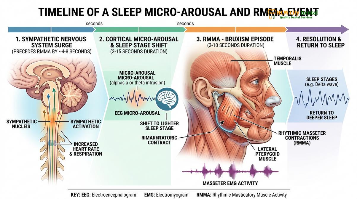
Other factors that increase the frequency of micro-arousals include the consumption of central nervous system stimulants close to bedtime. Caffeine, nicotine, and certain medications can fragment sleep architecture, leading to a higher density of arousals and, consequently, more frequent episodes of grinding. Alcohol, while initially acting as a depressant that aids in falling asleep, causes significant sleep fragmentation and rebound arousal during the second half of the night as it is metabolized. This rebound effect is strongly correlated with intense episodes of nocturnal clenching.
Sleep Stages and Bruxism: When Does Grinding Occur?
The majority of sleep bruxism episodes manifest during light non-rapid eye movement (NREM) sleep, specifically stages N1 and N2, rather than deep sleep.
Human sleep is not a uniform state; it is a dynamic process characterized by distinct stages that cycle throughout the night. These stages are broadly categorized into non-rapid eye movement (NREM) sleep and rapid eye movement (REM) sleep. NREM sleep is further subdivided into three stages: N1 (light sleep), N2 (intermediate sleep), and N3 (deep, slow-wave sleep). The occurrence of teeth grinding in sleep is not randomly distributed across these stages; it exhibits a strong predilection for specific phases of the sleep cycle, which provides further insight into its neurobiological origins.
Clinical polysomnography reveals that approximately 80% of all sleep bruxism episodes occur during the lighter stages of NREM sleep, specifically stages N1 and N2. Stage N1 is the transitional phase between wakefulness and sleep, characterized by a slowing of brain waves and muscle relaxation. Stage N2 constitutes the largest portion of the total sleep time and is marked by specific EEG features known as sleep spindles and K-complexes. It is during these stages, particularly in the moments leading up to a transition to another sleep stage or a brief awakening, that the brain is most susceptible to the micro-arousals that trigger rhythmic masticatory muscle activity [3].
Conversely, bruxism episodes are relatively rare during stage N3, the deep, restorative phase of sleep. During N3, the brain exhibits high-amplitude, slow delta waves, and the threshold for arousal is significantly higher. The autonomic nervous system is highly stable, and sympathetic activity is at its lowest, making the neurobiological cascade required for a grinding episode highly unlikely to initiate.
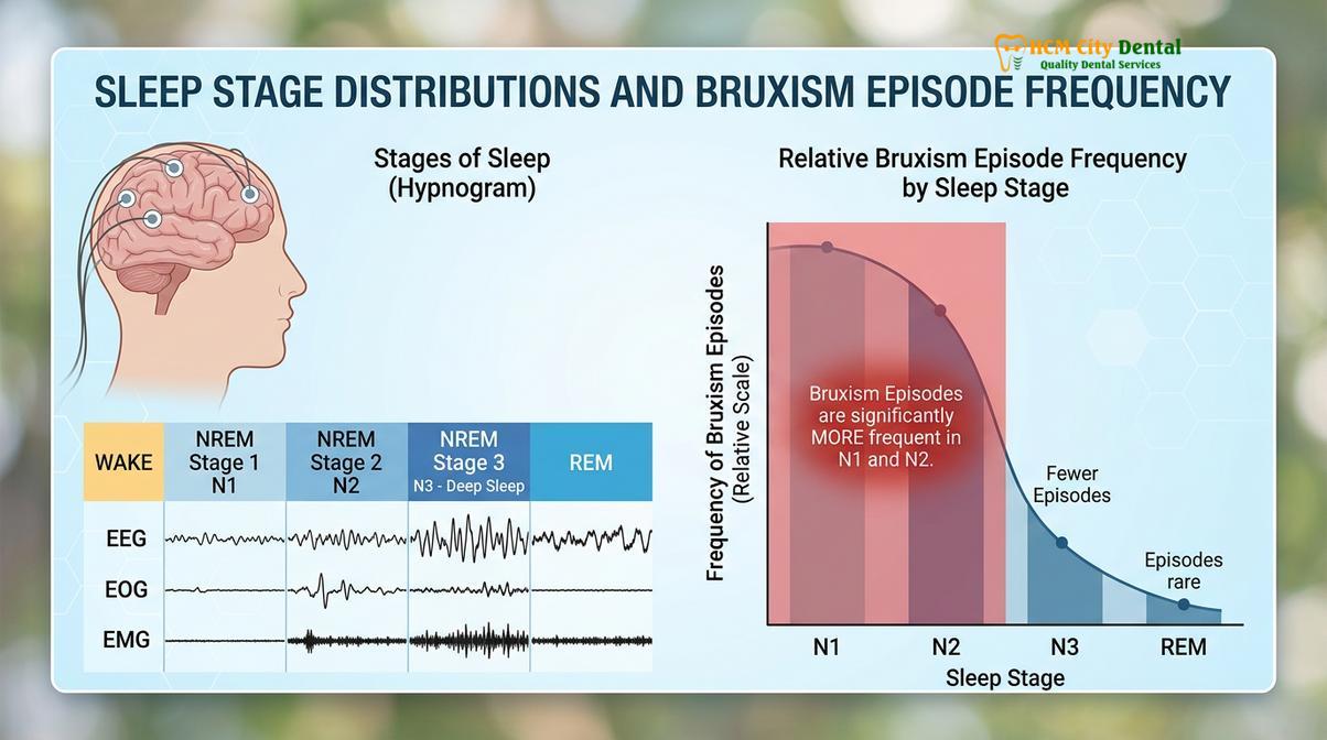
The relationship between REM sleep and bruxism is particularly intriguing. REM sleep is the stage associated with vivid dreaming. Under normal physiological conditions, the brain induces a state of profound muscle atonia (paralysis) during REM sleep to prevent individuals from physically acting out their dreams. Because of this protective paralysis, motor activity, including jaw clenching, is typically suppressed. However, in a small subset of patients, particularly those with REM sleep behavior disorder or severe neurological conditions, this atonia is incomplete or absent. In these rare cases, bruxism can occur during REM sleep and is often associated with intense, sometimes violent, jaw movements and a higher risk of severe dental trauma.
| Sleep Stage | Characteristics | Bruxism Frequency | Physiological State |
|---|---|---|---|
| NREM Stage 1 (N1) | Transition from wakefulness to light sleep | High | High susceptibility to micro-arousals; unstable autonomic tone. |
| NREM Stage 2 (N2) | Intermediate sleep; presence of sleep spindles | Very High | Accounts for the majority of RMMA episodes; frequent state transitions. |
| NREM Stage 3 (N3) | Deep, slow-wave sleep; restorative phase | Low | High arousal threshold; stable parasympathetic dominance. |
| REM Sleep | Vivid dreaming; rapid eye movements | Very Low (typically) | Profound muscle atonia usually prevents motor activity, unless pathology exists. |
How Partners Detect Night Grinding
Bed partners are typically the first to identify sleep bruxism by hearing distinct clicking, gnashing, or scraping sounds, or by noticing the patient’s restless sleep patterns and rhythmic jaw movements.
Because the patient is unconscious during the act, the diagnosis of nighttime teeth grinding often begins with a report from a distressed bed partner. The auditory manifestations of bruxism can be startlingly loud and highly disruptive to anyone sharing the sleeping environment. Partners frequently describe the sound as a harsh scraping, similar to stones rubbing together, or a sharp clicking and popping of the jaw joints. These sounds are generated by the immense friction of the upper and lower enamel surfaces sliding forcefully against one another under the power of the masseter muscles.
The frequency and intensity of these episodes vary throughout the night. Clinical sleep studies show that bruxism events occur most frequently during non-rapid eye movement (NREM) sleep, particularly in stages 1 and 2, which are the lighter stages of sleep. An episode typically lasts anywhere from a few seconds to over a minute, and a severe bruxer may experience dozens of these episodes per hour. The partner may also notice physical signs of distress, such as the patient tossing and turning, breathing heavily, or exhibiting rhythmic, pulsing movements of the jaw and temples.
Clinical Warning: If your bed partner informs you that you are grinding your teeth, do not dismiss it, even if you feel no pain upon waking. The absence of immediate pain does not mean damage isn’t occurring. Enamel wear is a slow, progressive process, and by the time tooth sensitivity or jaw pain becomes noticeable, irreversible structural damage has already taken place.
While the partner hears the noise, the patient experiences the silent aftermath upon waking. The most common morning symptoms include a dull, constant headache radiating from the temples, which is caused by the exhaustion of the temporalis muscle. Patients also frequently report a feeling of tightness or fatigue in the jaw, difficulty opening the mouth fully to eat breakfast, and unexplained tooth sensitivity to hot or cold temperatures. In severe cases, the temporomandibular joint (TMJ) may feel locked or produce a loud popping sound when the mouth is opened. The discrepancy between the partner’s auditory experience and the patient’s physical symptoms is a hallmark of the condition and a primary reason why clinical evaluation is essential.
Sleep Study and Diagnosis
Diagnosing sleep bruxism involves a comprehensive clinical dental examination to assess enamel wear, often supplemented by polysomnography (a sleep study) to monitor jaw muscle activity and rule out sleep apnea.
The formal diagnosis of sleep bruxism requires a meticulous clinical approach, combining intraoral examinations, patient history, and, when necessary, advanced sleep medicine diagnostics. At HCMC Dental Clinic in Ho Chi Minh City, the diagnostic protocol begins with a comprehensive evaluation of the hard and soft tissues of the mouth. Dr. Nguyen Van Cuong emphasizes that the teeth serve as a historical record of the forces placed upon them. The clinical signs of chronic grinding are unmistakable to a trained professional.
During the examination, the dentist looks for specific pathognomonic signs. The most obvious is severe attrition—the flattening of the biting surfaces of the teeth, often exposing the yellowish dentin layer beneath the translucent enamel. The dentist will also check for abfraction lesions at the gumline and inspect the soft tissues. Chronic bruxers frequently develop a “linea alba,” a raised, white line of callused tissue on the inside of the cheeks caused by constant negative pressure and friction. The tongue may also appear scalloped along its edges where it has been pressed forcefully against the lingual surfaces of the teeth. Furthermore, palpation of the masseter and temporalis muscles often reveals hypertrophy (enlargement) and severe tenderness, indicating chronic overuse.
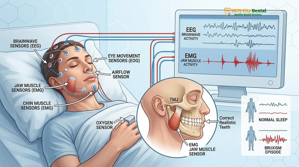
While a clinical dental exam can confirm the presence of damage, it cannot always determine the frequency, intensity, or underlying cause of the nocturnal events. For a definitive diagnosis, especially when sleep-disordered breathing is suspected, a polysomnography (PSG) or sleep study is the gold standard[4]. A PSG is conducted overnight in a controlled clinical environment or via an advanced home sleep testing device.
During a sleep study, multiple physiological parameters are monitored simultaneously. Electroencephalography (EEG) records brain wave activity to identify micro-arousals and sleep stage transitions. Electrocardiography (ECG) monitors heart rate spikes. Most importantly for bruxism, electromyography (EMG) sensors are placed directly over the masseter and temporalis muscles to record the exact frequency, duration, and intensity of jaw muscle contractions. Respiratory sensors also monitor airflow and oxygen saturation to detect episodes of sleep apnea.
| Diagnostic Method | Key Indicators Monitored | Clinical Findings |
|---|---|---|
| Clinical Dental Exam | Enamel wear, soft tissue trauma, muscle hypertrophy | Flattened cusps, exposed dentin, linea alba, scalloped tongue, enlarged masseters. |
| Patient/Partner History | Auditory reports, morning symptoms | Reports of loud grinding noises, morning headaches, jaw stiffness, TMJ pain. |
| Polysomnography (PSG) | Brain waves, muscle activity, respiration | Rhythmic masticatory muscle activity (RMMA) on EMG, micro-arousals on EEG, airway resistance. |
By correlating the EMG data with the respiratory and brain wave data, clinicians can determine whether the bruxism is a primary movement disorder or a secondary response to an airway collapse. This distinction is critical, as it dictates the entire trajectory of the treatment plan. Treating secondary bruxism caused by sleep apnea with a standard dental night guard alone can sometimes worsen the respiratory condition by pushing the jaw backward, highlighting the need for precise, data-driven diagnostics.
Diagnostic Criteria for Nocturnal Teeth Grinding
Diagnosis relies on clinical evidence of abnormal tooth wear, patient reports of morning jaw stiffness, and polysomnography to confirm sleep-related masticatory muscle activity.
Accurately diagnosing teeth grinding in sleep presents a unique clinical challenge because the patient is, by definition, unconscious during the act. Self-reporting is notoriously unreliable; many individuals grind their teeth for years without any conscious awareness until significant damage has occurred or a sleep partner brings the disruptive noise to their attention. Therefore, a definitive diagnosis requires a multi-faceted approach, combining thorough clinical examination, detailed patient history, and, in complex cases, objective instrumental sleep analysis.
The cornerstone of clinical diagnosis in a dental setting is the identification of pathognomonic signs within the oral cavity. The most obvious indicator is abnormal dental attrition—the mechanical wear of the biting surfaces of the teeth. Unlike normal physiological wear, which occurs slowly over decades, bruxism-induced attrition is rapid and often presents as flattened, shiny facets on the cusps of molars and the incisal edges of anterior teeth. In severe cases, the enamel may be completely worn away, exposing the underlying yellow dentin, which significantly increases tooth sensitivity and the risk of pulpal necrosis. Other intraoral signs include abfraction lesions (notches at the gumline caused by flexural forces), a scalloped appearance on the lateral borders of the tongue, and linea alba (a raised white line on the inner cheek mucosa corresponding to the occlusal plane) [4].
Beyond the teeth themselves, the masticatory musculature and the temporomandibular joints (TMJ) provide critical diagnostic clues. Patients frequently present with masseter muscle hypertrophy—an enlargement of the jaw muscles due to constant nocturnal overexertion, giving the face a widened, square appearance. Palpation of the masseter and temporalis muscles often elicits tenderness or referred pain patterns. Patients typically report a constellation of morning symptoms, including a dull, aching headache localized to the temples, jaw stiffness that makes it difficult to open the mouth fully upon waking, and unexplained earache-like pain that originates from the inflamed TMJ.
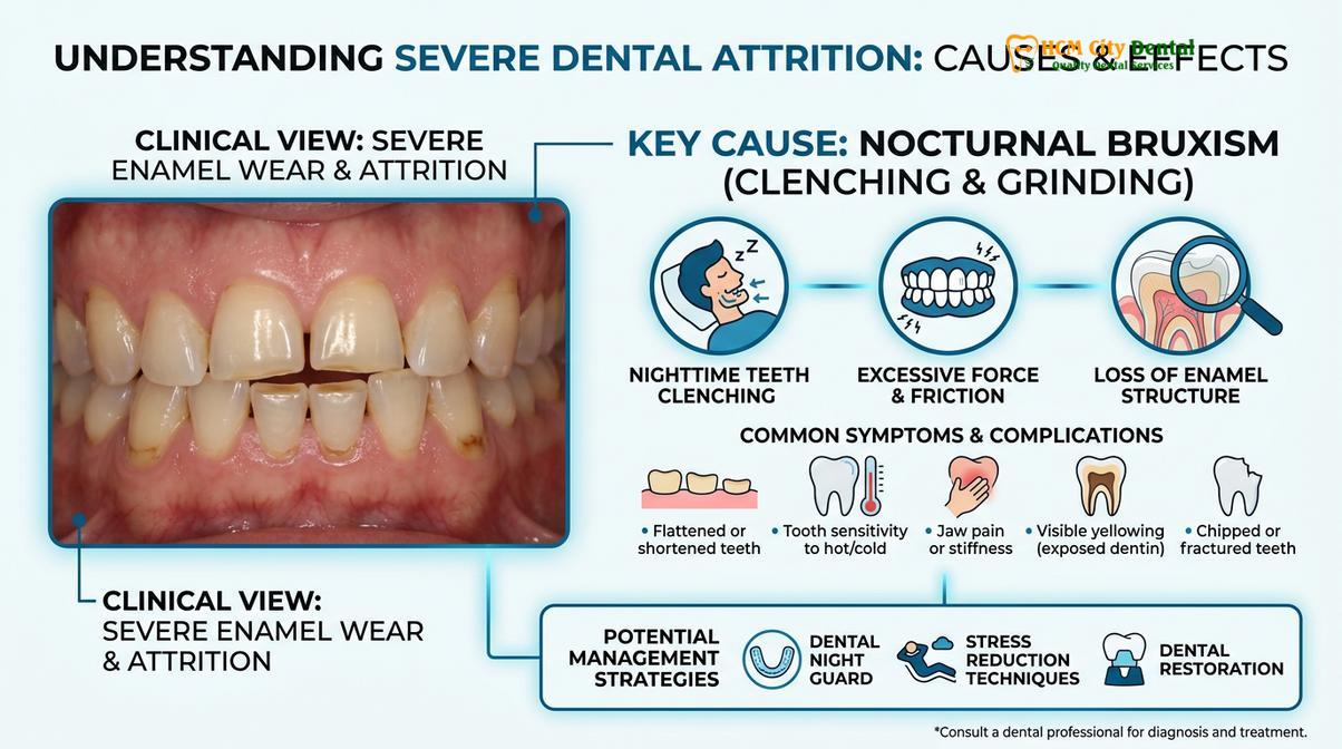
While clinical signs are often sufficient for a presumptive diagnosis and the initiation of protective therapy like custom-fitted occlusal splints, a definitive diagnosis according to the International Classification of Sleep Disorders requires polysomnography (PSG). A sleep study involves an overnight stay in a clinical laboratory where various physiological parameters are continuously monitored. For the specific diagnosis of sleep bruxism, the PSG must include audio-video recording to capture the grinding sounds and electromyography (EMG) of the masseter and temporalis muscles to quantify the frequency and intensity of the rhythmic masticatory muscle activity. This objective data is crucial for differentiating bruxism from other sleep-related movement disorders, such as facio-mandibular myoclonus or sleep-related epilepsy.
Night Guard as First-Line Treatment
Custom-fitted occlusal splints are the primary clinical intervention for sleep bruxism, designed to protect tooth enamel, distribute bite forces evenly, and reduce strain on the temporomandibular joint.
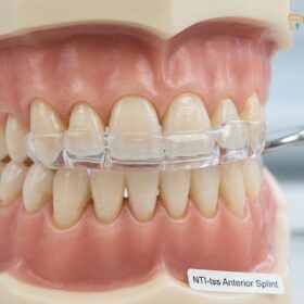
Once a diagnosis of primary sleep bruxism is established, the immediate clinical priority is to halt the destruction of the dentition and relieve the strain on the neuromuscular system. The universally accepted first-line treatment is the fabrication and continuous use of custom occlusal splints, commonly referred to as night guards. These intraoral appliances act as a physical barrier between the upper and lower teeth, absorbing the immense forces generated during a grinding episode and preventing the enamel surfaces from grinding against one another.
It is crucial to distinguish between professionally fabricated clinical night guards and over-the-counter (OTC) “boil-and-bite” mouthguards found in pharmacies. OTC guards are typically made of soft, compressible materials. While they may offer temporary protection, clinical studies have shown that the soft, rubbery texture can actually stimulate the brain to chew and clench more forcefully, exacerbating muscle fatigue and TMJ pain. Furthermore, OTC guards cannot be balanced to the patient’s specific bite, leading to uneven pressure distribution that can shift the teeth over time.
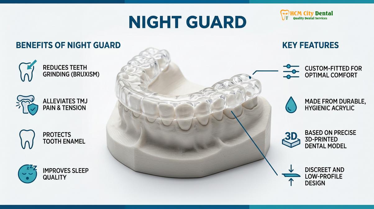
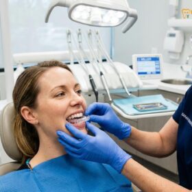
In contrast, professional protective dental appliances are precision-engineered medical devices. The fabrication process begins with highly accurate digital impressions of the patient’s teeth. The dentist then determines the optimal resting position of the jaw—often referred to as centric relation—where the TMJ is fully seated and the muscles are relaxed. The night guard is then custom-milled or 3D-printed from dense, medical-grade acrylic resin.
The occlusal (biting) surface of a professional night guard is meticulously adjusted by the dentist to ensure that all teeth contact the acrylic evenly and simultaneously. When the patient attempts to grind their teeth at night, the smooth, hard surface of the acrylic allows the opposing teeth to glide effortlessly without catching or locking. This lack of friction not only protects the enamel but also sends a biofeedback signal to the brain, often resulting in a significant reduction in the intensity of the muscle contractions[5].
Clinical Case Study: A 34-year-old patient presented to HCMC Dental Clinic in Ho Chi Minh City complaining of severe morning headaches and increasing tooth sensitivity. Clinical examination revealed moderate enamel attrition and hypertrophic masseter muscles. The patient was fitted with a custom, hard-acrylic maxillary occlusal splint. At the 4-week follow-up, the patient reported a complete cessation of morning headaches and a significant reduction in jaw tension. The night guard showed visible scuff marks, confirming that the destructive forces were successfully redirected from the teeth to the replaceable acrylic appliance.
Depending on the severity of the bruxism and the patient’s specific anatomy, the dentist may prescribe different types of splints. A hard acrylic splint is the standard for severe grinders, offering maximum durability. A dual-laminate splint, which features a hard outer shell for durability and a soft inner lining for comfort, is often prescribed for moderate bruxers. Regardless of the material, consistent nightly wear is mandatory to achieve long-term clinical success and prevent the need for extensive restorative dentistry, such as full-mouth crowns or implants, later in life.
Clinical Consequences of Parasomnia Teeth Grinding
Unmanaged sleep bruxism exerts extreme pathological forces on the dentition, leading to accelerated enamel attrition, structural tooth fractures, and chronic temporomandibular joint dysfunction.
The human masticatory system is designed to generate significant force for the purpose of breaking down food. During normal chewing, the teeth are in contact for only a few minutes per day, and the forces applied are carefully regulated by neurological feedback loops from the periodontal ligaments. However, during an episode of parasomnia teeth grinding, these protective reflexes are bypassed. The forces exerted during nocturnal clenching can be up to six times greater than maximum conscious biting force, sometimes exceeding 250 pounds of pressure per square inch. When these extreme, sustained forces are applied night after night, the clinical consequences are devastating and progressive.
The most immediate and visible casualty is the tooth structure itself. Enamel, though the hardest substance in the human body, is brittle and cannot withstand constant lateral grinding forces. Micro-fractures develop within the crystalline structure, eventually leading to macroscopic chipping and catastrophic tooth fractures. Dental restorations, including composite fillings, ceramic crowns, and porcelain veneers, are particularly vulnerable. The sheer mechanical stress can cause these restorations to debond, fracture, or completely shatter, necessitating extensive and costly prosthodontic rehabilitation. Furthermore, the constant pressure widens the periodontal ligament space, leading to increased tooth mobility and exacerbating underlying periodontal disease.
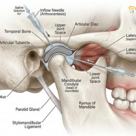
Beyond the dentition, the temporomandibular joints bear a massive burden. The TMJ is a complex, dual-hinging joint cushioned by a fibrocartilaginous disc. Chronic grinding compresses this joint space, squeezing out the synovial fluid that provides lubrication and nutrition to the tissues. Over time, this immense pressure can cause the articular disc to become displaced or perforated, leading to painful clicking, popping, or grating sounds (crepitus) upon opening and closing the mouth. In advanced stages, the constant trauma initiates degenerative joint disease (osteoarthritis) within the TMJ, resulting in chronic, debilitating facial pain and severely restricted jaw movement. This cascade of destruction underscores the necessity of early clinical TMJ evaluation before irreversible joint damage occurs.
Evidence-Based Treatment Workflows
Management focuses on mitigating damage through custom occlusal splints, pharmacological interventions like botulinum toxin, and addressing underlying sleep disordered breathing.
Because sleep bruxism is primarily a central nervous system phenomenon, there is currently no definitive “cure” that permanently stops the neurological triggers. Therefore, clinical treatment workflows are fundamentally focused on management: protecting the stomatognathic system from mechanical damage, alleviating muscle and joint pain, and reducing the frequency of episodes by addressing modifiable risk factors. A multidisciplinary approach is often required to achieve optimal patient outcomes.
The absolute gold standard and first line of defense in the management of nocturnal grinding is the fabrication of a custom occlusal appliance, commonly referred to as a nightguard or stabilization splint. Unlike soft, over-the-counter options, professional nightguards are meticulously fabricated from hard, durable acrylic resin. They are designed based on precise impressions or digital scans of the patient’s dentition. The critical feature of a proper clinical splint is its occlusal scheme; it provides a perfectly balanced, frictionless surface for the opposing teeth to glide against. This not only protects the enamel from direct wear but also helps to deprogram the masticatory muscles, reducing their hyperactive tone and alleviating strain on the temporomandibular joints [5].
In cases where mechanical protection via a splint is insufficient to control severe muscle pain and hypertrophy, pharmacological interventions may be considered. The off-label use of botulinum toxin type A (Botox) has gained significant traction in dental medicine for the management of refractory bruxism. By injecting precise doses of the neurotoxin directly into the masseter and temporalis muscles, the release of acetylcholine at the neuromuscular junction is temporarily blocked. This partially paralyzes the muscle, significantly reducing the force of the involuntary contractions without compromising normal chewing function, speech, or facial expression. The effects typically last for three to four months, providing profound relief for patients suffering from chronic facial pain.
Furthermore, addressing the root causes of sleep micro-arousals is a critical component of the treatment workflow. If polysomnography reveals concurrent obstructive sleep apnea, treating the airway collapse with Continuous Positive Airway Pressure (CPAP) therapy or a mandibular advancement device often leads to a dramatic reduction or complete resolution of the bruxism episodes. Additionally, cognitive behavioral therapy (CBT), sleep hygiene optimization, and stress management techniques are highly recommended to lower the overall sympathetic nervous system tone, thereby reducing the neurological excitability that triggers the grinding.
Complementary Treatments for Sleep Bruxism
Beyond occlusal splints, comprehensive management may include physical therapy, stress reduction techniques, muscle relaxants, or targeted botulinum toxin injections to decrease masseter muscle hyperactivity.
While a custom night guard is highly effective at protecting the teeth and managing the symptoms of sleep bruxism, it does not cure the underlying neurological or psychological triggers. Therefore, a comprehensive, multidisciplinary treatment plan often incorporates complementary therapies aimed at reducing the frequency and intensity of the grinding episodes themselves.
For patients whose bruxism is heavily driven by psychological stress and anxiety, Cognitive Behavioral Therapy (CBT) and stress management techniques are highly recommended. CBT helps patients identify and reframe the subconscious stressors that elevate their sympathetic nervous system activity. Incorporating relaxation protocols before bed—such as deep breathing exercises, progressive muscle relaxation, or meditation—can help lower cortisol levels and promote a smoother transition into deep, restorative sleep stages, thereby reducing the likelihood of micro-arousals.
“Managing severe bruxism requires us to look beyond the teeth. By integrating stress reduction protocols and physical therapy with our dental interventions, we treat the whole patient, significantly improving their overall quality of life and sleep architecture.”
Physical therapy is another vital component of comprehensive care, particularly for patients suffering from secondary temporomandibular joint dysfunction (TMD). A specialized physical therapist can teach the patient targeted stretching and massage techniques to release lactic acid buildup and tension in the masseter, temporalis, and lateral pterygoid muscles. Applying moist heat to the jaw before sleep can also increase blood flow and relax the musculature.
In severe, refractory cases where conservative treatments fail to provide adequate relief, pharmacological interventions may be considered. Short-term use of muscle relaxants taken before bed can help break the cycle of severe muscle spasms. More recently, targeted injections of Botulinum Toxin (Botox) into the masseter and temporalis muscles have shown remarkable clinical efficacy. Botox works by temporarily blocking the release of acetylcholine at the neuromuscular junction, effectively weakening the muscle’s ability to contract forcefully without interfering with normal chewing, speaking, or swallowing functions. This treatment provides profound relief from myofascial pain and significantly reduces the force exerted on the teeth, though the injections must be repeated every few months to maintain the effect.
When to See a Doctor
While occasional teeth grinding may not require immediate medical intervention, chronic sleep bruxism is a progressive condition that demands professional evaluation. You should schedule a comprehensive clinical examination with a dental professional if you experience any of the following red-flag symptoms:
- Severe Morning Pain: Waking up consistently with dull headaches, earaches, or intense pain in the jaw muscles and TMJ.
- Visible Tooth Damage: Noticing that your teeth appear shorter, flattened, chipped, or if you experience sudden, unexplained sensitivity to hot and cold foods.
- Locked Jaw: Experiencing difficulty opening or closing your mouth fully, accompanied by loud clicking, popping, or grating sounds in the jaw joint.
- Partner Complaints: Your bed partner frequently complains about loud grinding, scraping, or gnashing noises disrupting their sleep.
- Daytime Fatigue: Experiencing excessive daytime sleepiness, loud snoring, or waking up gasping for air, which strongly indicates concurrent obstructive sleep apnea.
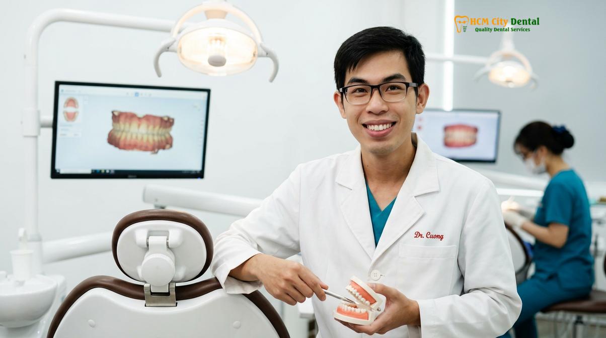
Dr. Cuong strongly advises that patients do not attempt to self-diagnose or rely solely on over-the-counter remedies. A proper clinical assessment is vital to rule out serious underlying conditions like sleep apnea and to design a personalized treatment protocol that protects your oral health and improves your overall well-being.
Frequently Asked Questions
Can you stop grinding teeth naturally?
While you cannot always stop it completely, natural methods like stress reduction, improved sleep hygiene, and avoiding caffeine before bed can significantly reduce the frequency and severity of sleep bruxism episodes. Clinical management is often still required to protect the teeth from long-term structural damage. Techniques such as progressive muscle relaxation, warm compresses on the jaw, and establishing a calming pre-sleep routine help lower the sympathetic nervous system arousal that triggers involuntary clenching.
Does sleeping position affect grinding?
Yes, sleeping on your back (supine position) can exacerbate airway resistance and sleep apnea, which are strong triggers for nocturnal bruxism. Switching to a side-sleeping position helps keep the airway open and may reduce the incidence of involuntary jaw clenching. When the airway is clear, the brain experiences fewer micro-arousals, thereby decreasing the neurological signals that command the jaw muscles to contract and grind.
Will a night guard cure sleep bruxism?
A night guard will not cure the underlying neurological triggers of sleep bruxism, but it is the most effective way to manage the condition. It protects tooth enamel from wear, distributes bite forces evenly, and reduces strain on the temporomandibular joint. By providing a smooth surface for the teeth to glide against, a custom splint prevents the destructive friction and often reduces the intensity of the muscle contractions through biofeedback.
How loud is teeth grinding at night?
The sound can range from completely silent clenching to loud, harsh scraping or gnashing noises that easily wake a bed partner. The volume depends on the friction between the upper and lower teeth and the force of the masseter muscles. Some patients only clench statically, producing no sound but still causing immense internal pressure on the teeth and joints, while others exhibit aggressive lateral movements that produce the characteristic grinding noise.
Should I do a sleep study for bruxism?
A sleep study (polysomnography) is highly recommended if your dentist suspects your bruxism is linked to obstructive sleep apnea, or if you experience severe daytime fatigue, loud snoring, and refractory jaw pain despite using a protective dental splint. The study monitors brain waves, muscle activity, and respiratory effort, allowing clinicians to determine if the grinding is a primary movement disorder or a secondary survival response to airway collapse.
Is sleep grinding a sleep disorder?
Yes, sleep grinding is officially classified as a sleep-related movement disorder by medical diagnostic manuals. It is characterized by involuntary rhythmic or sustained masticatory muscle activity during sleep, often linked to central nervous system arousals rather than simple anatomical jaw misalignment. This classification highlights that the condition originates in the brain’s sleep architecture, requiring a comprehensive diagnostic approach beyond just looking at the teeth.
Does stress make sleep bruxism worse?
Yes, psychological stress and anxiety significantly exacerbate sleep bruxism by increasing sympathetic nervous system activity. Elevated stress hormones lead to a higher frequency of sleep micro-arousals, which directly trigger the involuntary jaw muscle contractions associated with nocturnal grinding. While stress is not the sole cause, it acts as a powerful amplifier, making episodes more frequent and the biting forces much more intense during periods of high anxiety.
Can lifestyle changes stop teeth grinding at night?
While lifestyle changes cannot always completely stop teeth grinding at night, they can significantly reduce its frequency and severity. Implementing rigorous sleep hygiene, reducing alcohol and caffeine intake, and practicing stress-reduction techniques help lower the central nervous system arousals that trigger grinding episodes. Establishing a calming pre-sleep routine and maintaining a consistent sleep schedule are foundational steps in managing the neurological triggers of the condition.
How long does a custom nightguard last for heavy grinders?
A custom-fabricated hard acrylic nightguard typically lasts between two to five years for patients with severe bruxism. The longevity depends heavily on the intensity of the nocturnal clenching forces, the specific acrylic material used, and how meticulously the patient maintains and cleans the appliance. Regular clinical check-ups are necessary to adjust the occlusal surface of the guard as it wears down, ensuring it continues to provide balanced protection for the jaw joints.
Can sleep apnea cause nocturnal teeth grinding?
Yes, obstructive sleep apnea is a major risk factor and frequent co-occurring condition with nocturnal teeth grinding. When the airway collapses during sleep, the brain initiates a micro-arousal to resume breathing, and this sudden neurological shift frequently triggers a secondary bruxism episode to help open the airway. Treating the underlying sleep apnea with CPAP therapy or an oral appliance often results in a dramatic reduction in grinding activity.
References
- Journal of Clinical Sleep Medicine. Pathophysiology and management of sleep bruxism. (2021).
- American Academy of Sleep Medicine. International Classification of Sleep Disorders. (2014).
- Journal of Oral Rehabilitation. The relationship between sleep bruxism and obstructive sleep apnea. (2019).
- International Journal of Prosthodontics. Efficacy of occlusal splints in the management of bruxism. (2020).
- Journal of the American Dental Association. Clinical guidelines for the diagnosis of bruxism. (2022).
- Journal of Clinical Sleep Medicine. Neurobiological mechanisms of sleep bruxism and rhythmic masticatory muscle activity. (2021).
- Sleep Medicine Reviews. The role of sleep micro-arousals in the pathogenesis of nocturnal bruxism. (2020).
- Journal of Oral Rehabilitation. Polysomnographic evaluation of sleep stages and bruxism episodes. (2019).
- American Academy of Sleep Medicine. International Classification of Sleep Disorders: Diagnostic criteria for sleep-related movement disorders. (2022).
- Journal of Prosthetic Dentistry. Efficacy of hard stabilization splints in the management of severe sleep bruxism. (2023).