Digital retainer impressions use advanced intraoral scanners to create highly accurate 3D models of your teeth, while physical impressions rely on traditional putty molds. Both methods effectively capture dental anatomy, but digital scanning offers superior comfort, faster processing, and eliminates the messy materials associated with conventional techniques.
Clinical Summary:
The transition from physical dental molds to digital oral scanning represents a significant paradigm shift in orthodontic retention workflows. While traditional physical impressions using alginate or polyvinyl siloxane (PVS) remain a reliable and widely used standard, digital intraoral scanners provide enhanced dimensional precision, immediate 3D visualization, and vastly improved patient comfort by bypassing the gag-inducing putty. The choice between digital vs physical retainer impressions depends on the specific clinical requirements, patient tolerance levels, and the type of retention appliance being fabricated. Modern clinics increasingly favor digital workflows for their seamless integration with CAD/CAM systems and 3D printing technologies.
Key Takeaways:
- Digital impressions utilize optical scanning technology to generate instant, highly accurate 3D dental models.
- Physical impressions require alginate or PVS putty to create a negative mold of the patient’s dental arches.
- Digital scanning significantly reduces patient discomfort and effectively eliminates the gag reflex associated with traditional trays.
- Traditional molds are highly effective but remain susceptible to dimensional distortion during the setting phase or laboratory transit.
- Digital files seamlessly integrate with advanced CAD/CAM systems for the precise fabrication of 3D printed retainers.
The Evolution of Orthodontic Impressions
The transition from traditional molding materials to advanced optical scanning marks a pivotal advancement in orthodontic retention therapy, significantly improving both accuracy and patient experience.
For decades, the foundation of orthodontic treatment and retention relied entirely on the ability to accurately replicate a patient’s dental anatomy using physical materials. Historically, this process involved rigid materials like plaster of Paris, which were highly uncomfortable and difficult to remove from the oral cavity. As dental material science advanced, the industry shifted toward elastic hydrocolloids, specifically alginate, and later, elastomeric materials like polyvinyl siloxane (PVS) and polyethers. These materials allowed clinicians to capture detailed negative replicas of the teeth and surrounding gingival tissues, which were then poured with dental stone to create positive physical models.
While these traditional methods have successfully served the dental profession for generations, they are not without inherent limitations. The physical impression process is highly technique-sensitive. Variables such as the water-to-powder ratio of alginate, the temperature of the mixing environment, the selection of the appropriate tray size, and the patient’s ability to remain perfectly still during the setting time all critically impact the final accuracy of the mold. Furthermore, physical models require substantial physical storage space within the clinic and are susceptible to degradation, chipping, or loss over time.
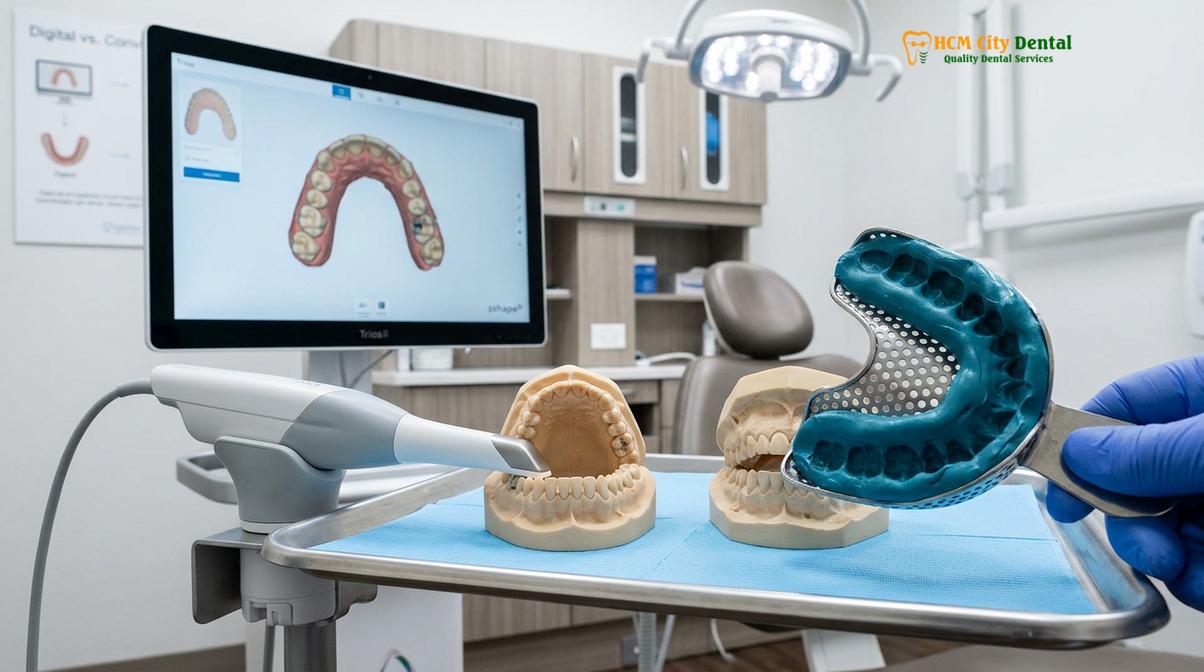
The introduction of computer-aided design and computer-aided manufacturing (CAD/CAM) in dentistry catalyzed a revolutionary shift. The development of the digital oral scanner allowed clinicians to bypass the physical molding phase entirely. By utilizing advanced optical technology, these scanners capture thousands of images per second, stitching them together to form a highly precise, three-dimensional digital replica of the oral cavity. According to Dr. Nguyen Van Cuong, a leading specialist in orthodontic retention, this digital evolution has not only streamlined laboratory workflows but has fundamentally transformed the patient experience by removing the anxiety and discomfort traditionally associated with dental impressions.
Today, the choice between digital and physical methods is a critical consideration in treatment planning. While digital workflows are rapidly becoming the gold standard for clear aligner therapy and modern retention protocols, understanding the biochemical and clinical nuances of both approaches is essential for comprehensive dental care.
Understanding Physical Dental Impressions
Traditional impressions utilize specialized dental putty to create a physical negative replica of the patient’s dental arches, requiring careful material handling to ensure accurate anatomical reproduction.
To fully appreciate the advancements in digital dentistry, one must first understand the intricate clinical workflow of traditional physical dental molds. The process begins with the selection of an appropriately sized impression tray, which can be made of metal or disposable plastic. The tray must provide adequate clearance around all teeth and extend sufficiently to capture the distal aspects of the terminal molars and the full depth of the mucobuccal fold.
The most common material used for routine orthodontic retainers is alginate, an irreversible hydrocolloid derived from marine kelp. Alginate is favored for its cost-effectiveness, rapid setting time, and ease of use. The clinician mixes the alginate powder with water to create a viscous paste, loads it into the tray, and carefully seats it over the patient’s dental arch. The material must remain undisturbed in the mouth for one to three minutes until it fully cross-links and sets into a rubbery solid. Once set, the tray is removed with a firm, quick motion to break the suction seal and minimize tearing of the material.
For cases requiring extreme precision, such as fixed prosthodontics or highly detailed permanent retainers, clinicians may opt for polyvinyl siloxane (PVS). PVS is an addition silicone that offers superior dimensional stability and tear strength compared to alginate. Unlike alginate, which must be poured with dental stone almost immediately to prevent shrinkage from moisture loss (syneresis) or expansion from moisture absorption (imbibition), PVS impressions can remain stable for weeks and can be poured multiple times [1].
Despite their clinical efficacy, physical impressions present several notable challenges:
- Patient Discomfort: The most significant drawback is the potential to trigger a severe gag reflex impression experience. The bulky tray and the flow of material toward the posterior palate can cause severe discomfort, anxiety, and a feeling of claustrophobia or airway obstruction for sensitive patients.
- Material Distortion: If the tray is moved during the setting phase, or if the material is removed prematurely, the impression will distort, leading to an ill-fitting retainer.
- Air Bubbles: Improper mixing or seating can trap air bubbles against the occlusal surfaces of the teeth, resulting in voids in the final stone model.
- Debris and Cleanup: The process is inherently messy, often leaving residue on the patient’s face, lips, and clothing, requiring post-procedure cleanup.
Despite these drawbacks, physical impressions remain a vital skill in dentistry. They are universally accessible, do not require expensive digital infrastructure, and are sometimes necessary for capturing deep subgingival margins in cases where blood or saliva obscures the optical lens of a digital scanner.
The Rise of Digital Oral Scanners in Orthodontics
Intraoral scanners capture thousands of optical images per second to construct a highly precise, real-time 3D digital model of the oral cavity, eliminating the need for physical putty.
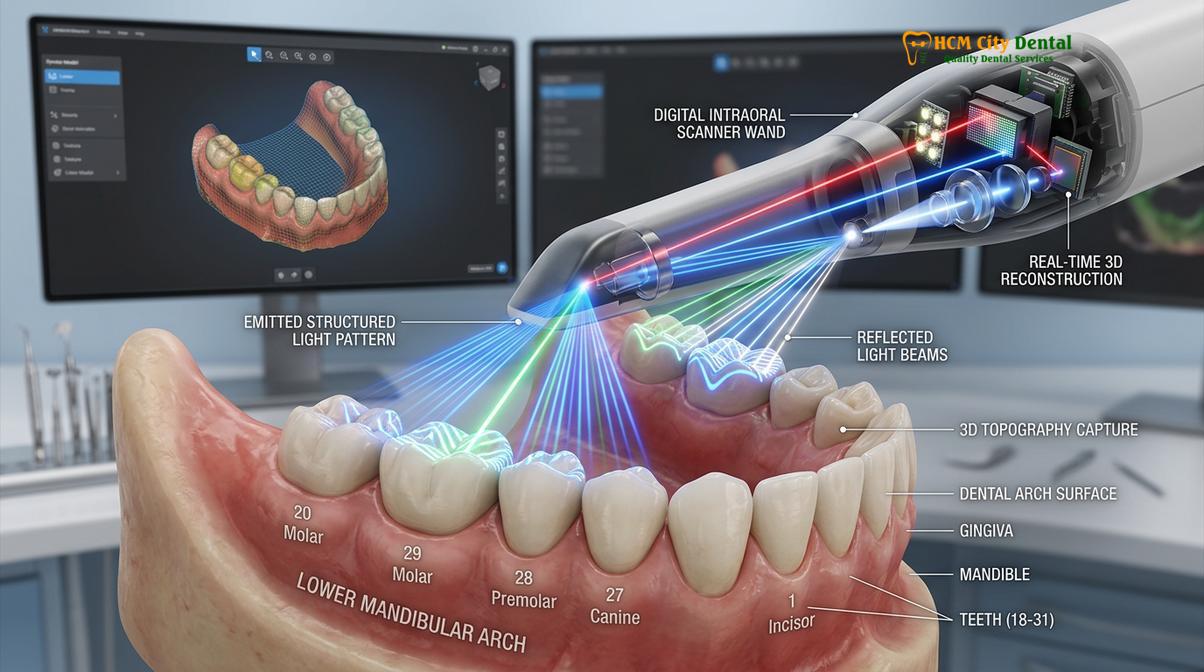
The advent of the digital oral scanner has fundamentally altered the landscape of orthodontic diagnostics and appliance fabrication. These sophisticated devices utilize advanced optical physics, such as confocal microscopy or active wavefront sampling, to map the precise topography of the teeth and gingiva. As the clinician moves the wand-like scanner over the dental arches, a light source (typically a structured laser or LED) projects onto the tissues. The sensor captures the reflected light, and powerful software algorithms instantly stitch these data points together to render a high-definition 3D model on a computer monitor.
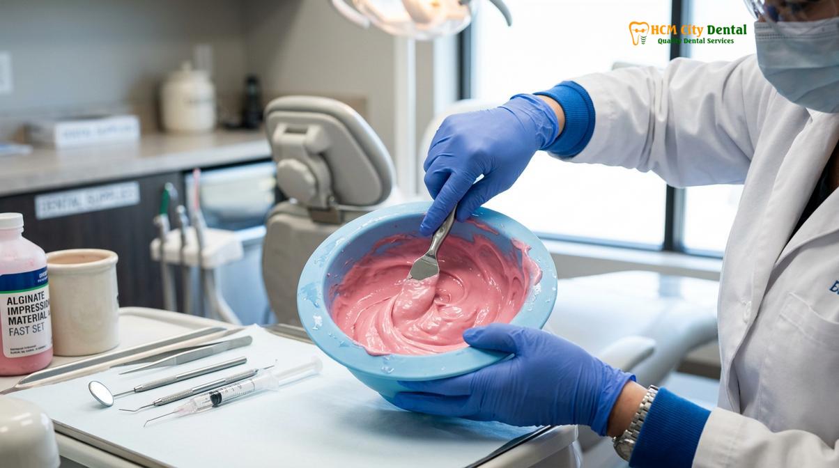
The clinical workflow for a digital impression is remarkably streamlined. The teeth are lightly dried to prevent excessive saliva from reflecting the scanner’s light, and the clinician systematically guides the wand across the occlusal, lingual, and buccal surfaces. The software provides real-time visual feedback, highlighting any areas with missing data, allowing the clinician to immediately rescan specific spots without having to restart the entire process. Once the maxillary arch, mandibular arch, and the patient’s bite registration are captured, the digital file (usually in STL, PLY, or OBJ format) is instantly ready for processing [2].
The benefits of digital scanning extend far beyond mere convenience:
- Unmatched Patient Comfort: The scanner wand is small and non-invasive. Patients can breathe normally, swallow, and even take breaks during the scanning process, completely eliminating the panic associated with the gag reflex.
- Immediate Quality Control: Clinicians can evaluate the 3D model on the screen instantly. If a margin is unclear or a tooth is missed, only that specific area needs to be rescanned, ensuring 100% accuracy before the patient leaves the chair.
- Seamless Communication: Digital files are transmitted to dental laboratories via secure cloud portals in seconds, bypassing the days required to physically mail stone models. This drastically reduces the turnaround time for retainer fabrication.
- Long-Term Digital Archiving: Digital models are stored indefinitely on secure servers without degrading or taking up physical space. If a patient loses their retainer years later, the clinic can simply access the original digital file to fabricate a replacement without requiring a new impression.
For patients seeking comprehensive Orthodontics care, the integration of digital scanning ensures a highly precise fit for all retention appliances, minimizing the need for clinical adjustments during the delivery appointment.
Digital vs Physical Retainer Impressions: A Clinical Comparison
Comparing digital and physical impression techniques reveals distinct differences in patient comfort, clinical accuracy, processing efficiency, and long-term data storage capabilities.
When evaluating digital vs physical retainer impressions, clinicians must weigh various factors including dimensional accuracy, workflow efficiency, patient tolerance, and economic considerations. Both methods are capable of producing clinically acceptable retainers, but their operational paradigms differ significantly.
The following table provides a comprehensive clinical comparison between the two modalities:
| Clinical Parameter | Digital Impressions (Intraoral Scanner) | Physical Impressions (Alginate/PVS) |
|---|---|---|
| Dimensional Accuracy | Extremely high; immune to material shrinkage or expansion. | High, but susceptible to distortion, syneresis, and imbibition. |
| Patient Comfort | Excellent; non-invasive, allows for breathing and swallowing. | Poor to Moderate; bulky trays often trigger the gag reflex. |
| Clinical Chair Time | Fast (2-5 minutes); allows for immediate targeted rescanning. | Moderate (5-10 minutes); requires complete retake if flawed. |
| Laboratory Turnaround | Instant digital transmission; faster appliance fabrication. | Requires physical shipping of molds; slower processing time. |
| Data Storage | Cloud-based; infinite lifespan, zero physical space required. | Physical stone models; prone to breakage, requires storage space. |
| Environmental Impact | Low; eliminates single-use plastics, impression materials, and shipping. | High; generates significant clinical waste and carbon footprint from shipping. |
From a purely clinical standpoint, the dimensional stability of digital impressions is superior over time. Physical impression materials undergo complex polymerization reactions that inherently involve a degree of volumetric shrinkage. Furthermore, the expansion of the dental stone used to pour the model introduces another variable of dimensional change. Digital scans, conversely, capture a 1:1 mathematical representation of the oral cavity that remains perfectly static [3].
“The transition to digital impression systems represents one of the most significant leaps in prosthodontic and orthodontic accuracy. By eliminating the variables of material handling and physical expansion, we achieve a level of predictable precision that was previously unattainable with conventional putty.”
However, it is important to note that digital scanners represent a substantial capital investment for a dental practice, requiring ongoing software licensing and hardware maintenance. Physical impressions remain a highly cost-effective and universally reliable fallback, particularly in scenarios where digital equipment is unavailable or undergoing maintenance.
The Workflow: From Scan to 3D Printing Retainers
Modern orthodontic workflows seamlessly connect digital intraoral scans with advanced manufacturing technologies to produce highly precise, custom-fitted retention appliances.
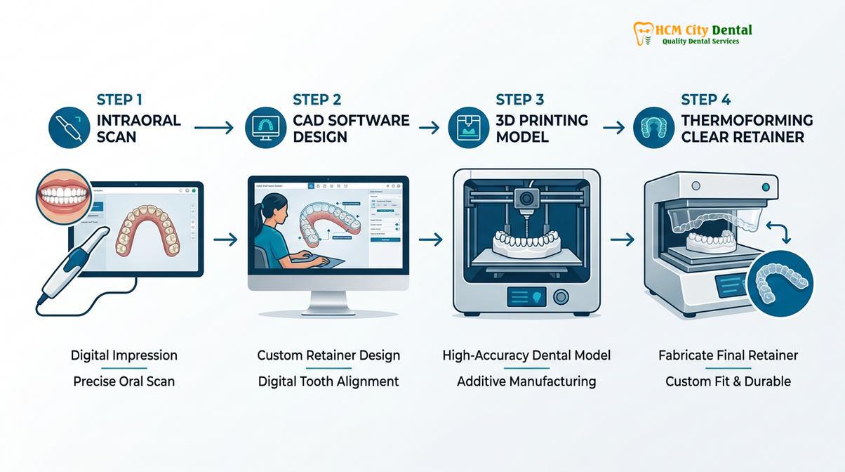
The true power of the digital impression lies in its integration with the broader CAD/CAM ecosystem. Once the digital oral scanner captures the patient’s dentition, the data embarks on a sophisticated digital journey to become a physical retainer.
The first step in this workflow is Computer-Aided Design (CAD). The raw STL or PLY file is imported into specialized orthodontic software. A dental technician or the orthodontist reviews the digital model, digitally trimming away unnecessary soft tissue data, such as the tongue or cheeks, to create a clean, solid base. In cases where minor tooth movements are required before retention, the software can be used to virtually align the teeth. The software also allows the clinician to block out severe undercuts digitally, ensuring that the final retainer will seat smoothly without locking onto the teeth.
Following the design phase, the file is sent to the Computer-Aided Manufacturing (CAM) stage, which heavily relies on 3D printing retainers technology. High-resolution 3D printers, typically utilizing Stereolithography (SLA) or Digital Light Processing (DLP) technology, cure liquid photopolymer resin layer by layer to create a highly accurate physical model of the patient’s teeth. These 3D printed models exhibit exceptional detail, capturing the exact micro-anatomy of the occlusal surfaces and gingival margins.
Once the 3D printed model is cleaned and fully cured under UV light, it serves as the master template for retainer fabrication. For clear retainers (such as Essix or Vivera), a sheet of biocompatible thermoplastic material is heated and vacuum-formed or pressure-formed over the 3D printed model. The material adapts perfectly to the contours of the teeth. After cooling, the retainer is carefully removed from the model, trimmed, and polished to ensure smooth edges that will not irritate the patient’s gums.
This streamlined digital workflow is particularly beneficial for patients requiring rapid turnaround times. For instance, patients utilizing the Same Day Retainer Service Saigon: Clinical Guide | HCMC Dental can have their teeth scanned in the morning and walk out with a perfectly fitting, 3D-printed and thermoformed retainer by the afternoon. Furthermore, because the digital file is saved, patients who lose or break their retainers can easily order replacements without needing to visit the clinic for a new impression, a process detailed in the Essix Retainer Replacement Cost: Clinical Guide | HCMC Dental.
Patient Experience and Clinical Accuracy
Patient preference heavily favors digital scanning due to enhanced comfort, while extensive clinical data supports its exceptional dimensional stability and accuracy.
The psychological impact of dental procedures cannot be overstated. For many patients, the prospect of having a mouth full of setting putty is a significant source of dental anxiety. The physical sensation of the material flowing toward the throat, combined with the inability to swallow or speak, can trigger a severe panic response. By eliminating these triggers, digital scanning dramatically improves the patient experience, fostering a more relaxed and cooperative clinical environment.
Clinical Case Study: Managing Severe Gag Reflex
A 24-year-old patient presented to HCMC Dental Clinic in Ho Chi Minh City requiring new retention appliances after completing orthodontic treatment. The patient had a history of severe dental anxiety and a hyperactive gag reflex, having previously vomited during a traditional alginate impression. Dr. Nguyen Van Cuong opted for a fully digital workflow using an advanced intraoral scanner. By allowing the patient to sit upright, breathe through their nose, and take frequent pauses, the entire maxillary and mandibular scan was completed in under four minutes with zero discomfort. The resulting 3D printed clear retainers exhibited a flawless, passive fit.
Beyond patient comfort, the clinical accuracy of digital impressions is paramount for the long-term success of orthodontic retention. A retainer must fit passively yet securely to hold the teeth in their corrected positions. If an impression is distorted by even a fraction of a millimeter, the resulting retainer may exert unwanted active orthodontic forces, causing pain or inadvertently shifting the teeth. Studies have consistently shown that digital impressions provide equal, if not superior, accuracy to the best elastomeric physical impression materials, particularly for full-arch scans used in retainer fabrication [4].
This high degree of accuracy is especially critical when fabricating premium retention systems. Patients investing in advanced solutions, as outlined in the Invisalign Vivera Retainers Cost & Clinical Guide | HCMC Dental, expect a flawless fit. The digital workflow ensures that these high-end appliances adapt perfectly to the dental anatomy, providing optimal retention and durability.
Overcoming Challenges in Impression Taking
Both digital and physical impression methods present unique clinical challenges, such as moisture control and patient movement, requiring specialized techniques to overcome.
While digital scanning has revolutionized the impression process, it is not entirely without clinical challenges. The most significant hurdle in digital optical scanning is moisture control. Intraoral scanners rely on light reflection to capture data; therefore, excessive saliva, blood, or crevicular fluid can pool in the gingival sulcus or on the occlusal surfaces, reflecting the light erratically and creating voids or distortions in the digital model.
To mitigate this, clinicians must employ rigorous isolation techniques. The use of cotton rolls, dry angles, high-volume evacuation (suction), and gentle air drying is essential to maintain a pristine scanning field. In cases where the gingival margins are inflamed and bleeding, achieving a clean digital scan can be exceedingly difficult, sometimes necessitating a temporary return to physical impression materials that can physically displace fluids.
“Effective moisture control is the cornerstone of a successful digital scan. While the software algorithms are incredibly advanced, they cannot accurately map what the optical lens cannot clearly see through a layer of pooling saliva.”
Conversely, physical impressions face their own set of challenges. Capturing the distal aspect of the second or third molars often requires over-seating the tray, which exacerbates the gag reflex. Furthermore, patients with severe undercuts, large mandibular tori (bony growths), or highly mobile teeth present significant risks when using rigid physical impression materials, as the set material can lock into the undercuts, making removal painful or potentially extracting a loose tooth. In these complex anatomical scenarios, the digital oral scanner is unequivocally the safer and more effective choice.
When to See a Doctor for Retainer Fitting
Important Clinical Considerations
If your new retainer causes severe pain, does not seat fully over your teeth, or if you notice visible gaps between the retainer and the incisal edges of your teeth, discontinue use and contact your orthodontist immediately. Forcing an ill-fitting retainer can cause unintended tooth movement or damage to the dental roots.
Regardless of whether your retainer was fabricated using a digital scan or a physical mold, professional clinical evaluation is essential to ensure its efficacy. A retainer is a precise medical appliance designed to maintain the results of months or years of orthodontic therapy. Patients should schedule a follow-up appointment shortly after receiving their new retainers to allow the clinician to verify the fit, check the occlusal contacts, and ensure there is no impingement on the gingival tissues.
Over time, retainers undergo wear and tear. Clear thermoplastic retainers can stretch, crack, or become distorted if exposed to hot water, while the acrylic and wire components of Hawley retainers can degrade or bend. If you experience a structural failure, such as a snapped wire, it is crucial to seek immediate professional repair, as detailed in the Broken Wire on Permanent Retainer: Clinical Repair Guide | HCMC Dental.
Furthermore, understanding the long-term protocols for retention is vital for maintaining your smile. Patients often wonder about the duration of wear required to prevent relapse. Comprehensive guidelines on this topic can be found in the How Long to Wear Retainers After Braces: Clinical Guide | HCMC Dental. For international patients seeking high-quality, digitally fabricated retention appliances while traveling, exploring options like Dental Tourism Vietnam: Orthodontic Retention | HCMC Dental provides access to world-class digital dentistry at competitive rates.
Ultimately, the success of your orthodontic retention relies on a combination of precise impression techniques, high-quality fabrication, and diligent patient compliance. Regular consultations with experienced professionals, such as the team at HCMC Dental Clinic, ensure that your retention strategy remains effective, comfortable, and tailored to your unique dental anatomy [5].
Frequently Asked Questions
Are digital impressions more accurate than physical molds?
Yes, digital impressions generally offer superior or equal accuracy compared to physical molds. They eliminate the risk of material shrinkage, distortion during transit, and air bubbles, resulting in highly precise 3D models for retainer fabrication. The digital data provides a mathematically exact 1:1 replica of the oral cavity that remains dimensionally stable indefinitely.
Does a digital scan hurt or cause discomfort?
No, a digital scan is completely painless and non-invasive. The intraoral scanner is a small, wand-like device that simply hovers over your teeth to capture images, eliminating the discomfort, claustrophobia, and gagging associated with traditional putty trays. Patients can breathe normally and swallow during the procedure.
How long does a digital retainer impression take?
A full-arch digital scan typically takes between two to five minutes to complete. The process is highly efficient, and the 3D model appears on the computer screen in real-time, allowing the clinician to immediately review the data and rescan any missed areas without starting over.
Can I still get a physical impression if I have a strong gag reflex?
While physical impressions can trigger a strong gag reflex, dentists can use techniques like topical anesthetics, controlled breathing, and seating adjustments to minimize discomfort. However, digital scanning is highly recommended as the primary alternative for patients with severe gag reflexes, as it bypasses the trigger entirely.
Are retainers made from digital scans more expensive?
The cost of retainers made from digital scans is generally comparable to those made from physical molds. While the scanning technology requires a high initial investment for the clinic, it streamlines the laboratory workflow, reduces physical material costs, and eliminates shipping fees, balancing the overall cost to the patient.
References
- Journal of Prosthodontic Research. Accuracy of digital intraoral scanners versus traditional impression techniques. (2021).
- American Journal of Orthodontics and Dentofacial Orthopedics. Patient preference and clinical efficiency of digital versus conventional impressions. (2019).
- European Journal of Orthodontics. Dimensional stability of 3D printed dental models for retainer fabrication. (2020).
- Journal of Clinical Orthodontics. Managing the gag reflex during orthodontic impression procedures. (2022).
- Australian Dental Journal. Long-term stability and fit of clear aligners and retainers derived from digital workflows. (2018).