A piezotome dental implant procedure utilizes advanced ultrasonic micro-vibrations to precisely cut jawbone while preserving surrounding soft tissues, nerves, and blood vessels. This minimally invasive technology significantly reduces post-operative pain, swelling, and surgical trauma compared to traditional rotary drills, ensuring a safer and faster healing process.
Clinical Summary:
Piezotome technology represents a paradigm shift in modern implantology, replacing conventional rotary burs with piezoelectric ultrasonic frequencies (typically between 28 and 36 kHz). By leveraging the cavitation effect and selective tissue cutting, piezosurgery allows clinicians to perform highly precise osteotomies without damaging adjacent neurovascular structures or the delicate Schneiderian membrane. This approach ensures optimal bone preservation, accelerates the biological cascade of osseointegration, and minimizes post-surgical morbidity. It is highly indicated for complex anatomical scenarios, including ridge expansion, sinus augmentation, and immediate implant placement, offering a superior safety profile compared to traditional mechanical drilling.
Key Takeaways:
- Piezotome devices use specific ultrasonic frequencies to cut bone selectively without harming adjacent soft tissues.
- The technology significantly lowers the risk of inferior alveolar nerve damage during mandibular implant surgeries.
- Cavitation effects from the ultrasonic coolant provide a nearly blood-free surgical field, enhancing clinical visibility.
- Post-operative swelling, pain, and overall recovery times are drastically reduced compared to traditional rotary drilling.
- Piezosurgery is highly effective for delicate procedures like sinus lifts, narrow ridge splitting, and atraumatic extractions.
The Evolution of Dental Surgical Instruments: Drills vs. Piezoelectricity
Traditional rotary drills have long been the standard for bone preparation, but they carry inherent risks of thermal trauma and soft tissue damage. The advent of piezoelectric bone surgery introduces ultrasonic precision, mitigating these risks and revolutionizing modern implantology.
The history of oral and maxillofacial surgery has been dominated by the use of mechanical rotary instruments. For decades, high-speed and low-speed drills equipped with carbide or diamond burs have been the primary tools for cutting bone, extracting teeth, and preparing sites for Dental Implants. While effective, these traditional instruments operate on the principle of macro-vibrations and mechanical friction. This frictional force inherently generates significant heat. Bone tissue is highly susceptible to thermal trauma; research indicates that exposing alveolar bone to temperatures exceeding 47°C for more than one minute can lead to irreversible thermal necrosis, compromising osteocyte viability and leading to early implant failure [1].
To counteract this heat generation, copious external saline irrigation is required during traditional drilling. However, even with adequate cooling, the mechanical shearing action of a rotary bur is non-selective. A drill will cut through anything in its path—mineralized bone, delicate mucosal membranes, blood vessels, and nerve bundles alike. This lack of tissue selectivity requires the surgeon to maintain a wide safety margin, sometimes compromising the ideal biomechanical positioning of the implant to avoid catastrophic neurovascular injury.
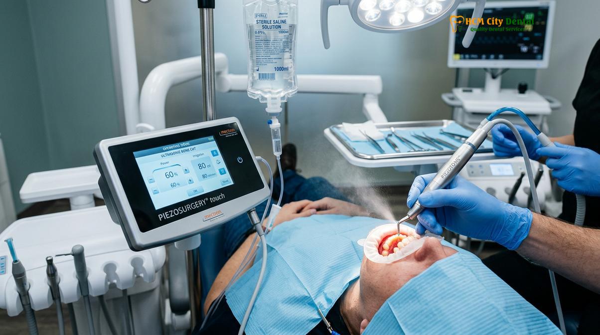
The paradigm shifted dramatically with the introduction of piezoelectricity into the dental field. The piezoelectric effect, originally discovered by the Curie brothers in the late 19th century, describes the ability of certain materials (like quartz crystals or specialized ceramics) to generate an electrical charge in response to applied mechanical stress. Conversely, the reverse piezoelectric effect—where an alternating electrical current applied to these materials causes them to rapidly expand and contract—forms the basis of modern piezosurgery.
By harnessing this reverse piezoelectric effect, modern piezotome devices convert electrical energy into precise, high-frequency mechanical micro-vibrations. Instead of spinning like a drill, the surgical tip oscillates back and forth at a microscopic level. This transition from mechanical macro-shearing to ultrasonic micro-vibration represents one of the most significant advancements in surgical safety and precision. Dr. Nguyen Van Cuong, a leading implantologist, frequently emphasizes that the transition to piezoelectric bone surgery allows clinicians to approach anatomically challenging cases with a level of confidence and biological respect that was previously unattainable with conventional rotary instruments.
How Piezotome Technology Works: Soft Tissue Preservation and Bone Cutting
Piezotome devices operate on specific ultrasonic frequencies (28–36 kHz) that exclusively target mineralized bone. This selective cutting mechanism ensures that adjacent soft tissues, such as nerves and membranes, absorb the vibrations without sustaining structural damage.
Understanding the clinical superiority of a piezotome dental implant procedure requires a deep dive into the physics of ultrasonic tissue interaction. The core principle that makes piezosurgery revolutionary is its tissue selectivity. The device is calibrated to operate strictly within an ultrasonic frequency range of 28 to 36 kilohertz (kHz). At this specific frequency, the micro-vibrations are only capable of cutting highly mineralized, dense structures—namely, cortical and cancellous bone [2].
When the oscillating metallic tip comes into contact with non-mineralized soft tissue—such as the gingiva, the Schneiderian membrane lining the maxillary sinus, or the inferior alveolar nerve—the tissue’s inherent elasticity allows it to absorb and dampen the micro-vibrations. Instead of being lacerated or severed, the soft tissue simply vibrates along with the tip. This phenomenon is clinically referred to as select soft tissue protection. It provides an unprecedented safety net for the surgeon, allowing them to cut bone immediately adjacent to critical anatomical landmarks without the constant fear of causing iatrogenic soft tissue trauma.
Beyond selective cutting, the piezotome operates with micrometric amplitude. The surgical inserts vibrate with an amplitude ranging from 20 to 200 micrometers. This microscopic movement provides the surgeon with exceptional tactile feedback and absolute control over the osteotomy. Unlike a rotary drill, which can “walk” or slip across the bone surface due to rotational torque, the piezotome tip stays exactly where it is placed, allowing for geometrically precise bone cuts that are essential for complex grafting and implant site preparation.
Another critical mechanism of action is the cavitation effect. During piezosurgery, sterile saline solution is continuously pumped to the vibrating surgical tip. The high-frequency ultrasonic oscillations create rapid pressure changes in the fluid, leading to the formation and subsequent implosion of microscopic vapor bubbles. This implosion generates localized shockwaves—a process known as cavitation. The cavitation effect serves multiple vital functions during the surgery:
- Enhanced Cooling: The atomization of the saline coolant ensures highly efficient heat dissipation, keeping the bone temperature well below the critical threshold for thermal necrosis.
- Debris Removal: The acoustic microstreaming created by cavitation actively flushes bone chips, debris, and bacteria out of the osteotomy site, maintaining a pristine surgical field.
- Hemostasis: The shockwaves temporarily seal small capillary vessels in the surrounding bone, resulting in a nearly blood-free surgical site that drastically improves the surgeon’s visibility.
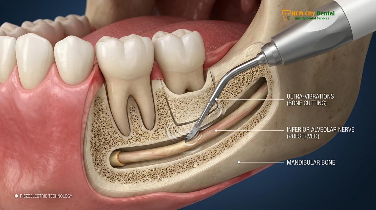
Histological studies comparing bone healing after rotary drilling versus piezoelectric osteotomy have consistently shown that piezosurgery preserves a higher number of viable osteocytes at the cutting edge. The absence of thermal damage and mechanical smearing of the bone trabeculae allows for a more rapid onset of angiogenesis and osteogenesis, laying the ideal biological foundation for the integration of premium dental implants.
Clinical Benefits: Less Swelling, Bleeding, and Nerve Damage Risks
Utilizing ultrasonic micro-vibrations minimizes surgical trauma, leading to a profound reduction in post-operative edema and hemorrhage. Furthermore, the selective nature of the piezotome virtually eliminates the risk of severing critical neurovascular bundles during implant placement.
The theoretical advantages of piezoelectric physics translate into highly tangible clinical benefits for the patient. When evaluating the success of a dental implant procedure, clinicians must consider not only the long-term osseointegration of the titanium fixture but also the immediate post-operative morbidity experienced by the patient. In this regard, piezotome technology offers a vastly superior patient experience.
One of the most immediate benefits observed during surgery is the profound reduction in bleeding. As mentioned, the cavitation effect of the ultrasonic coolant creates a micro-coagulation effect on the severed intraosseous capillaries. While a traditional drill often leaves the surgical site obscured by pooling blood, requiring constant suction and interrupting the workflow, the piezotome maintains a clean, highly visible field. This enhanced visibility is crucial when navigating complex anatomy or when placing implants in areas with limited bone volume.
The most significant safety benefit of the piezotome is the mitigation of nerve damage. The inferior alveolar nerve (IAN) runs through the mandible, supplying sensation to the lower lip, chin, and teeth. Accidental injury to this nerve during drilling can result in paresthesia (altered sensation), dysesthesia (painful sensation), or permanent anesthesia (complete numbness) of the affected area. Because the piezotome utilizes select soft tissue protection, the risk of severing the IAN is virtually eliminated, even if the surgical tip inadvertently makes direct contact with the nerve sheath [3].
“The integration of piezoelectric technology in implant dentistry has fundamentally altered our approach to complex anatomical challenges. By prioritizing biological preservation over mechanical force, we can achieve predictable outcomes while drastically reducing the surgical burden on our patients.”
— Dr. Nguyen Van Cuong
Furthermore, the reduction in mechanical trauma directly influences the body’s inflammatory response. Traditional drilling causes micro-fractures and crushing of the bone trabeculae, triggering a robust release of pro-inflammatory cytokines and prostaglandins. This aggressive inflammatory cascade is the primary cause of post-operative swelling (edema) and pain. Because piezosurgery cuts bone cleanly without crushing or thermal damage, the subsequent inflammatory response is significantly blunted. Patients routinely report less facial swelling, reduced reliance on post-operative analgesics, and a faster return to their normal daily activities.
This minimally invasive approach is particularly beneficial for older adults or patients with compromised healing capacities. When considering Dental Implants for Seniors, minimizing surgical trauma is paramount to reducing the risk of post-operative complications and ensuring a smooth recovery phase.
Guided Bone Preparation (Osteotomy) with High Precision
Piezotome technology allows for micrometric surgical control during bone preparation, enabling highly accurate implant site development. When combined with an ultrasonic surgical guide, clinicians can achieve unparalleled precision in complex anatomical regions.
The preparation of the bone cavity to receive the implant—known as the osteotomy—is the most critical phase of the surgical procedure. The long-term stability of the implant relies on achieving high primary stability, which requires the osteotomy to be prepared with exact dimensions corresponding to the implant’s geometry. Piezotome technology excels in facilitating a minimally invasive osteotomy, particularly in compromised bone sites.
One of the most challenging scenarios in implantology is the severely atrophic (resorbed) alveolar ridge. When a tooth is lost, the surrounding bone naturally shrinks over time, often leaving a ridge that is too narrow to accommodate a standard-diameter implant. Traditionally, this required extensive block bone grafting, a highly invasive procedure with significant morbidity. With piezosurgery, clinicians can perform a technique known as “ridge splitting” or “ridge expansion.” Using ultra-thin ultrasonic saws, the surgeon can delicately divide the narrow bone crest longitudinally and gently expand it, creating a space for immediate implant placement. The micrometric precision of the piezotome prevents the fragile buccal bone plate from fracturing during expansion—a common complication when using traditional chisels or rotary discs.
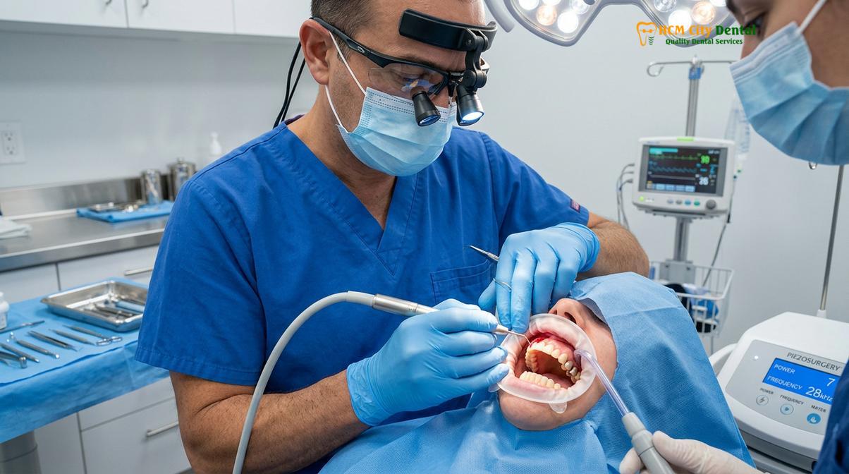
Another area where piezosurgery has become the gold standard is maxillary sinus augmentation (sinus lifting). In the posterior upper jaw, bone height is often limited by the presence of the maxillary sinus cavity. To place an implant, the surgeon must access the sinus, gently elevate the delicate Schneiderian membrane lining the cavity, and place bone graft material underneath it. Using a rotary drill to create the lateral access window carries a high risk (up to 30%) of tearing the membrane, which can lead to graft infection and failure. The piezotome, however, can cut the bony window and safely bounce off the membrane without tearing it, reducing the perforation rate to near zero [4].
Clinical Case Review: Complex Maxillary Rehabilitation
A 58-year-old patient presented to HCMC Dental Clinic in Ho Chi Minh City with severe posterior maxillary bone loss, requiring bilateral sinus lifts prior to implant placement. Due to the extremely thin residual bone (less than 3mm) and a highly convoluted sinus anatomy, traditional rotary drilling posed a severe risk of membrane perforation. The surgical team utilized a piezotome to perform a minimally invasive lateral window approach. The ultrasonic micro-vibrations allowed for precise bone removal while the cavitation effect kept the surgical field completely clear of blood. The Schneiderian membrane was successfully elevated without a single micro-tear, allowing for simultaneous bone grafting and implant placement. The patient experienced minimal post-operative swelling and reported zero pain by the second day, highlighting the atraumatic nature of piezoelectric surgery.
Modern implantology also heavily relies on digital planning and guided surgery. Clinicians can now integrate piezotome technology with an ultrasonic surgical guide. These 3D-printed templates fit over the patient’s teeth or gums and direct the ultrasonic inserts with pinpoint accuracy, ensuring the osteotomy is prepared exactly according to the virtual pre-surgical plan. This synergy of digital planning and ultrasonic execution represents the pinnacle of surgical precision.
Post-Op Recovery Comparisons: Traditional Drill vs. Piezotome Surgery
Patients undergoing piezotome-assisted implant surgery experience a significantly accelerated healing cascade compared to those treated with conventional rotary instruments. The reduction in surgical trauma translates directly to less reliance on analgesics and a faster return to normal function.
The biological response to surgical trauma dictates the speed and comfort of the patient’s recovery. Bone healing around a dental implant follows a specific cascade: hemostasis, inflammation, proliferation, and remodeling. The severity of the initial trauma directly impacts the intensity and duration of the inflammatory phase.
When a conventional rotary drill is used, the mechanical shearing and frictional heat create a zone of necrotic bone at the osteotomy interface. The body’s immune system must first resorb this dead bone before new, healthy bone can be deposited against the implant surface (a process known as distance osteogenesis). This prolonged inflammatory and resorptive phase delays the overall integration process and increases post-operative discomfort.
Conversely, the clean, micrometric cut produced by a piezotome leaves the bone trabeculae intact and highly viable. The absence of a necrotic layer allows osteoblasts (bone-forming cells) to immediately begin depositing new bone matrix directly onto the implant surface (contact osteogenesis). This accelerated biological response not only speeds up the timeline for placing the final prosthetic crown but also drastically reduces the clinical symptoms of inflammation [5].
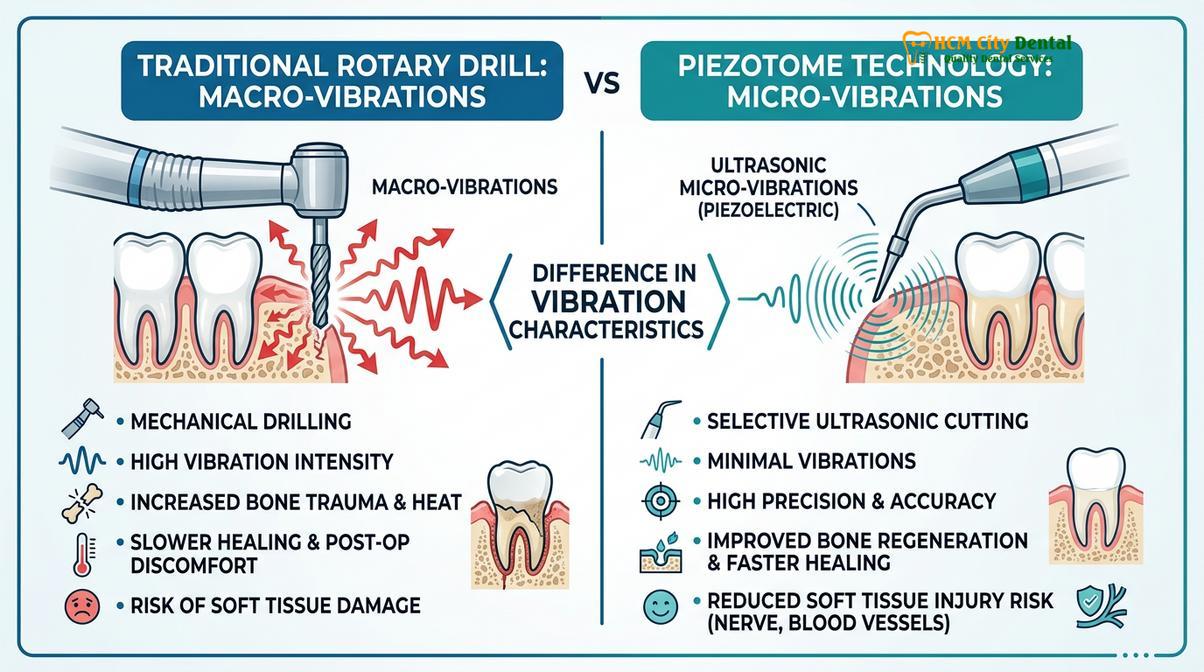
To clearly illustrate the clinical differences, the following table compares the key parameters of traditional rotary drilling versus piezoelectric bone surgery:
| Clinical Parameter | Traditional Rotary Drill | Piezotome Ultrasonic Surgery |
|---|---|---|
| Cutting Mechanism | Mechanical macro-shearing and friction | Ultrasonic micro-vibrations (28-36 kHz) |
| Tissue Selectivity | None (cuts bone, nerves, and soft tissue equally) | High (cuts only mineralized bone, protects soft tissue) |
| Heat Generation | High risk of thermal necrosis without heavy cooling | Minimal heat, highly efficient cavitation cooling |
| Intraoperative Bleeding | Moderate to high, often obscuring the surgical field | Minimal, cavitation provides a nearly blood-free field |
| Risk of Nerve Damage | Moderate (requires strict safety margins) | Extremely low (due to soft tissue protection) |
| Post-Operative Swelling | Moderate to severe (depending on procedure extent) | Mild to minimal |
| Bone Healing Cascade | Delayed (requires resorption of necrotic bone layer) | Accelerated (intact osteocytes promote rapid osteogenesis) |
For patients evaluating their restorative options, understanding these recovery differences is crucial. While the initial cost of dental implants may be a primary consideration, the value of a safer, less painful, and more predictable surgical experience cannot be overstated. The reduction in lost work days and the minimized need for prescription pain medication make piezosurgery a highly attractive option for modern dental patients.
When to See a Doctor (Important Clinical Considerations)
While piezotome technology offers exceptional safety, a thorough clinical evaluation is necessary to determine patient candidacy. Certain systemic conditions and anatomical factors must be assessed by a qualified implantologist before proceeding with ultrasonic surgery.
Despite its profound advantages, piezotome dental implant surgery is a medical procedure that requires careful patient selection and comprehensive diagnostic planning. Not every patient or every anatomical site strictly requires the use of ultrasonic technology, and in some specific instances, its use may be contraindicated.
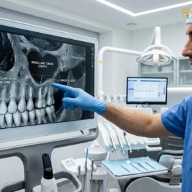
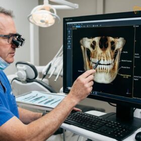
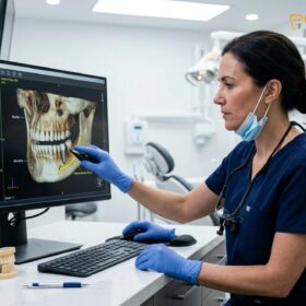
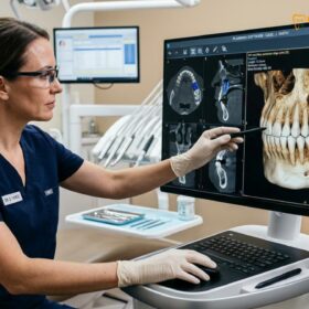
Patients should schedule a consultation with a specialized implantologist if they are missing one or more teeth and are seeking a permanent, fixed replacement. During the consultation, the clinician will utilize 3D Cone Beam Computed Tomography (CBCT) to evaluate the volume, density, and architecture of the jawbone. This imaging is critical for identifying the proximity of the maxillary sinuses in the upper jaw and the inferior alveolar nerve in the lower jaw. If the CBCT reveals severe bone atrophy, a narrow alveolar ridge, or an implant site in dangerously close proximity to these vital structures, the surgeon will likely recommend piezosurgery to mitigate the surgical risks.
Important Clinical Contraindications:
While highly safe for the vast majority of patients, piezotome devices emit electromagnetic fields and ultrasonic frequencies. Therefore, their use is strictly contraindicated in patients with unshielded cardiac pacemakers or certain implantable cardioverter-defibrillators (ICDs), as the electromagnetic interference could potentially disrupt the device’s function. Always disclose your complete medical history, including all implanted medical devices, to your dental surgeon prior to any procedure.
Furthermore, patients who have previously been told they are not candidates for implants due to “lack of bone” should seek a second opinion from a clinic equipped with piezotome technology. The ability to perform minimally invasive ridge splitting or highly predictable sinus lifts often expands the scope of candidacy, allowing patients who were previously relegated to removable dentures to finally receive fixed implant prosthetics. For a broader understanding of how implants compare to traditional removable options, patients can review the clinical differences between Dental Implants vs Dentures.
“The true art of implantology lies not just in placing a titanium screw, but in managing the surrounding biology with the utmost respect. Piezosurgery gives us the technological means to execute our surgical vision without compromising the patient’s natural anatomy.”
— Dr. Nguyen Van Cuong
Ultimately, the decision to utilize traditional rotary instruments or a piezotome rests on the clinical judgment of the surgeon, based on the specific anatomical challenges of the individual case. Patients are encouraged to ask their provider about the technologies utilized in their practice and how those tools align with their specific surgical needs. For those seeking comprehensive evaluations, the team at HCMC Dental Clinic in Saigon routinely utilizes advanced diagnostic protocols to tailor the safest and most effective surgical approach for each individual patient.
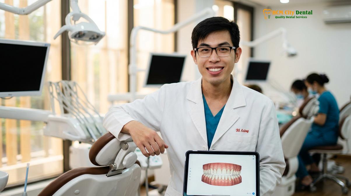
If you are considering restoring your smile and oral function, understanding the pros and cons of dental implants and the technologies used to place them is the first step toward a successful outcome. A thorough clinical examination will provide the clarity needed to proceed with confidence.
Frequently Asked Questions
Does HCMC Dental use Piezotome for all implant cases?
No, piezotome technology is not mandatory for every single implant case. While it is highly beneficial for complex anatomical situations, such as sinus lifts or surgeries near the inferior alveolar nerve, standard osteotomies in dense, healthy bone with ample clearance may still be efficiently performed using conventional guided rotary instruments based on the surgeon’s clinical assessment. The choice of instrument is always tailored to maximize safety and efficiency for the specific anatomical site.
Is Piezotome surgery more expensive than traditional drill surgery?
Generally, procedures utilizing piezotome technology may incur a slightly higher cost due to the advanced equipment and specialized surgical inserts required. However, the investment is often offset by the reduced risk of complications, decreased need for extensive bone grafting in certain cases, and a significantly more comfortable, accelerated recovery period for the patient. Many patients find the added safety and reduced post-operative pain well worth the marginal difference in initial cost.
Does ultrasonic bone cutting reduce the risk of nerve damage?
Yes, ultrasonic bone cutting drastically reduces the risk of nerve damage. The piezotome operates at a specific frequency that cuts only mineralized tissue (bone). When the ultrasonic tip encounters soft tissue, such as the inferior alveolar nerve or blood vessels, it simply bounces off without causing structural laceration or severing the tissue. This selective cutting ability provides a massive safety margin when operating in the posterior mandible.
How long does a piezotome dental implant procedure take?
A piezotome dental implant procedure may take slightly longer during the bone preparation phase compared to traditional drilling, typically adding 10 to 15 minutes to the surgery. The micrometric cutting action is intentionally slower to ensure maximum precision, thermal control, and absolute preservation of the surrounding delicate anatomical structures. However, this slight increase in surgical time is heavily outweighed by the reduction in post-operative healing time.
Can piezosurgery be used for tooth extractions before implant placement?
Absolutely, piezosurgery is highly indicated for atraumatic tooth extractions prior to immediate implant placement. The ultrasonic micro-vibrations gently sever the periodontal ligament and expand the socket without fracturing the delicate buccal bone plate, thereby preserving the maximum amount of native bone required for successful implant anchoring. This atraumatic approach is crucial for maintaining the aesthetic contours of the gum tissue, especially in the anterior aesthetic zone.
References
- Journal of Periodontology. Piezoelectric bone surgery in implant dentistry: clinical applications and biological rationale. (2020).
- International Journal of Oral and Maxillofacial Implants. Thermal changes during osteotomy: a comparison between conventional drilling and piezosurgery. (2019).
- Clinical Oral Implants Research. Histological and histomorphometric evaluation of bone healing after piezoelectric osteotomy. (2021).
- Journal of Oral and Maxillofacial Surgery. The efficacy of piezosurgery in lateral window sinus floor elevation: a systematic review. (2022).
- European Journal of Dentistry. Patient morbidity and healing outcomes following ultrasonic vs. rotary bone preparation. (2018).