Dental implant complications encompass a range of surgical and prosthetic challenges, from mechanical failures like screw loosening to biological issues such as peri-implantitis and sinus perforations. Prompt identification and evidence-based clinical management are essential to restore oral function and ensure the long-term survival of the implant prosthesis.
Clinical Summary:
While implantology boasts exceptionally high success rates, dental implant complications can arise during the surgical phase, the healing period, or years after prosthetic loading. Intraoperative challenges frequently involve the maxillary sinus, where the delicate Schneiderian membrane may inadvertently tear during site preparation or sinus augmentation. The management of such perforations depends heavily on the size of the defect; small tears are routinely repaired using a collagen membrane patch, whereas larger ruptures may necessitate the postponement of the implant procedure. Beyond sinus-related issues, long-term complications include biomechanical failures—such as abutment screw loosening or fixture fracture due to occlusal overload—and biological inflammatory conditions like peri-implantitis. Successful mitigation of these risks requires meticulous 3D radiographic planning, precise surgical execution, strict adherence to post-operative pharmacological protocols, and a commitment to lifelong periodontal maintenance.
Key Takeaways:
- Sinus perforations are common intraoperative complications during upper jaw implant surgery, requiring immediate assessment and repair.
- Small membrane tears (under 5mm) can be effectively sealed using resorbable collagen membranes without compromising the final implant outcome.
- Post-operative adherence to sinus precautions, including avoiding nose-blowing and taking prescribed decongestants, is critical for preventing acute infections.
- Late-stage complications often involve peri-implantitis, a bacteria-induced inflammatory condition that causes progressive bone loss around the implant.
- Biomechanical overloading from bruxism or poor prosthesis design can lead to mechanical failures, such as screw loosening or implant fracture.
What is a Sinus Perforation (Schneiderian Membrane Tear)?
A sinus perforation is an unintended rupture of the Schneiderian membrane, the delicate lining of the maxillary sinus cavity, often occurring during upper jaw implant site preparation or sinus augmentation procedures.
The maxillary sinus is a pneumatic cavity located within the body of the maxilla, positioned directly above the roots of the upper premolars and molars. This cavity is lined by a specialized mucosal layer known as the Schneiderian membrane. Histologically, this membrane is composed of ciliated pseudostratified columnar epithelium, which plays a vital role in the mucociliary clearance system, sweeping mucus and foreign particles toward the nasal ostium for elimination[1]. In a healthy state, the membrane is incredibly thin, typically measuring between 0.3 to 0.8 millimeters in thickness, making it highly susceptible to surgical trauma.
When patients lose their upper posterior teeth, the alveolar bone undergoes rapid resorption, while the maxillary sinus simultaneously expands downward—a process known as sinus pneumatization. To place Dental Implants in this region, surgeons frequently perform a sinus lift (augmentation) to create sufficient vertical bone height. A sinus perforation occurs when surgical instruments inadvertently breach this delicate lining during the elevation process or during the drilling of the implant osteotomy.
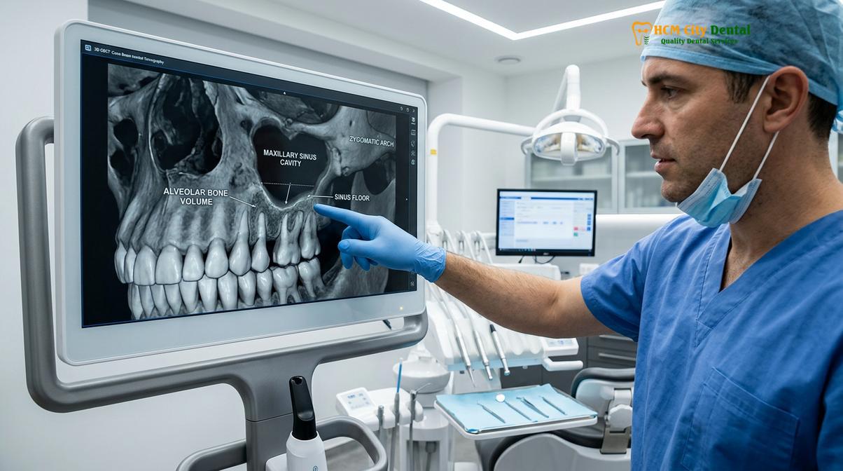
Understanding the implications of a Schneiderian membrane tear is fundamental to managing dental implant complications in the posterior maxilla. If a perforation occurs and goes unnoticed or untreated, the particulate bone graft material intended to build the new foundation can migrate into the sinus cavity. This ectopic graft material acts as a foreign body, potentially obstructing the natural drainage pathways of the sinus and leading to acute or chronic sinusitis[2]. Furthermore, the loss of graft containment compromises the volume of bone available for implant integration, directly threatening the primary stability and long-term survival of the titanium fixture.
Causes of Membrane Tears During Sinus Lifts or Implant Drilling
Membrane tears typically result from anatomical complexities such as sinus septa, an exceptionally thin mucosal lining, or iatrogenic factors like excessive drilling pressure and inadequate surgical instrumentation.
The etiology of sinus perforations is multifactorial, encompassing both patient-specific anatomical variations and surgical technique. One of the most prominent anatomical risk factors is the presence of Underwood’s septa. These are bony ridges or walls that project from the floor or walls of the maxillary sinus, dividing the cavity into multiple compartments. Studies indicate that sinus septa are present in approximately 25% to 30% of patients[3]. When a surgeon attempts to elevate the membrane over these sharp bony projections, the tension applied to the mucosa increases exponentially, making a tear highly probable.
Another critical factor is the inherent thickness and health of the Schneiderian membrane. Patients with a history of chronic sinus infections may present with a thickened, fibrotic membrane that is relatively resilient. Conversely, patients with pristine sinus health often possess a translucent, friable membrane that can tear under minimal mechanical stress. Additionally, the angle between the buccal and palatal walls of the sinus plays a role; a narrow, V-shaped sinus floor creates a constricted working space, increasing the difficulty of membrane elevation and the likelihood of perforation.
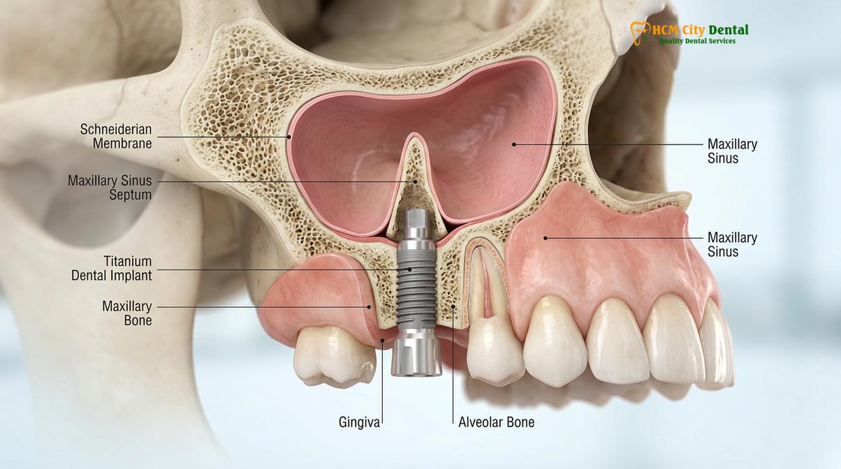
Iatrogenic causes—those related to the surgical procedure itself—are also significant contributors to this specific subset of dental implant complications. The use of high-speed rotary instruments near the sinus floor requires exceptional tactile sensitivity. If the surgeon applies excessive apical pressure or fails to accurately gauge the depth of the osteotomy, the drill can easily puncture the membrane. To mitigate these risks, modern implantology increasingly relies on piezoelectric surgery. Piezosurgery utilizes ultrasonic micro-vibrations that selectively cut hard bone tissue while sparing soft tissues, dramatically reducing the incidence of membrane tears during lateral window preparations[4].
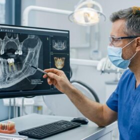
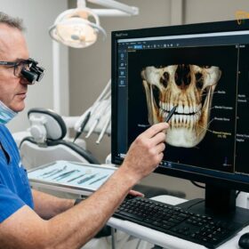
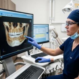
At HCMC Dental Clinic, comprehensive preoperative planning is mandatory to minimize these risks. Dr. Nguyen Van Cuong emphasizes the necessity of high-resolution 3D Cone Beam Computed Tomography (CBCT) scans to map the exact topography of the sinus floor, identify the presence of septa, and measure the residual bone height before any surgical intervention begins.
How Surgeons Repair Small Tears: Collagen Membranes and Sutures
Small sinus membrane tears are effectively managed using a resorbable collagen membrane patch to seal the defect, sometimes combined with delicate suturing techniques to prevent bone graft migration.
When a perforation occurs, the immediate clinical priority is to assess the size and location of the tear, as this dictates the appropriate schneiderian membrane repair protocol. The Vlassis classification system is widely used to categorize these defects. Class I and Class II tears, which are generally smaller than 5 to 10 millimeters, are considered highly manageable and rarely require the abandonment of the augmentation procedure[5]. The primary objective of the repair is to re-establish a continuous barrier that will contain the particulate bone graft and prevent bacterial ingress from the nasal cavity.
The gold standard for repairing small to medium-sized defects is the application of a resorbable collagen membrane patch. These membranes, typically derived from porcine or bovine sources, are highly biocompatible and possess excellent handling characteristics. When moistened with sterile saline or the patient’s own blood, the collagen membrane becomes pliable and adheres naturally to the surrounding moist tissues of the sinus wall.
| Tear Classification | Size of Defect | Clinical Management Protocol | Implant Placement Status |
|---|---|---|---|
| Class I | < 2 mm | Spontaneous folding of the membrane; no additional materials needed. | Proceed as planned. |
| Class II | 2 mm – 5 mm | Application of a resorbable collagen membrane patch over the defect. | Proceed as planned. |
| Class III | 5 mm – 10 mm | Loma Linda pouch technique; possible use of fibrin sealants or sutures. | Proceed with caution; consider delaying implant. |
| Class IV & V | > 10 mm | Procedure aborted. Defect allowed to heal naturally for 4-6 months. | Delayed placement mandatory. |
For slightly larger tears (Class III), surgeons often employ the “Loma Linda pouch technique.” This advanced maneuver involves placing a large, slow-resorbing collagen membrane inside the sinus cavity, carefully adapting it against the bony walls to create an artificial pouch. The bone graft material is then packed inside this collagen pouch, completely isolating it from the torn Schneiderian membrane. In cases where the tear is located near the lateral window access, delicate suturing using fine resorbable threads (such as 5-0 or 6-0 Vicryl) may be utilized to anchor the membrane to the surrounding periosteum, ensuring it does not dislodge during the healing phase.
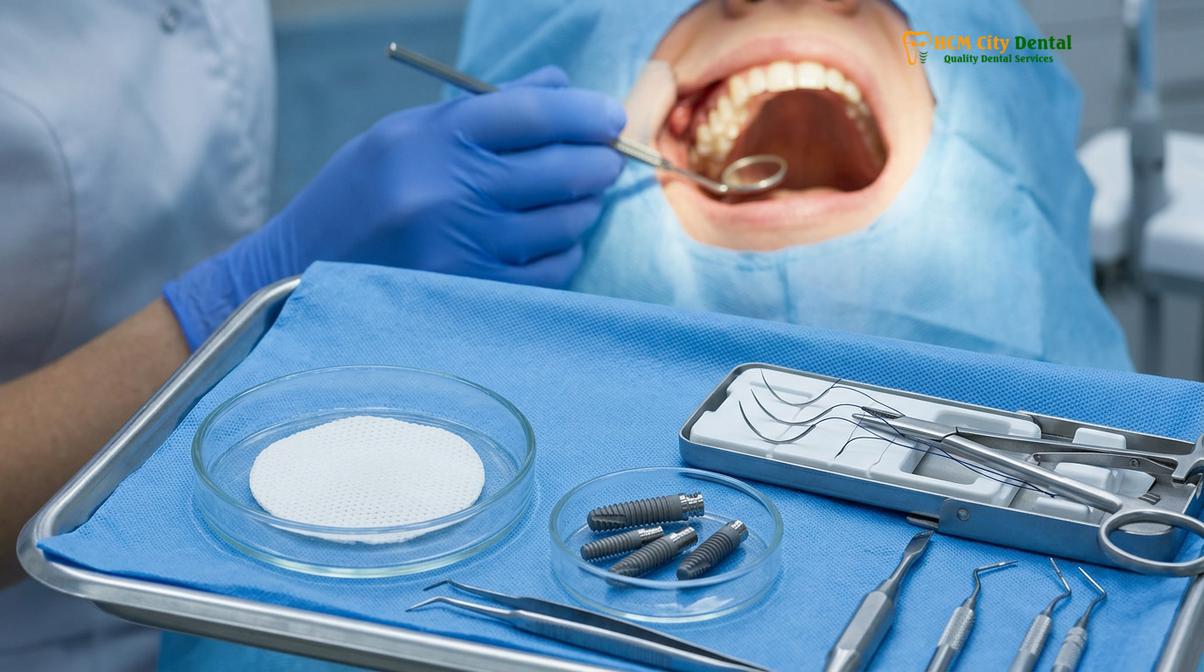
In addition to barrier membranes, some clinicians utilize biological modifiers such as Platelet-Rich Fibrin (PRF) or fibrin glue to enhance the seal and accelerate soft tissue healing. PRF, derived from the patient’s own centrifuged blood, is rich in growth factors and leukocytes, providing an excellent biological matrix that promotes rapid epithelialization of the torn mucosa.
Post-Operative Management: Preventing Infection and Sinus Pressure
Successful recovery relies on strict adherence to sinus precautions, including avoiding nose-blowing, alongside a prescribed regimen of broad-spectrum antibiotics and systemic decongestants to minimize sinus pressure.
The intraoperative repair of a sinus perforation is only the first step in managing this complication; rigorous post-operative care is equally critical to ensure the survival of the bone graft and the eventual success of the dental implant. The primary goal during the immediate healing phase is sinus infection prevention. Because the natural barrier between the oral cavity and the sinus has been temporarily compromised, the risk of bacterial contamination is elevated.
Pharmacological management is aggressive and highly specific. Patients are typically prescribed a course of broad-spectrum antibiotics, such as Amoxicillin combined with Clavulanate (Augmentin), to cover the diverse flora of the upper respiratory tract. For patients with penicillin allergies, alternatives like Azithromycin or Clindamycin are utilized. In addition to antibiotics, managing the internal pressure of the maxillary sinus is paramount. Systemic and topical nasal decongestants (e.g., Oxymetazoline nasal spray) are prescribed to keep the ostium—the natural drainage port of the sinus—open and functioning. This prevents the accumulation of fluid and the subsequent buildup of pressure that could dislodge the collagen membrane patch or push the bone graft out of position[6].
“The success of a repaired sinus perforation is heavily dependent on maintaining a patent ostium and preventing positive pressure within the sinus cavity. Pharmacological intervention with decongestants and strict patient compliance with sinus precautions are non-negotiable elements of the post-operative protocol.”
Patient compliance with “sinus precautions” is arguably the most critical factor in the healing process. Patients must be explicitly instructed to avoid any actions that create positive or negative pressure in the nasal and sinus cavities. This includes a strict prohibition against blowing the nose for at least two to three weeks. If the patient needs to sneeze, they must do so with their mouth wide open to dissipate the air pressure. Furthermore, activities such as playing wind instruments, inflating balloons, scuba diving, or even drinking through a straw must be completely avoided during the initial healing phase. Smoking is strictly contraindicated, as it paralyzes the cilia of the sinus membrane, severely impairing the mucociliary clearance mechanism and drastically increasing the risk of graft failure and infection.
When to Delay Implant Placement After a Sinus Perforation
Implant placement must be aborted and delayed for several months if the membrane tear exceeds 10 millimeters, if graft containment is impossible, or if acute purulent sinusitis is present.
While modern surgical techniques allow for the immediate repair of most minor perforations, there are definitive clinical scenarios where proceeding with bone grafting and implant placement is contraindicated. The decision to abort the procedure requires clinical maturity and a deep understanding of the biological limits of wound healing. If a tear is classified as Class IV or V (exceeding 10 to 15 millimeters in length), or if the tear occurs along the delicate ostium drainage pathway, the risk of massive graft displacement into the sinus cavity is unacceptably high.
In such instances, the most prudent course of action is to halt the augmentation procedure. The surgeon will carefully clean the surgical site, ensure hemostasis, and close the lateral window or crestal access with a barrier membrane and primary closure of the gingival flap. The patient is then placed on a standard post-operative pharmacological regimen and monitored closely. The Schneiderian membrane possesses a remarkable capacity for regeneration. Over a period of 4 to 6 months, the torn mucosa will naturally heal and re-epithelialize, restoring the integrity of the sinus lining[7].
Clinical Case Review: Managing a Complex Sinus Anatomy
A 55-year-old patient presented to HCMC Dental Clinic in Ho Chi Minh City seeking posterior maxillary implants. During the 3D CBCT evaluation, Dr. Nguyen Van Cuong identified a prominent Underwood’s septum dividing the sinus floor. During the lateral window approach, a 12mm tear occurred due to the severe angulation of the septum. Recognizing the high risk of graft migration, Dr. Cuong aborted the grafting phase and closed the site. After a 5-month healing period, a secondary CBCT confirmed complete regeneration of the membrane. The sinus lift was then successfully re-attempted, and the implants were placed without further complications, demonstrating the importance of clinical patience and proper complication management.
Attempting to force a repair on a massive tear often leads to catastrophic failure, resulting in acute sinusitis, complete loss of the bone graft, and the need for invasive functional endoscopic sinus surgery (FESS) by an ENT specialist. By choosing to delay the procedure, the implantologist prioritizes patient safety and ensures that the foundation for the future Dental Implants will be biologically sound upon re-entry.
Other Common Dental Implant Complications
Beyond sinus issues, implant therapy can be compromised by biological complications like peri-implantitis, mechanical failures such as screw loosening, and neurosensory deficits.
While sinus perforations are a primary concern in the upper jaw, a comprehensive understanding of dental implant complications requires examining issues that can affect any implant site. These complications are broadly categorized into biological and biomechanical failures.
Biological Complications: Peri-Implant Mucositis and Peri-Implantitis
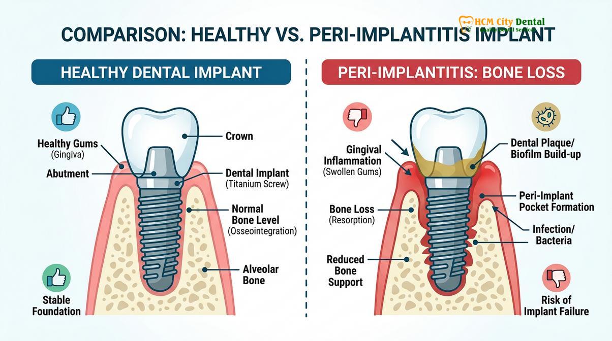
Biological complications are primarily driven by bacterial biofilm. Peri-implant mucositis is the reversible inflammation of the soft tissues surrounding the implant, analogous to gingivitis around natural teeth. If left untreated, this condition can progress to peri-implantitis, a severe inflammatory disease characterized by the progressive destruction of the supporting alveolar bone[8]. The microbiome associated with peri-implantitis shifts from healthy Gram-positive aerobic bacteria to highly pathogenic Gram-negative anaerobes. Treatment requires aggressive mechanical debridement of the contaminated titanium surface, localized antimicrobial therapy, and often surgical intervention to regenerate lost bone. Patients considering Dental Implants must commit to rigorous daily oral hygiene and professional maintenance to mitigate this risk.
Biomechanical Complications: Screw Loosening and Fracture
Biomechanical complications arise when the forces exerted on the implant prosthesis exceed the physical tolerance of the components. The most frequent mechanical issue is the loosening of the abutment screw. This occurs due to a phenomenon known as the “settling effect,” where microscopic rough spots on the mating surfaces of the implant and abutment flatten under occlusal load, causing a loss of screw preload. If a loose screw is not promptly retightened to the manufacturer’s specified torque, the continuous micro-movement can lead to metal fatigue and eventual fracture of the screw or the implant fixture itself. These complications are particularly prevalent in patients with untreated bruxism (teeth grinding) or those lacking sufficient posterior support. Proper occlusal adjustment and the use of night guards are essential preventive measures.
“Biomechanical overload is a silent destroyer of implant prosthetics. Ensuring passive fit of the framework, precise occlusal calibration, and managing parafunctional habits are critical steps in preventing late-stage mechanical failures.”
When evaluating Dental Implants systems, clinicians often consider the connection design—such as internal conical connections versus external hexes—as certain designs offer superior resistance to micro-movement and bacterial leakage, thereby reducing the incidence of both mechanical and biological complications.
When to See a Doctor
While minor discomfort and swelling are expected following any oral surgery, certain symptoms indicate a developing complication that requires immediate professional intervention. Early diagnosis is the key to salvaging an implant and preventing systemic spread of infection.
Important Clinical Considerations & Red Flags:
Seek immediate clinical evaluation if you experience any of the following symptoms after implant surgery or a sinus lift:
- Severe, throbbing facial pain that is not relieved by prescribed analgesics.
- Purulent (pus) discharge from the surgical site or a foul taste in the mouth.
- Persistent nosebleeds or the discharge of granular bone graft material from the nasal cavity.
- A sensation of air passing from the nose into the mouth when breathing or speaking.
- Prolonged numbness or tingling in the lips, chin, or tongue lasting more than 24 hours post-surgery, which may indicate nerve compression.
- Noticeable mobility or looseness of the implant crown.
If you are experiencing any of these warning signs, do not wait for your scheduled follow-up appointment. Contact your surgical provider immediately. For patients in Vietnam, the clinical team at HCMC Dental Clinic is equipped to handle complex diagnostic evaluations and emergency complication management. Understanding the Dental Implants process, including its risks, empowers patients to seek timely care. For those planning their treatment journey, reviewing the Dental Implants cost and ensuring you select a highly qualified specialist is the first step in complication prevention.
Frequently Asked Questions
What happens if the sinus membrane is torn during surgery?
If the sinus membrane is torn during surgery, the surgeon will immediately assess the size of the perforation to determine the appropriate repair protocol. Small tears are typically patched with a resorbable collagen membrane to prevent bone graft material from entering the sinus cavity. If managed correctly by an experienced clinician, the complication rarely impacts the long-term success of the implant, though it requires strict adherence to post-operative sinus precautions to prevent infection.
How do I know if my sinus was punctured during implant placement?
Signs of a punctured sinus may include mild nosebleeds on the side of the surgery, a sensation of air passing through the surgical site, or fluid draining into the nasal cavity. In some cases, patients may notice small granules of bone graft material discharging from the nose. If you experience severe facial pain, persistent nasal discharge, or throbbing pressure, it is critical to seek immediate clinical evaluation to rule out acute sinusitis.
Are sinus lifts safe in the long term?
Yes, sinus lifts are highly predictable and safe in the long term when performed by a skilled implantologist following strict surgical protocols. The augmented bone matures over several months, creating a stable, robust foundation for dental implants. Long-term clinical studies demonstrate that implants placed in grafted sinus bone exhibit survival rates comparable to those placed in native, non-grafted maxillary bone, provided the patient maintains excellent oral hygiene and attends regular professional cleanings.
Can a dental implant fail years after placement?
Yes, a dental implant can fail years after placement, most commonly due to a biological condition known as peri-implantitis or mechanical overloading. Peri-implantitis is an inflammatory disease triggered by bacterial plaque that progressively destroys the supporting jawbone around the titanium fixture. Mechanical failures, such as screw loosening or implant fracture, can occur if the prosthesis is subjected to excessive occlusal forces, particularly in patients with untreated bruxism or inadequate posterior tooth support.
How is peri-implantitis treated?
Peri-implantitis is treated through a combination of mechanical debridement, antimicrobial therapy, and sometimes surgical intervention to halt bone loss. The clinician will thoroughly clean the contaminated implant surface using specialized titanium or carbon curettes, air-abrasion devices, and localized antibiotics. In advanced cases, surgical flap debridement combined with bone grafting procedures may be necessary to regenerate lost tissue and re-establish a healthy biological seal around the implant collar.
References
- Journal of Oral and Maxillofacial Surgery. Management of Schneiderian membrane perforations during sinus augmentation procedures. (2021).
- Clinical Oral Implants Research. Incidence and management of intraoperative complications in dental implantology. (2020).
- Clinical Implant Dentistry and Related Research. Anatomical variations of the maxillary sinus and their impact on implant surgery. (2020).
- European Journal of Oral Implantology. The efficacy of collagen membranes in repairing sinus mucosal tears. (2018).
- Journal of the American Dental Association. Pharmacological management of sinus floor elevation complications. (2021).
- Journal of Periodontology. Peri-implantitis: Etiology, pathogenesis, and clinical management protocols. (2022).
- International Journal of Oral and Maxillofacial Implants. Biomechanical complications in implant-supported prostheses: A systematic review. (2019).
- Journal of Prosthetic Dentistry. Abutment screw loosening: Causes, prevention, and clinical management. (2019).