A sinus lift dental implant procedure is a specialized bone-grafting surgery designed to increase bone volume in the upper jaw. By carefully elevating the delicate sinus membrane, surgeons create sufficient vertical depth to securely anchor titanium implants in areas affected by severe bone loss.
Clinical Summary:
The maxillary sinus lift (sinus augmentation) is a foundational pre-implant procedure utilized when the posterior upper jaw lacks the necessary bone height to support dental implants. This deficiency typically arises from natural bone resorption following tooth extraction or the physiological expansion (pneumatization) of the sinus cavity. Depending on the patient’s existing bone volume, clinicians employ either a crestal (internal) approach for minor elevations or a lateral window (open) approach for extensive grafting. The procedure involves gently lifting the Schneiderian membrane and packing a biocompatible bone graft into the newly created space. Following a dedicated healing period of several months, the graft osseointegrates, forming a dense, stable foundation. Strict adherence to post-operative protocols—specifically avoiding actions that alter intra-sinus pressure—is critical to preventing complications and ensuring the long-term success of the subsequent implant placement.
Key Takeaways:
- A sinus lift restores lost vertical bone height in the upper premolar and molar regions, enabling secure implant placement.
- The procedure involves elevating the Schneiderian membrane and inserting bone graft material to stimulate new bone growth.
- Surgeons utilize either a minimally invasive crestal approach or a more comprehensive lateral window technique based on anatomical needs.
- Simultaneous implant placement is possible only if there is sufficient native bone to provide immediate mechanical stability.
- Post-operative compliance is vital; patients must strictly avoid sneezing with a closed mouth, using straws, or flying during early recovery.
Bone density and volume are critical parameters, especially for patients with systemic conditions; learn more about jawbone osteoporosis and implants.
The Sinus Cavity Limitation: Maxillary Bone Loss Challenges
The posterior upper jaw frequently presents a challenge for implantology due to the natural expansion of the sinus cavity and progressive bone resorption after tooth loss, necessitating surgical intervention to restore adequate vertical height.
The anatomy of the human maxilla presents unique challenges in the field of restorative dentistry. The maxillary sinuses are large, air-filled cavities located just above the posterior teeth (premolars and molars). In a healthy, dentate adult, the roots of these upper teeth often extend up into the sinus cavity, separated only by a paper-thin layer of cortical bone and the mucosal lining known as the Schneiderian membrane. When these posterior teeth are lost or extracted, a dual process of bone degradation begins, fundamentally altering the landscape of the upper jaw.
First, the alveolar ridge undergoes centripetal resorption. Without the mechanical stimulation previously provided by the tooth roots during chewing, the surrounding jawbone begins to atrophy, shrinking in both width and height. Second, a physiological phenomenon known as sinus pneumatization occurs. The air pressure within the empty sinus cavity causes it to gradually expand downward, encroaching upon the space where the tooth roots once resided. Over time, these two processes can leave a patient with merely a few millimeters of bone separating the oral cavity from the sinus space. Standard Dental Implants typically require a minimum vertical bone height of 8 to 10 millimeters to achieve primary stability and withstand the immense occlusal forces of chewing. Attempting to place an implant into a deficient maxilla without augmentation would result in the titanium fixture protruding directly into the sinus cavity, leading to chronic infections, sinusitis, and inevitable implant failure.
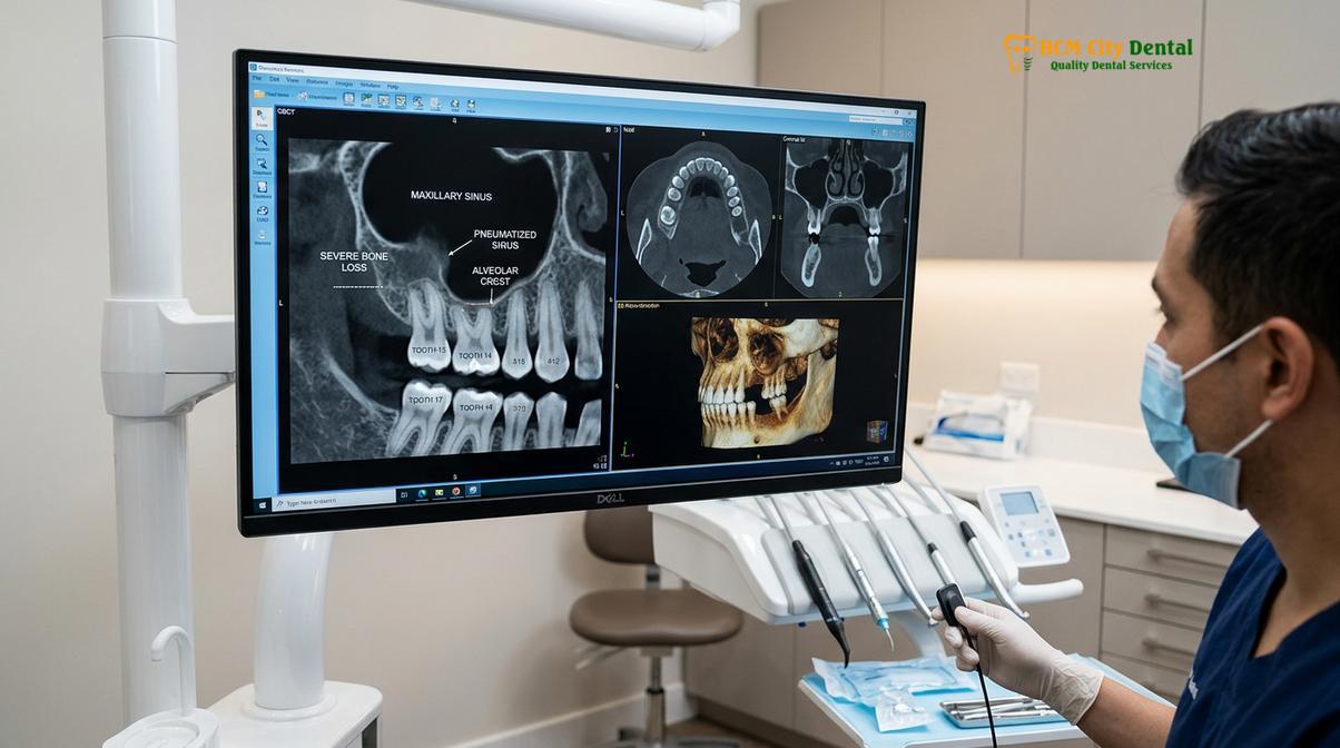
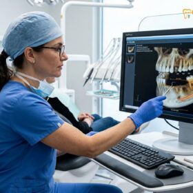
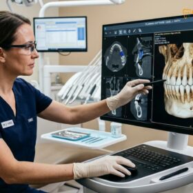
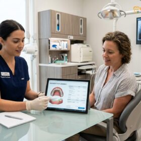
To overcome this anatomical limitation, the sinus lift procedure was developed. By surgically accessing the sinus cavity and performing a precise sinus membrane elevation, clinicians can create a protected space above the jawbone. This space is then filled with regenerative grafting materials. According to Dr. Nguyen Van Cuong, a leading specialist in complex oral rehabilitation, the success of this procedure relies heavily on meticulous pre-surgical planning. High-resolution 3D Cone Beam Computed Tomography (CBCT) scans are mandatory to evaluate the exact thickness of the remaining bone, the health of the sinus membrane, and the presence of any anatomical anomalies, such as Underwood’s septa (bony ridges within the sinus), which could complicate the elevation process.[1]
“The maxillary sinus is a dynamic anatomical structure. Our goal in sinus augmentation is not merely to add bone, but to carefully manipulate the sinus floor biology to create a robust, vascularized foundation that will seamlessly integrate with the titanium implant for decades to come.”
Understanding the vascular supply is also critical. The maxillary sinus is primarily supplied by the posterior superior alveolar artery and the infraorbital artery. During the surgical planning phase, the clinician must map these blood vessels to avoid intraoperative hemorrhage. By respecting the delicate anatomy and utilizing advanced imaging, modern sinus augmentation has become a highly predictable and routine procedure for patients requiring posterior maxillary implants, offering a superior alternative when comparing implant solutions to traditional dentures.
Internal vs. Lateral Window Sinus Lifts (Nâng xoang kín vs. Nâng xoang hở)
Surgeons select between a minimally invasive internal (crestal) approach for minor bone deficiencies and a comprehensive lateral window (open) approach for severe bone loss, tailoring the technique to the patient’s specific anatomical requirements.
In clinical practice, there are two primary surgical techniques utilized to achieve sinus floor elevation: the internal (crestal) approach and the lateral window approach. The decision of which technique to employ is dictated almost entirely by the patient’s residual bone height (RBH), as measured on the pre-operative CBCT scan. Both methods aim to achieve the same biological outcome—increasing vertical bone volume—but they differ significantly in their surgical execution, invasiveness, and indications.
The Crestal Approach (Closed Sinus Lift)
The crestal sinus lift, also known as the osteotome technique or closed approach, is generally indicated when the patient has a moderate amount of native bone remaining—typically between 5 to 8 millimeters. Because there is enough existing bone to provide initial mechanical stability, this technique is frequently performed simultaneously with the placement of the dental implant.[2]
During a crestal sinus lift, the surgeon does not make an incision on the side of the jaw. Instead, access to the sinus floor is achieved directly through the osteotomy—the small vertical hole drilled into the crest of the gum line to accommodate the implant. Once the drill reaches just below the sinus floor, specialized instruments called osteotomes are used to gently tap and fracture the thin remaining bone upward. Hydraulic pressure or specialized balloon systems may also be employed to carefully lift the Schneiderian membrane without tearing it. Bone graft material is then pushed up through the drill hole to fill the newly created space, and the implant is immediately threaded into position. This approach is highly favored for its minimal surgical trauma, reduced post-operative swelling, and shorter overall treatment timeline.
The Lateral Window Approach (Open Sinus Lift)
When a patient presents with severe bone atrophy—often possessing less than 4 millimeters of residual bone height—the crestal approach is contraindicated. In these complex cases, the lateral window approach (open sinus lift) becomes the mandatory standard of care. This technique allows for maximum visibility and the placement of a large volume of graft material.
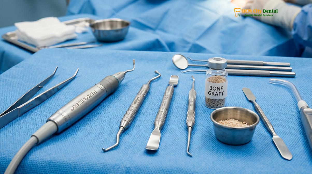
The procedure begins with the surgeon reflecting the gum tissue to expose the lateral (outer) wall of the maxillary bone. Using specialized piezosurgery instruments—which utilize ultrasonic vibrations to cut hard bone while preserving soft tissue—a small, oval-shaped window is created in the bone. Through this window, the surgeon gains direct visual access to the Schneiderian membrane. Using delicate curettes, the membrane is meticulously detached from the bony floor and elevated upward. The void created beneath the lifted membrane is then densely packed with bone grafting material. Finally, a protective collagen membrane is placed over the lateral window, and the gums are sutured closed.
| Clinical Parameter | Crestal Approach (Closed) | Lateral Window Approach (Open) |
|---|---|---|
| Residual Bone Height (RBH) | Typically 5mm to 8mm | Typically less than 4mm |
| Surgical Access | Vertical, through the implant drill hole | Horizontal, through the side of the jawbone |
| Implant Placement | Usually simultaneous | Usually delayed (two-stage protocol) |
| Volume of Graft | Small to moderate (1-3mm lift) | Large (can achieve 5-10mm+ lift) |
| Invasiveness & Recovery | Minimally invasive, faster recovery | More invasive, requires longer healing time |
Because the lateral window technique is utilized in cases of severe bone deficiency, it is almost always performed as a two-stage procedure. The graft must be allowed to heal and consolidate for six to nine months before the jaw is strong enough to receive premium implant systems like Straumann and Nobel Biocare. Attempting simultaneous placement in less than 4mm of bone carries an unacceptably high risk of implant micromovement and subsequent failure.
The Bone Placement Phase: Creating Depth for Upper Implants
Biocompatible grafting materials are strategically packed beneath the elevated membrane to act as a scaffold, stimulating the body’s natural regenerative processes to form dense, living bone capable of supporting an implant.
The core objective of any sinus augmentation is guided bone regeneration (GBR). The surgical elevation of the membrane merely creates the necessary space; it is the introduction of the bone graft material that dictates the biological success of the procedure. The graft serves as a three-dimensional scaffold, maintaining the elevated position of the membrane while simultaneously encouraging the patient’s own osteoblasts (bone-forming cells) to migrate into the area and synthesize new, living bone tissue.[3]
Clinicians have access to several categories of bone grafting materials, each with distinct histological properties. The selection of the graft depends on the size of the defect, the patient’s medical history, and the surgeon’s preference:
- Autografts: Bone harvested from the patient’s own body (often from the lower jaw or chin). Autografts are considered the “gold standard” because they are highly osteogenic, containing living bone cells that actively participate in regeneration. However, this requires a second surgical site, increasing morbidity.
- Allografts: Bone derived from a human tissue donor. This material is rigorously processed and sterilized. It is highly osteoconductive, providing an excellent scaffold for the patient’s own bone to grow into, without the need for a secondary harvest site.
- Xenografts: Bone derived from another species, most commonly bovine (cow) or porcine (pig). Xenografts are extensively purified to remove all organic proteins, leaving only the mineral matrix. They resorb very slowly, making them exceptional space maintainers, which is particularly useful in the large voids created during a lateral window approach.
- Alloplasts: Synthetic materials, such as hydroxyapatite or beta-tricalcium phosphate. These are entirely biocompatible and eliminate any risk of disease transmission, though they may not integrate as rapidly as biological grafts.
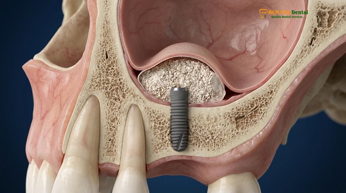
In modern clinical practice, surgeons frequently utilize a composite graft—a mixture of autogenous bone chips and xenograft particles. The autogenous bone provides the necessary cellular activity and growth factors, while the xenograft provides long-term structural stability, preventing the graft from resorbing before the implant can be placed. Once the graft is meticulously packed into the sinus cavity, a resorbable collagen barrier membrane is often placed over the lateral window. This barrier prevents fast-growing soft tissue cells (fibroblasts) from invading the graft site, ensuring that only slower-growing bone cells populate the area.
Clinical Case Review: Severe Maxillary Atrophy
A 62-year-old patient presented to HCMC Dental Clinic in Ho Chi Minh City with a desire to replace missing upper molars. CBCT imaging revealed severe pneumatization, leaving only 2mm of native bone. Dr. Cuong performed a lateral window sinus lift utilizing a 50/50 mixture of bovine xenograft and autogenous bone scrapings. A collagen membrane was secured over the osteotomy. After an 8-month consolidation period, follow-up imaging confirmed the successful regeneration of 12mm of dense vertical bone. Two titanium implants were subsequently placed with excellent primary stability, demonstrating the efficacy of staged implant procedures for senior patients.
The biological transformation of the graft material takes time. During the initial weeks, blood vessels infiltrate the graft (angiogenesis). Over the following months, osteoclasts break down the graft particles while osteoblasts lay down new native bone—a process known as creeping substitution. Rushing this healing phase can compromise the entire foundation, which is why patience is a critical component of the treatment workflow.
Recovery Protocols: Avoiding Sinus Pressure, Sneezing, and Flying
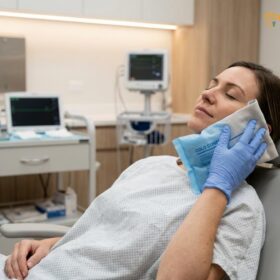
Strict adherence to post-operative instructions is paramount; patients must actively manage intra-sinus pressure to prevent membrane rupture, graft displacement, and surgical site infections during the critical early healing phase.
The success of a sinus lift is not determined solely in the operating chair; the patient’s post-operative compliance plays an equally vital role. The newly elevated Schneiderian membrane is incredibly delicate, and the particulate bone graft is initially held in place only by blood clots and the surrounding anatomy. Any sudden fluctuation in intra-sinus pressure can have catastrophic consequences, potentially tearing the membrane or forcing the graft material out of position. Therefore, patients must strictly avoid sinus pressure post-op.[4]
The most immediate risk during the recovery period is sneezing. A forceful sneeze with a closed mouth generates an immense spike in pressure within the nasal and sinus cavities. If a patient feels the urge to sneeze, they must be instructed to do so with their mouth completely open. This allows the explosive force of the air to vent safely through the oral cavity rather than being directed upward into the fragile surgical site. Similarly, patients must avoid blowing their nose for at least two to three weeks. If nasal congestion occurs, it should be managed with prescribed saline sprays or chemical decongestants, never by forceful blowing.
Critical Post-Operative Restrictions
To protect the integrity of the bone graft and the sinus membrane, patients must strictly adhere to the following rules for the first 14 to 21 days:
- Do not blow your nose: Wipe gently if necessary.
- Sneeze with your mouth open: Vent the pressure away from the sinuses.
- Avoid using straws: The suction creates negative pressure in the mouth, which can dislodge blood clots and disturb the graft.
- No heavy lifting or strenuous exercise: Physical exertion raises blood pressure and can induce throbbing or bleeding at the surgical site.
- Avoid playing wind instruments or inflating balloons.
Air travel presents another significant risk factor due to the principles of Boyle’s Law, which states that the volume of a gas expands as ambient pressure decreases. Commercial aircraft cabins are pressurized, but not to sea-level equivalents. As the plane ascends, the air trapped within the maxillary sinus expands. In a healthy sinus, this pressure equalizes through the ostium (the natural drainage pathway). However, post-surgical swelling can temporarily block the ostium. If a patient flies too soon after a sinus lift, the expanding trapped air can exert severe pressure on the healing membrane, causing excruciating pain (barotrauma) and potentially rupturing the surgical site. Most oral surgeons mandate a strict “no flying” rule for a minimum of 10 to 14 days post-surgery.
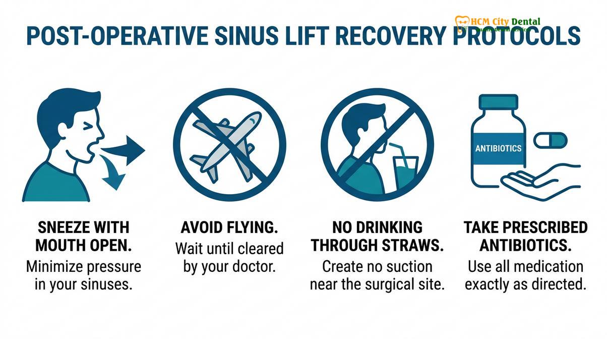
Pharmacological management is also a cornerstone of the recovery protocol. Because the sinus cavity is connected to the respiratory tract, the risk of infection is higher than in standard oral surgeries. Patients are typically prescribed a broad-spectrum antibiotic (such as Amoxicillin/Clavulanate) to prevent bacterial colonization of the graft. Anti-inflammatory medications (NSAIDs) and pain relievers are prescribed to manage post-operative discomfort and swelling, which usually peaks around 48 to 72 hours after the procedure. Chlorhexidine gluconate mouth rinses are often recommended to maintain oral hygiene without the need for aggressive brushing near the surgical incisions.
Clinical Success Rates and Sinus Health Considerations
When executed with precise surgical technique and proper patient compliance, sinus augmentation boasts exceptionally high long-term success rates, providing a durable and stable foundation for functional dental implants.
Extensive clinical literature and decades of longitudinal studies confirm that sinus augmentation is a highly predictable and successful procedure. Implants placed in grafted maxillary sinuses demonstrate survival rates that are comparable to, and in some studies slightly higher than, implants placed in native, non-grafted bone—consistently exceeding 95% over a 10-year period. The dense, regenerated bone provides an excellent biomechanical foundation, capable of withstanding the complex occlusal forces generated during mastication.[5]
However, the procedure is not without potential complications. The most frequently encountered intraoperative complication is the perforation of the Schneiderian membrane. The membrane is exceptionally thin—often measuring less than 1 millimeter in thickness—and can tear during the elevation process, particularly if the anatomy is complicated by sharp bony septa or previous sinus infections that have caused the tissue to become fibrotic and adherent. If a small perforation occurs, the surgeon can typically repair it immediately using a resorbable collagen patch or specialized suturing techniques, allowing the procedure to continue safely. If a large tear occurs, the procedure may need to be aborted. The site is allowed to heal for several months, after which the membrane naturally repairs itself, and the sinus lift can be reattempted.
Pre-existing sinus health is a critical determinant of candidacy. Patients with acute sinusitis, chronic severe allergic rhinitis, or nasal polyps may not be immediate candidates for a sinus lift. These conditions compromise the health of the membrane and the natural drainage pathways of the sinus cavity, significantly increasing the risk of post-operative infection and graft failure. In such cases, a collaborative approach with an Ear, Nose, and Throat (ENT) specialist is often required to resolve the underlying sinus pathology before any dental grafting can be considered. This comprehensive diagnostic approach is a hallmark of the care provided at HCMC Dental Clinic, ensuring that all biological variables are optimized prior to surgery.
“The integration of advanced 3D imaging and piezosurgery has revolutionized the safety profile of sinus augmentation. By minimizing trauma to the soft tissues and precisely managing the anatomical variables, we can offer patients a highly predictable pathway to restoring their upper dentition, even in cases of extreme bone atrophy.”
Ultimately, understanding the evaluating the pros and cons of dental implants involves recognizing the necessity of preparatory procedures like the sinus lift. While it extends the overall treatment timeline and adds to the cost of dental implants in Vietnam, the creation of a robust, biologically sound foundation is non-negotiable for long-term implant survival. Cutting corners on bone volume inevitably leads to biomechanical failure.
When to See a Doctor Post-Surgery
While post-operative swelling, mild bruising, and minor nasal bleeding are expected during the first few days following a sinus lift, certain symptoms warrant immediate clinical evaluation. You should contact your oral surgeon or visit a medical professional promptly if you experience any of the following warning signs:
- Persistent or Heavy Bleeding: Continuous epistaxis (nosebleeds) or bleeding from the surgical site that does not subside with gentle pressure.
- Signs of Infection: The presence of purulent (pus-like) discharge from the nose or the surgical incision, often accompanied by a foul odor or taste in the mouth.
- Severe, Unrelenting Pain: Throbbing pain that worsens after the first 72 hours and is not relieved by prescribed analgesic medications.
- Fever: A sustained body temperature above 101°F (38.3°C), which may indicate a systemic infection.
- Graft Displacement: The sensation of small, gritty particles falling into your mouth or nose, which may suggest that the incision has opened or the membrane has ruptured.
Early intervention is critical in managing complications. Do not attempt to self-diagnose or alter your prescribed medication regimen without consulting your dental care provider.
Frequently Asked Questions
What is the difference between open and closed sinus lifts?
An open sinus lift (lateral window approach) accesses the sinus through the side of the jawbone for extensive grafting, while a closed lift (crestal approach) accesses it through the implant drill hole for minor elevations. The choice depends entirely on your existing vertical bone height. If you have less than 4mm of native bone, the open approach is generally required to safely place a sufficient volume of graft material. Conversely, if you have 5mm or more, the less invasive closed approach may be utilized, often allowing for simultaneous implant placement.
How long after a sinus lift can I fly home?
Patients should generally wait at least 10 to 14 days after a sinus lift before flying to prevent barotrauma and graft displacement. Changes in cabin pressure can severely impact the healing sinus cavity. Because the sinus is an air-filled space, altitude changes cause gas expansion and contraction, which can put immense stress on the newly grafted bone and the delicate Schneiderian membrane. Your surgeon will evaluate your healing progress and provide specific clearance before you undertake any air travel.
Is it dangerous to sneeze after a sinus lift?
Yes, forcefully sneezing with your mouth closed can create extreme intra-sinus pressure, potentially rupturing the elevated membrane or displacing the bone graft. It is a critical post-operative risk. If you feel the urge to sneeze, you must do so with your mouth completely open to allow the air pressure to escape through your oral cavity rather than being forced up into the nasal and sinus passages. Managing allergies and avoiding irritants is highly recommended during the initial healing phase.
Can a dental implant be placed at the same time as a sinus lift?
Simultaneous implant placement is possible if there is enough native bone (usually 4-5mm) to provide primary mechanical stability for the titanium fixture. If the remaining bone is too thin to anchor the implant securely, a two-stage approach is mandatory. In a two-stage procedure, the bone graft is placed first and allowed to heal and solidify for six to nine months before the implant surgery is performed, ensuring a robust foundation for long-term success.
What happens if the sinus membrane tears during surgery?
If a minor perforation occurs in the Schneiderian membrane during elevation, the surgeon can typically repair it immediately using a resorbable collagen barrier or specialized suturing techniques. Membrane perforation is the most common intraoperative complication. While small tears are easily managed and do not compromise the final outcome, a severe tear may require the surgeon to abort the procedure, allow the membrane to heal naturally for a few months, and reattempt the sinus lift at a later date.
References
- Journal of Oral and Maxillofacial Surgery. Maxillary sinus augmentation: anatomical and clinical considerations. (2021).
- International Journal of Implant Dentistry. Comparative outcomes of lateral window and transalveolar sinus floor elevation. (2020).
- Clinical Oral Implants Research. Long-term survival rates of dental implants placed in grafted maxillary sinuses. (2019).
- Journal of Periodontology. The role of Schneiderian membrane thickness in sinus augmentation complications. (2022).
- European Journal of Oral Implantology. Post-operative management and barotrauma prevention following sinus lift procedures. (2018).