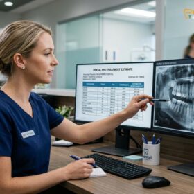
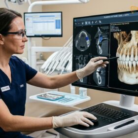
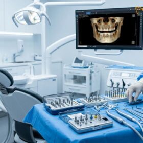
Peri implantitis prevention requires a strict, lifelong regimen of daily mechanical plaque removal and routine professional clinical maintenance. Because dental implants lack the natural periodontal ligament’s blood supply and immune defense, preventing bacterial biofilm accumulation is the most critical factor in ensuring long-term implant survival and surrounding bone health.
Clinical Summary:
Peri-implantitis is an inflammatory condition affecting the soft and hard tissues surrounding a dental implant, leading to progressive bone loss. Prevention hinges on disrupting the bacterial biofilm before it triggers a destructive immune response. Dr. Nguyen Van Cuong emphasizes that unlike natural teeth, implants have a fragile mucosal seal that is highly susceptible to bacterial invasion. Effective prevention combines meticulous home care—utilizing interdental brushes and oral irrigators—with professional scaling using implant-safe instruments. Early detection of peri-implant mucositis is fully reversible, making routine clinical monitoring and radiographic assessment at HCMC Dental Clinic essential for long-term implant success.
Key Takeaways:
- Peri-implantitis is primarily caused by bacterial plaque accumulation leading to inflammation and progressive bone loss.
- The condition often begins as peri-implant mucositis, a reversible inflammatory stage without bone loss.
- Implants lack a periodontal ligament, making them more susceptible to rapid inflammatory destruction than natural teeth.
- Daily mechanical cleaning using specialized interdental brushes and water flossers is mandatory for prevention.
- Professional maintenance every 3 to 6 months using titanium or plastic curettes prevents surface scratching and biofilm retention.
- Smoking and a history of severe periodontitis significantly increase the risk of implant failure.
What is Peri-Implantitis? The Silent Thread of Implant Failure
Peri-implantitis is a destructive inflammatory disease triggered by bacterial plaque, characterized by the progressive loss of supporting alveolar bone around a fully integrated dental implant.
To fully grasp the importance of peri implantitis prevention, one must first understand the unique anatomical and histological differences between a natural tooth and a Dental Implants restoration. While both function similarly in the oral cavity, their connection to the surrounding jawbone and gingival tissues is fundamentally different. A natural tooth is suspended in the bone by the periodontal ligament (PDL), a complex network of connective tissue fibers (Sharpey’s fibers) that insert directly into the root cementum. This ligament not only acts as a shock absorber but also provides a rich vascular supply and a robust immune defense mechanism against bacterial invasion.[1]
In contrast, a dental implant achieves stability through osseointegration—a direct structural and functional connection between the living bone and the surface of the load-bearing titanium implant. There is no periodontal ligament. The soft tissue surrounding the implant, known as the peri-implant mucosa, forms a “biological seal” to protect the underlying bone. However, the connective tissue fibers in this seal run parallel to the implant surface rather than inserting into it. This creates a mechanically weaker and less vascularized barrier. Dr. Nguyen Van Cuong frequently reminds patients that this fragile mucosal seal is the primary reason why implants are highly susceptible to bacterial infiltration and why meticulous oral hygiene is non-negotiable.
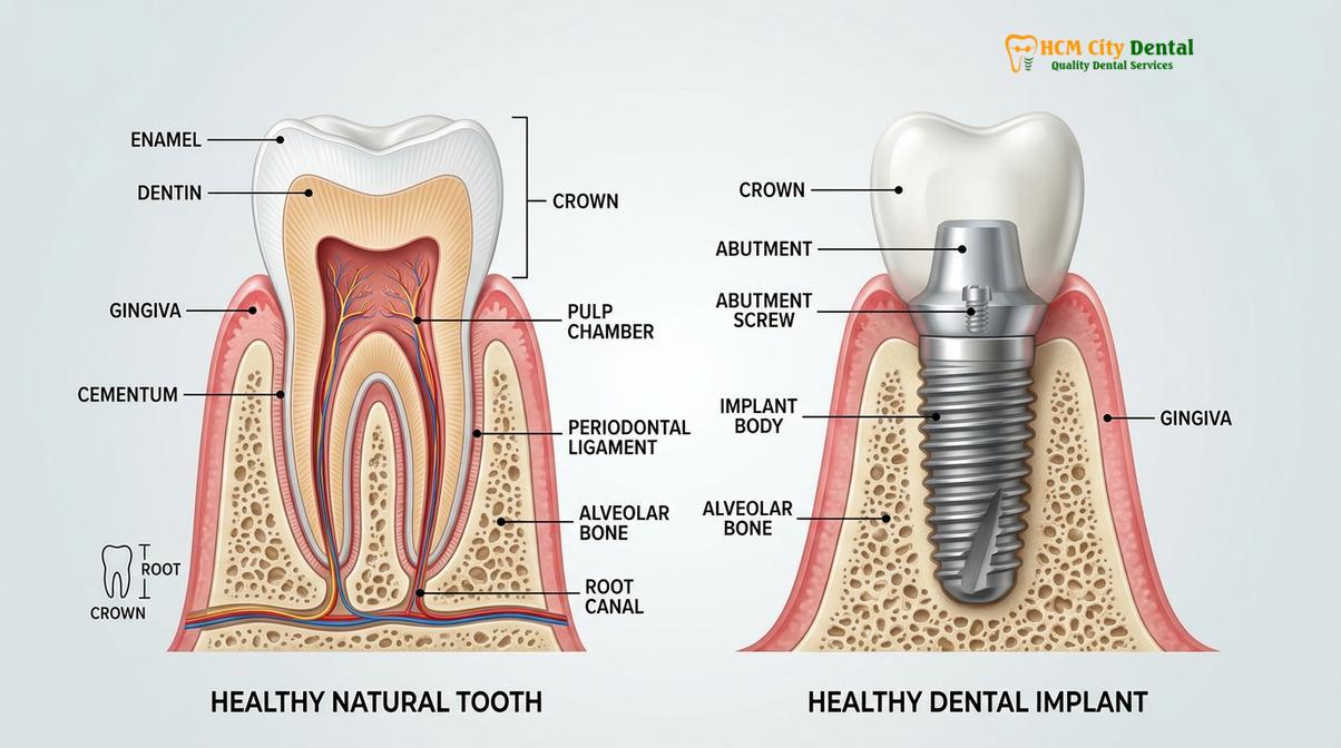
When oral hygiene is inadequate, bacterial biofilm accumulates at the gingival margin. This initial accumulation leads to peri-implant mucositis, which is the localized inflammation of the soft tissues surrounding the implant without any signs of bone loss. Peri-implant mucositis is the implant equivalent of gingivitis and is entirely reversible with appropriate intervention. However, if left untreated, the inflammation can penetrate deeper, breaching the biological seal and initiating an aggressive immune response that destroys the surrounding alveolar bone. This advanced stage is diagnosed as peri-implantitis. Unlike periodontitis around natural teeth, which often progresses slowly, peri-implantitis can advance rapidly due to the lack of the protective PDL barrier.[2]
Preventing this progression is the cornerstone of implant dentistry. Whether a patient has received a single implant, immediate loading implants, or a full-arch restoration, the biological principles of disease progression remain the same. The goal of prevention is to maintain the integrity of the peri-implant mucosa and prevent the shift from a healthy, symbiotic oral microbiome to a pathogenic, dysbiotic state.
Plaque Bacteria Accumulation and the Inflammatory Response
The primary etiology of peri-implant disease is the accumulation of a pathogenic bacterial biofilm on the implant surface, which provokes a localized, destructive immune response.
The oral cavity is a complex ecosystem harboring hundreds of bacterial species. In a state of health, these microorganisms exist in harmony with the host. However, the titanium surface of a dental implant, particularly the micro-roughened areas designed to enhance osseointegration, can serve as an ideal substrate for bacterial colonization if exposed to the oral environment. The process begins within minutes of cleaning, as salivary glycoproteins form a conditioning film known as the acquired pellicle on the implant crown and abutment.
Primary colonizers, mostly Gram-positive aerobic bacteria, adhere to this pellicle. If not mechanically disrupted through daily brushing and flossing, these early colonizers multiply and create an extracellular matrix, forming a mature biofilm. As the biofilm thickens, the environment at its base becomes oxygen-depleted, favoring the proliferation of highly pathogenic Gram-negative anaerobic bacteria, such as Porphyromonas gingivalis and Aggregatibacter actinomycetemcomitans. This shift in the microbial flora is the critical turning point in the development of peri-implant gum inflammation.[3]
“The transition from a state of peri-implant health to active disease is driven by a dysbiotic shift in the biofilm microbiome, which overwhelms the localized host defenses and triggers a cascade of tissue-destructive inflammatory cytokines.”
The host’s immune system detects the presence of these pathogenic bacteria, specifically their toxic byproducts like lipopolysaccharides (LPS). In response, the body dispatches immune cells, including neutrophils and macrophages, to the site of infection. While the intention of the immune response is to eliminate the bacterial threat, the continuous presence of the biofilm leads to chronic inflammation. The immune cells release pro-inflammatory cytokines, such as Interleukin-1 (IL-1) and Tumor Necrosis Factor-alpha (TNF-alpha). These biochemical signals inadvertently activate osteoclasts—the cells responsible for breaking down bone tissue.[4]
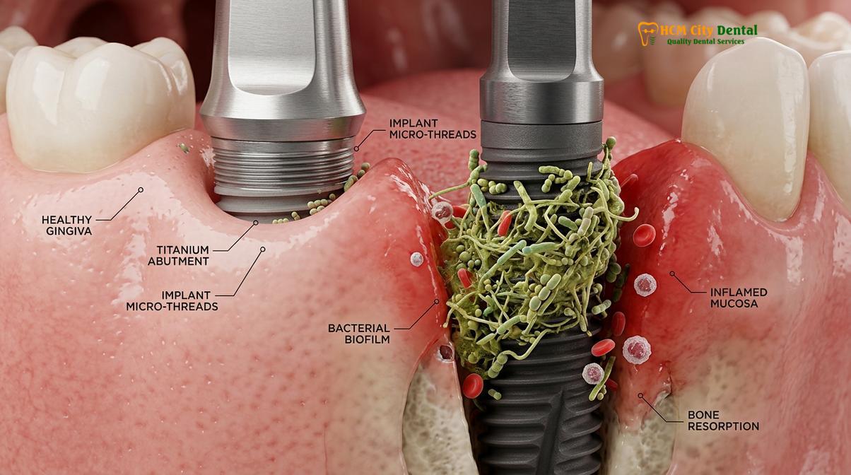
This localized osteoclastic activity results in the progressive resorption of the alveolar bone supporting the implant. Because the inflammatory infiltrate in peri-implantitis extends closer to the bone crest compared to periodontitis around natural teeth, the rate of bone destruction is often significantly faster. This biological mechanism underscores why routine dental plaque scaling and professional debridement are absolutely critical. By physically disrupting the biofilm architecture, the bacterial load is reduced to a level that the host immune system can manage without triggering bone resorption. Patients who have undergone complex procedures, such as a bone graft for dental implant placement, must be particularly vigilant, as the newly regenerated bone can be highly vulnerable to inflammatory breakdown.
Symptoms to Watch For: Bleeding Gums, Redness, and Bone Loss
Early clinical signs of peri-implant disease include mucosal redness, swelling, and bleeding on probing, which can progress silently to deep pocketing and radiographic bone loss.
One of the most challenging aspects of peri-implantitis is its insidious nature. In its early stages, the disease is often entirely painless, leading many patients to unknowingly harbor active inflammation. Recognizing the clinical signs early is paramount for successful intervention and implant bone loss prevention. The transition from health to disease is marked by specific clinical parameters that dental professionals monitor closely during recall visits.
The earliest and most reliable indicator of peri-implant inflammation is Bleeding on Probing (BOP). When a dentist or hygienist gently inserts a periodontal probe into the sulcus (the shallow crevice between the gum and the implant), healthy tissue will not bleed. If bleeding occurs, it signifies that the epithelial lining of the sulcus is ulcerated and inflamed. This is the hallmark of peri-implant mucositis. Patients may also notice this bleeding at home while brushing or using interdental cleaners. Additionally, the peri-implant mucosa may appear erythematous (red), swollen, and glossy, losing its healthy, firm, pink stippled appearance.[5]
Clinical Warning: Never ignore bleeding gums around a dental implant. While natural teeth may occasionally bleed due to aggressive brushing, bleeding around an implant is a definitive sign of active bacterial infection and requires immediate professional evaluation to prevent irreversible bone destruction.
As the disease progresses to peri-implantitis, more severe clinical signs manifest. Suppuration, or the presence of pus exuding from the implant sulcus, is a clear indication of a severe, active bacterial infection. Probing depths will increase significantly. A healthy implant typically has probing depths of 2 to 4 millimeters. In peri-implantitis, the destruction of the connective tissue attachment and underlying bone creates deep pathological pockets, often exceeding 5 or 6 millimeters. These deep pockets become anaerobic reservoirs, further accelerating bacterial growth and making home care virtually impossible.
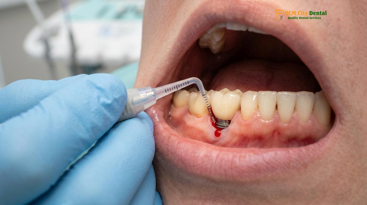
Radiographic assessment is the definitive diagnostic tool for confirming peri-implantitis. A baseline radiograph taken at the time of prosthesis delivery is essential for future comparison. In active disease, subsequent radiographs will reveal a characteristic crater-like or “saucer-shaped” pattern of bone loss around the coronal portion of the implant fixture. If the bone loss progresses unchecked, the implant may eventually become mobile. However, implant mobility is a very late-stage sign; by the time an implant is noticeably loose, complete failure has usually occurred, and removal is the only viable option. Patients with a history of periodontal disease or those who have required a sinus lift for upper jaw implants must be monitored with heightened scrutiny, as their anatomical and biological risk factors are elevated.
Home Care Protocols: Dental Flossing, Interdental Brushing, and Water Flossers
Effective daily home care requires mechanical disruption of the biofilm using soft-bristled brushes, appropriately sized interdental cleaners, and targeted oral irrigators to protect the peri-implant seal.
The foundation of peri implantitis prevention rests squarely on the patient’s daily oral hygiene routine. Because the biological seal around an implant is delicate, the goal of home care is to thoroughly remove bacterial plaque without causing mechanical trauma to the soft tissues or scratching the restorative components. A standard toothbrushing routine is insufficient for implant patients; specialized tools and techniques are required to navigate the unique contours of implant-supported prostheses.
Brushing should be performed at least twice daily using a soft-bristled manual or electric toothbrush. The Modified Bass technique is highly recommended: the brush bristles are angled at 45 degrees toward the gumline, and a gentle vibratory motion is used to sweep plaque away from the sulcus. Abrasive toothpastes, such as those marketed for heavy stain removal or whitening, should be avoided, as they can scratch the polished surface of the implant abutment or the ceramic crown, creating micro-retentive areas for future plaque accumulation.
Interproximal cleaning—cleaning between the teeth—is where most implant hygiene regimens fail. Standard dental floss can be difficult to adapt around the cylindrical shape of an implant abutment. Instead, spongy or tufted floss is often recommended. The floss should be wrapped around the implant in a “C-shape” and gently moved in a shoe-shine motion to disrupt the biofilm. However, care must be taken not to snap the floss aggressively into the gumline, which could sever the fragile mucosal attachment.
Clinical Case Review: A 55-year-old patient presented to HCMC Dental Clinic in Ho Chi Minh City with generalized bleeding around a full-arch implant bridge. Clinical examination revealed heavy plaque accumulation beneath the prosthesis. After professional debridement, the patient was instructed on a customized water flosser cleaning guide and the use of specific interdental brushes. At the three-month recall, the peri-implant tissues were firm, pink, and completely free of bleeding, demonstrating the profound impact of targeted home care.
Interdental brushes are often more effective than floss for cleaning the embrasure spaces between implants or between an implant and a natural tooth. It is crucial to select a brush with a nylon-coated or Teflon-coated central wire; exposed metal wires can scratch the titanium implant surface. The brush should fit snugly but comfortably into the space without requiring excessive force.
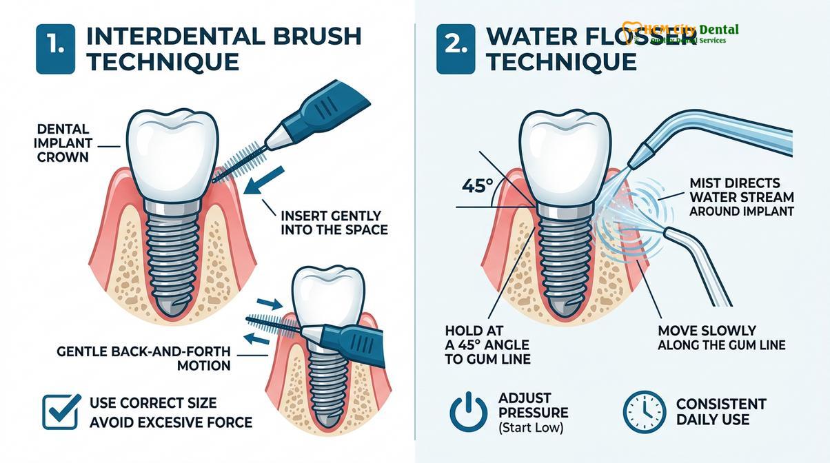
Oral irrigators, commonly known as water flossers, are an invaluable adjunct for implant patients, particularly those with complex restorations like implant supported overdentures or multi-unit bridges. A proper water flosser cleaning guide dictates that the device should be used on a low to medium pressure setting. The pulsating stream of water should be directed horizontally through the interdental spaces, perpendicular to the long axis of the tooth. Directing the high-pressure stream vertically down into the peri-implant pocket is contraindicated, as it can force bacteria deeper into the tissues or physically detach the mucosal seal. When used correctly, water flossers effectively flush out unattached plaque and food debris, significantly reducing the inflammatory burden on the peri-implant tissues.
Professional Scaling and Cleaning Frequencies for Implant Patients
Professional maintenance every 3 to 6 months utilizes specialized, non-abrasive instruments to safely remove calculus without scratching the titanium surface, ensuring long-term tissue health.
While meticulous home care is the first line of defense, it cannot completely prevent the formation of calculus (tartar). Over time, residual plaque biofilm mineralizes into hard calculus deposits that adhere stubbornly to the implant components. These deposits cannot be removed by brushing or flossing and require professional intervention. Routine clinical maintenance is a mandatory component of implant therapy, serving both as a preventive measure and a diagnostic checkpoint.
The frequency of professional recall visits is highly individualized, based on the patient’s specific risk profile. Factors such as a history of severe periodontitis, smoking status, systemic conditions like poorly controlled diabetes, and the complexity of the prosthetic design all influence the recall interval. Generally, a 3 to 4-month recall is recommended for the first year following implant placement. If the peri-implant tissues remain stable and the patient demonstrates excellent home care, the interval may be extended to 6 months. However, high-risk patients must remain on a strict 3-month maintenance schedule indefinitely.
“The longevity of a dental implant is directly proportional to the quality and frequency of professional maintenance. Neglecting routine clinical recall is the leading cause of late-stage implant complications.”
The instrumentation used during professional implant maintenance differs significantly from that used on natural teeth. Traditional stainless steel curettes and ultrasonic scaler tips are strictly contraindicated for use on titanium implants. Stainless steel is harder than titanium and will inevitably scratch the implant surface or the dental implant abutment. These microscopic scratches increase the surface roughness, creating an ideal environment for accelerated bacterial colonization. Instead, dental professionals utilize specialized instruments made of softer materials, such as solid titanium, carbon fiber, or PEEK (polyetheretherketone) plastic. These instruments effectively remove calculus without altering the topography of the implant components.
| Clinical Parameter | Natural Teeth | Dental Implants |
|---|---|---|
| Attachment Type | Periodontal Ligament (PDL) | Osseointegration & Mucosal Seal |
| Probing Force | Standard pressure (approx. 0.25 N) | Light pressure to protect fragile seal |
| Scaling Instruments | Stainless steel curettes, standard ultrasonics | Titanium, PEEK plastic, carbon fiber curettes |
| Polishing Method | Prophy paste, sodium bicarbonate air-polishing | Non-abrasive paste, glycine powder air-polishing |
| Disease Progression | Generally slower, contained by PDL | Rapid bone loss due to lack of PDL barrier |
In addition to hand instrumentation, modern implant maintenance often incorporates air-polishing technology. However, traditional sodium bicarbonate powder is too abrasive for titanium. Instead, glycine-based or erythritol-based powders are utilized. These low-abrasive powders are delivered via a specialized subgingival nozzle, safely and efficiently eradicating the biofilm from the micro-threads of the implant without causing surface damage. For patients who have undergone advanced procedures like Piezotome dental implant surgery, maintaining pristine surface conditions is vital to protect the delicate bone-to-implant interface.
When to See a Doctor: Important Clinical Considerations
Immediate clinical evaluation is required if you experience persistent bleeding, swelling, or discomfort around an implant, as early intervention can reverse inflammation and prevent bone loss.
Patient awareness is a critical component of peri implantitis prevention. Because the early stages of peri-implant disease are often asymptomatic, patients must be vigilant in monitoring their oral health between scheduled maintenance visits. You should schedule an immediate clinical evaluation if you notice any of the following warning signs:
- Persistent Bleeding: Any bleeding during brushing, flossing, or eating is abnormal and indicates active mucosal inflammation.
- Swelling or Redness: Changes in the color or contour of the gums around the implant, particularly if the tissue appears engorged or purplish.
- Suppuration: The presence of pus or a bad taste originating from the implant site is a sign of advanced bacterial infection.
- Discomfort or Dull Ache: While severe pain is rare, a persistent dull ache or sensitivity upon chewing warrants investigation.
- Visible Thread Exposure: If the gum tissue recedes and the metallic threads of the implant become visible, bone loss has likely occurred.
- Implant Mobility: Any perceived looseness of the implant crown or the fixture itself is a critical emergency requiring immediate attention.
Dr. Cuong advises that early intervention is the key to successful management. If diagnosed at the mucositis stage, the inflammation is entirely reversible with professional debridement and optimized home care. However, once the disease progresses to peri-implantitis and bone loss occurs, the treatment becomes significantly more complex, often requiring surgical intervention, surface decontamination, and guided bone regeneration procedures. Regular monitoring at a specialized facility like HCMC Dental Clinic in Ho Chi Minh City ensures that any deviations from health are detected and managed promptly, safeguarding your investment in your dental health.
Frequently Asked Questions
How do I clean my new dental implants at home?
You must clean your dental implants daily using a soft-bristled toothbrush, non-abrasive toothpaste, and specialized interdental brushes or water flossers. Because implants lack a natural protective ligament, meticulous mechanical plaque removal around the gumline is essential to prevent bacterial accumulation and subsequent inflammation. Ensure you use the Modified Bass brushing technique and select interdental brushes with coated wires to avoid scratching the titanium surface.
Is bleeding around implants normal?
No, bleeding around a dental implant is never normal and is a primary clinical indicator of active inflammation. It typically signifies peri-implant mucositis, an early and reversible stage of infection that requires immediate professional evaluation to prevent irreversible bone loss. If you notice bleeding while brushing or flossing, do not ignore it; schedule a clinical assessment promptly.
How is peri-implantitis treated at HCMC Dental?
Treatment at HCMC Dental involves a stepwise approach, starting with non-surgical mechanical debridement using titanium instruments and glycine air-polishing. If bone loss is advanced, surgical intervention, surface decontamination, and guided bone regeneration may be required to stabilize the implant. The specific protocol depends on the severity of the bone defect and the morphological characteristics of the lesion.
Can a water flosser damage my dental implants?
A water flosser is highly beneficial and safe for dental implants when used correctly on a low to medium pressure setting. You should direct the pulsating water stream horizontally between the teeth rather than vertically into the gum pocket to avoid disrupting the fragile mucosal seal. When utilized as part of a comprehensive daily routine, it effectively flushes out unattached biofilm and debris.
How often should I get my implants professionally cleaned?
Implant patients should undergo professional clinical maintenance every three to six months, depending on their individual risk profile. Frequent professional scaling using specialized, non-scratching instruments is critical to remove calcified deposits that cannot be managed with home care alone. High-risk patients, such as smokers or those with a history of periodontitis, require more frequent monitoring.
References
- Journal of Clinical Periodontology. Peri-implant diseases and conditions: Consensus report of workgroup 4 of the World Workshop. (2018).
- Clinical Oral Implants Research. The pathogenesis of peri-implantitis: A comprehensive review of the literature. (2019).
- Journal of Periodontology. Mechanical and chemical protocols for the prevention of peri-implant mucositis. (2020).
- International Journal of Oral & Maxillofacial Implants. Efficacy of air-polishing in the maintenance of dental implants. (2021).
- Clinical Implant Dentistry and Related Research. Risk indicators for peri-implant disease: A systematic review. (2022).