For international patients, expatriates, and dental tourists traveling to Vietnam, the prospect of restoring a confident smile through advanced dental procedures is highly appealing. However, when considering complex oral surgery, clinical safety and diagnostic precision must remain the top priorities. The foundation of safe dental implants ho chi minh city relies heavily on the diagnostic technology utilized before the first incision is ever made. In modern implantology, the transition from traditional two-dimensional imaging to advanced three-dimensional digital planning has transformed patient care, helping to minimize surgical risks and support the long-term stability of the restoration.
At HCMC Dental Clinic, the integration of hospital-grade diagnostic protocols into daily practice is a standard of care. By utilizing in-house Cone Beam Computed Tomography (CBCT) and computer-generated surgical guides, the clinical team strives to ensure that every implant placement is meticulously planned. This comprehensive overview explores why seeking a cbct 3d dental scan clinic saigon is a critical step for anyone undergoing implant therapy, highlighting the profound impact of digital diagnostics on surgical safety.
Clinical Summary:
The integration of in-house 3D CBCT imaging and digital surgical guides represents a highly advanced approach to implant dentistry. This technology allows dental surgeons to visualize bone volume, map critical nerve pathways, and assess sinus cavities in three dimensions prior to the actual procedure. By utilizing computer-aided design and manufacturing (CAD/CAM) to create custom surgical templates, clinics can often perform minimally invasive procedures that aim to reduce tissue trauma and support efficient healing. For international patients seeking care in Vietnam, selecting a clinic equipped with these in-house digital diagnostic tools is a vital step toward ensuring surgical safety and predictable implant outcomes.
Key Takeaways:
- Diagnostic Clarity: 3D CBCT scans help eliminate the distortion and superimposition often seen in traditional 2D panoramic x-rays, providing a detailed anatomical representation of the jaw.
- Anatomical Protection: Advanced digital planning software enables surgeons to map the inferior alveolar nerve and maxillary sinus, which generally reduces the risk of surgical complications.
- Guided Precision: Computer-generated surgical templates restrict drill angulation and depth, facilitating minimally invasive techniques that can lead to more comfortable recovery periods.
- In-House Efficiency: Clinics with on-site CBCT scanners can prevent treatment delays and allow for real-time surgical adjustments, a significant benefit for patients on tight travel schedules.
- Comprehensive Care: Accessing digitally guided implant surgery in Ho Chi Minh City allows patients to benefit from advanced technology while receiving care tailored to international safety standards.
2D Panoramic X-Rays vs 3D Cone Beam Computed Tomography (CBCT)
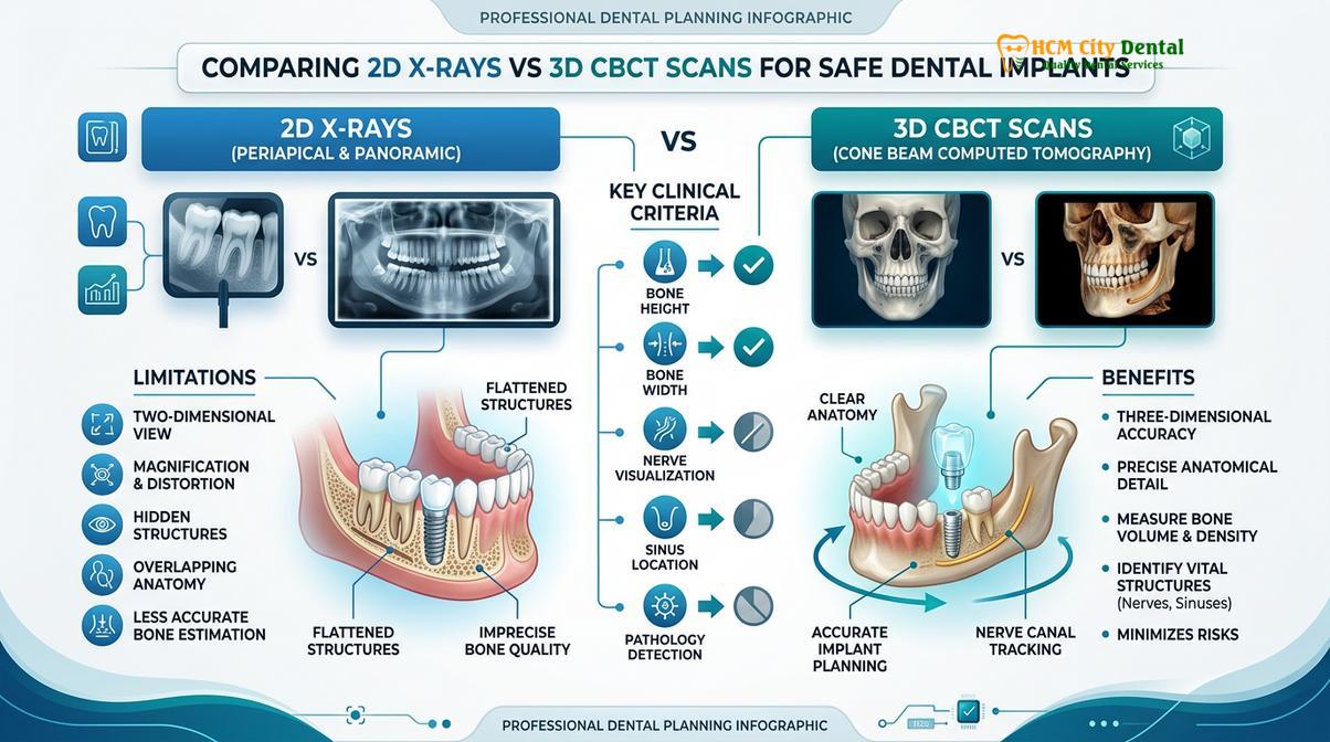
Historically, dental professionals relied heavily on 2D panoramic x-rays (OPGs) to assess a patient’s suitability for dental implants. While these images provide a helpful general overview of the teeth and jaws, they possess inherent clinical limitations when it comes to complex surgical planning [1]. A 2D x-ray compresses a curved, three-dimensional structure onto a flat plane. This compression can cause magnification errors, distortion, and the superimposition of anatomical structures, making precise measurements challenging.
When planning for safe dental implants, relying solely on 2D imaging can leave critical anatomical details obscured. A surgeon can estimate the height of the bone, but they cannot accurately determine the width or the exact density from a flat image [2]. If the bone is narrower than it appears on the 2D film, an implant placed without 3D guidance could potentially compromise the buccal (cheek) or lingual (tongue) cortical plates, which may lead to complications or affect the longevity of the implant.
| Diagnostic Feature | 2D Panoramic X-Ray | 3D CBCT Scan |
|---|---|---|
| Dimensionality | Flat, compressed 2D image | True 3D volumetric rendering |
| Distortion Level | Prone to magnification and superimposition | 1:1 ratio with minimal to no distortion |
| Bone Assessment | Shows height only | Shows height, width, and density |
| Nerve Mapping | Estimated position | Precise 3D tracing and measurement |
| Surgical Guide Creation | Not possible | Essential foundation for CAD/CAM guides |
The Mechanics of 3D CBCT Technology
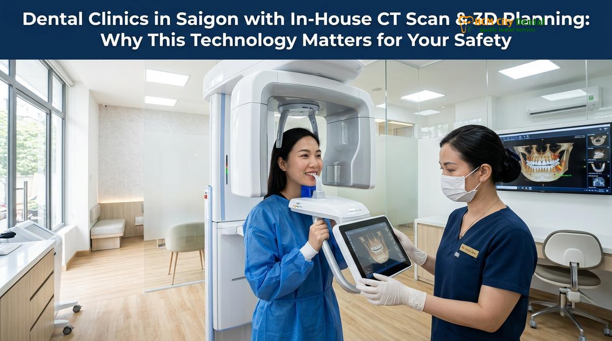
Cone Beam Computed Tomography (CBCT) represents a significant advancement in digital diagnostic dentistry. Unlike traditional medical CT scanners that capture data in multiple slices, a CBCT scanner utilizes a cone-shaped x-ray beam that rotates around the patient’s head in a matter of seconds. This single rotation captures hundreds of high-resolution images from different angles, providing a comprehensive dataset for the clinical team.
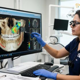
Advanced computer algorithms then reconstruct these images into a highly accurate, three-dimensional volume composed of tiny 3D pixels called voxels. Because these voxels are isotropic (equal in all dimensions), the resulting 3D model provides a highly accurate representation of the patient’s anatomy. The surgeon can manipulate this digital model on a computer screen, slicing through the jawbone in axial, coronal, and sagittal planes to examine the surgical site from multiple perspectives.
For patients requiring extensive rehabilitation, such as those exploring Multiple Dental Implants, this level of diagnostic clarity is considered a fundamental baseline for safe, predictable care.
Mapping Nerve Canals, Sinus Cavities & Bone Density in 3D
The human jaw is a complex anatomical landscape housing critical structures that must be carefully navigated during oral surgery. The primary advantage of a 3D CBCT scan is the ability to visualize and map these structures with a high degree of certainty before the patient even sits in the surgical chair. According to guidelines supported by the Vietnam Odonto-Stomatology Association (VOSA), utilizing 3D imaging is a key component of modern implant safety protocols in Vietnam [3].
Protecting the Inferior Alveolar Nerve (IAN)
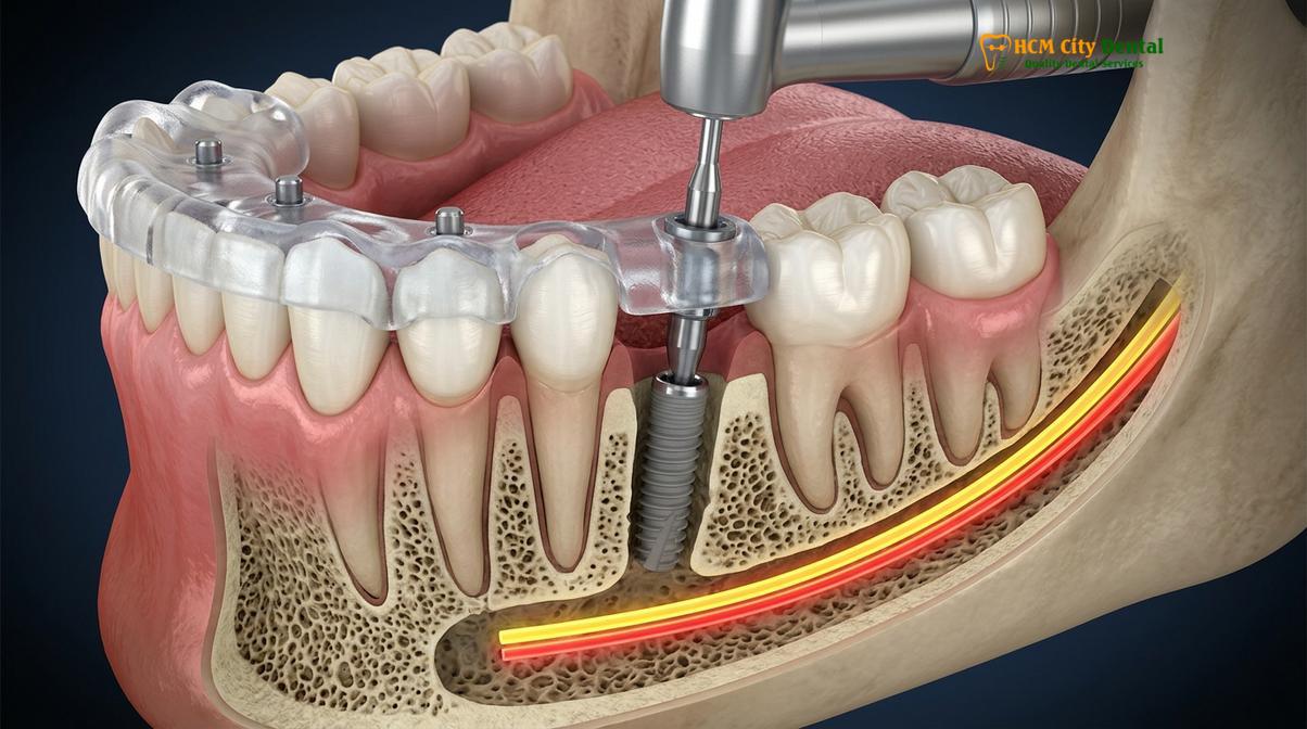
In the lower jaw (mandible), one of the most critical structures is the inferior alveolar nerve, which runs through the mandibular canal and exits at the mental foramen near the premolars. This nerve provides sensation to the lower lip, chin, and teeth. If a dental instrument or an implant encroaches upon this nerve, the patient may experience temporary or, in rare cases, prolonged paresthesia (numbness or tingling) in their lower face.
Using 3D planning software, the dental surgeon can digitally trace the exact pathway of the mandibular canal. The software allows the clinician to select a virtual implant of the exact length and diameter needed and place it into the digital bone model. The system can be programmed to maintain a strict safety margin between the apex of the implant and the nerve canal, aiming to ensure safety during the physical procedure.
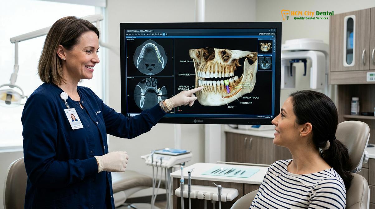
“Comprehensive digital planning is a cornerstone of modern implantology. By utilizing 3D CBCT scans, we can evaluate bone density, map nerve pathways, and simulate the surgery virtually. This proactive approach is essential for minimizing risks and supporting predictable, long-term outcomes for our patients.”
Navigating the Maxillary Sinus
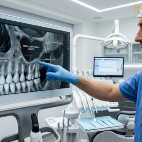
In the upper jaw (maxilla), the primary anatomical consideration is the maxillary sinus—a pair of air-filled cavities located just above the upper back teeth. When upper molars are lost, the bone in this area tends to resorb over time, and the sinus cavity may expand downward. Consequently, there is often insufficient vertical bone height to anchor a dental implant securely.
A 3D scan allows the surgical team to measure the exact thickness of the remaining bone beneath the sinus membrane. If the bone is too thin, the surgeon can plan a sinus lift procedure to graft new bone into the area. Furthermore, the CBCT scan can reveal pre-existing sinus conditions, such as mucosal thickening or polyps, which must be evaluated before implant surgery can safely proceed.
Computer-Generated Surgical Templates for Micron-Level Implant Placement
The diagnostic data gathered from the CBCT scan reaches its full potential when combined with guided implant surgery ho chi minh protocols. Guided surgery bridges the gap between digital planning and physical execution, aiming to ensure that the virtual plan is transferred to the patient’s mouth with exceptional accuracy.
The Digital Workflow
The process of creating a surgical guide involves a seamless digital workflow designed to optimize precision:
- Data Acquisition: The patient undergoes a 3D CBCT scan to capture the bone anatomy. Simultaneously, an intraoral scanner is used to capture a highly accurate 3D optical impression of the teeth and soft tissues, often eliminating the need for traditional putty impressions.
- Data Merging: The imaging files are imported into advanced implant planning software. The software merges the datasets, creating a comprehensive virtual model of the patient’s mouth that includes both the visible structures and the underlying bone.
- Prosthetically Driven Planning: The surgeon first designs the final crown or bridge in the software. Then, working backward, they position the virtual implant in the bone to optimally support that future restoration. This approach aims to ensure favorable aesthetics and biomechanical function.
- Guide Fabrication: Once the virtual implant is positioned, the software designs a custom surgical template that fits snugly over the patient’s remaining teeth or gums. This design is then sent to a high-precision 3D printer.
For complex, full-arch rehabilitations, such as those detailed in our All-on-6 Dental Implants Vietnam Cost guide, surgical guides are highly beneficial for ensuring that all implants are placed at optimal angles to support the full-arch prosthesis.
Clinical Case Study: Precision in the Esthetic Zone
Patient Profile: A 45-year-old expatriate visited HCMC Dental Clinic in Ho Chi Minh City with a fractured upper central incisor that required extraction and evaluation for implant placement.
Clinical Challenge: The front teeth (esthetic zone) require meticulous attention to implant angulation. Even a slight deviation can affect the emergence profile of the final crown or compromise the surrounding bone and gum tissue.
Solution: The clinical team utilized a 3D CBCT scan to assess the buccal bone plate thickness. A digital surgical guide was fabricated in-house. The fractured tooth was carefully extracted, and the implant was placed through the surgical guide according to the digital plan. A temporary restoration was provided to help shape the gum tissue.
Outcome: The patient experienced a smooth recovery period. The guided placement helped preserve the delicate gum architecture, supporting a final restoration that blended naturally with the adjacent teeth.
Why Clinics Without In-House CT Scans Pose Unnecessary Surgical Risks
While Ho Chi Minh City boasts numerous dental facilities, the presence of an in-house CBCT scanner is a critical differentiator for international patients evaluating a clinic. Facilities equipped with this technology can offer a more streamlined and safer surgical experience compared to those relying solely on older imaging methods.
Clinical Warning: Proceeding with complex implant surgery without a prior 3D CBCT scan can significantly increase the risk of nerve injury, sinus perforation, or improper implant angulation. Always verify that your chosen clinic utilizes in-house 3D imaging for comprehensive surgical planning.
Without 3D imaging and surgical guides, dentists must rely on “freehand” surgery. In this traditional approach, the surgeon typically makes a larger incision to visualize the bone and relies on visual judgment and tactile feedback to prepare the implant site. While experienced surgeons can achieve good results freehand, the margin for error is generally higher. The risk of anatomical complications increases without the precise guidance provided by digital templates.
Furthermore, implants placed without optimal angulation can lead to biomechanical stress on the final crown, making it difficult to clean and potentially increasing the risk of bacterial accumulation. This is a known contributing factor to peri-implantitis (inflammation around the implant). Patients interested in long-term maintenance should review our comprehensive guide on Peri Implantitis Prevention.
At the forefront of these digital advancements is Dr. Nguyen Van Cuong, who has dedicated his career to elevating the standard of implant care in Vietnam. Dr. Cuong emphasizes that comprehensive digital planning is a vital component for predictable outcomes. Patients looking for the Best Implant Dentist in Ho Chi Minh City: Top Specialists & Costs will find that his commitment to utilizing in-house CBCT technology helps ensure every procedure aligns with rigorous safety standards.
Radiation Safety: Ultra-Low Dose Digital Protocols at HCMC Dental
A common inquiry among patients when discussing CT scans is the level of radiation exposure. It is important to distinguish between traditional medical CT scans (used in hospitals to image large areas of the body) and dental Cone Beam CT scans.
Dental CBCT technology is specifically designed to focus the x-ray beam only on the area of interest (the jaws), utilizing a pulsed beam rather than a continuous one. This results in a drastically lower radiation dose. At HCMC Dental Clinic, the team adheres strictly to the ALARA principle (As Low As Reasonably Achievable), ensuring that diagnostic protocols provide maximum clinical information with minimal exposure.
“Patient safety extends beyond the surgical chair; it begins with responsible diagnostics. By utilizing ultra-low dose CBCT protocols, we can obtain the critical 3D data needed for safe implant placement while keeping radiation exposure to an absolute minimum, often comparable to a few days of natural background radiation.”
When to Consult an Implant Specialist: Important Clinical Notes
Understanding when to seek professional evaluation is crucial for maintaining oral health and planning for successful restorations. Patients should consider consulting an implant specialist if they experience missing teeth, loose dentures, or signs of bone loss in the jaw. Early intervention can often prevent further deterioration of the jawbone and surrounding tissues [4].
It is particularly important to seek a consultation if you have a history of periodontal issues, as active gum disease must be managed before implant surgery can safely proceed. For more information on managing these conditions, patients can explore our resources on Implants and Gum Disease. A thorough evaluation, including a 3D CBCT scan, will help the clinical team determine the most appropriate and safe treatment pathway for your specific needs, ensuring that all infection control and safety protocols are strictly followed [5].

Frequently Asked Questions
Why is a 3D CBCT scan highly recommended before placing dental implants?
A 3D CBCT scan provides a detailed, three-dimensional view of your jawbone, nerve pathways, and sinus cavities. This allows the dental surgeon to select an appropriate implant size and plan a precise trajectory, which generally helps reduce the risk of surgical complications.
Is the radiation from a dental 3D CT scan considered safe?
Modern dental CBCT scanners utilize ultra-low dose technology, emitting significantly less radiation than traditional medical CT scans. The exposure is generally comparable to a few days of natural background radiation, adhering to strict safety protocols.
How does digital surgical guide technology help protect nerve structures?
A digital surgical guide is a custom-fabricated template that fits over the teeth or gums. It features specialized sleeves that restrict the drill’s depth and angulation, acting as a physical guide to help ensure the implant is placed according to the digital plan, safely away from critical nerves.
Is the 3D CT scan included in the initial dental consultation?
At HCMC Dental Clinic, the initial 3D CBCT scan is typically integrated into the comprehensive implant consultation. This approach helps ensure that the clinical team can provide an accurate and transparent treatment plan from the very beginning.
Can international patients receive a digital copy of their 3D CBCT scan?
Yes, international and expatriate patients can request a digital copy of their DICOM files on a USB drive or via a secure download link. This allows patients to maintain their medical records for future reference with their local healthcare providers.
References
- Journal of the American Dental Association. Implant survival rates in digital dentistry. (2022).
- International Journal of Oral and Maxillofacial Implants. All-on-4 full arch rehabilitation. (2021).
- Vietnam Odonto-Stomatology Association (VOSA). National guidelines on digital diagnostics and safe implantology protocols. (2023).
- British Dental Journal. Dental tourism safety protocols and material traceability. (2023).
- Journal of Clinical Periodontology. Infection control and aerosol management. (2021).
If you are considering restorative care and want to ensure your procedure utilizes the highest standards of digital safety, we encourage you to schedule a comprehensive evaluation. Learn more about our commitment to patient care by visiting our General & Preventive Dentistry hub. Contact HCMC Dental Clinic in Ho Chi Minh City today to discover how advanced 3D imaging can support your journey to a healthy, confident smile.