When a tooth is removed, the surrounding jawbone rapidly deteriorates. A socket preservation bone graft is a proactive surgical procedure performed immediately after extraction to fill the empty void with grafting material, preventing bone loss and maintaining a strong foundation for future dental implants.
Clinical Summary:
Socket preservation is an essential interceptive therapy following tooth extraction, designed to mitigate the natural alveolar ridge resorption that occurs once the periodontal ligament is severed. By placing an osteoconductive or osteoinductive graft material directly into the fresh extraction site—often protected by a barrier membrane—clinicians can maintain the three-dimensional architecture of the jawbone. This technique significantly reduces the need for extensive, costly, and highly invasive ridge augmentation surgeries later, ensuring optimal bone volume and soft tissue contours for predictable, aesthetically pleasing implant placement.
Key Takeaways:
- Without intervention, up to 50% of alveolar bone width can be lost within the first year after extraction.
- The procedure involves placing biocompatible graft material into the socket immediately after tooth removal.
- It preserves both hard tissue volume and the overlying soft tissue architecture.
- Preserving the socket simplifies future implant surgery and increases long-term success rates.
- Investing in immediate preservation is generally more cost-effective than complex ridge augmentation later.
The Immediate Bone Shrinkage After Tooth Extraction
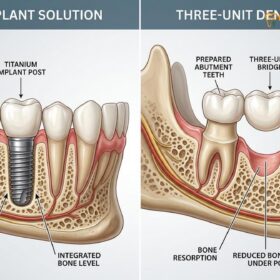
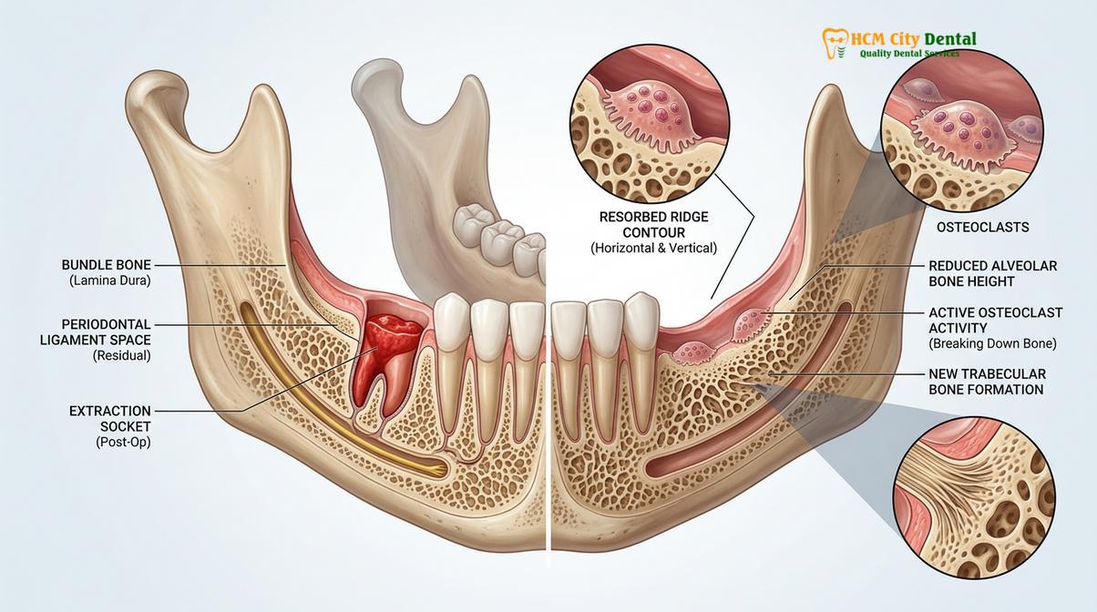
Following tooth removal, the jawbone undergoes rapid and irreversible dimensional changes, primarily losing horizontal width and vertical height due to the absence of mechanical stimulation.
To fully appreciate the necessity of a socket preservation bone graft, one must first understand the biological cascade of events that occurs the moment a tooth is extracted. The alveolar bone—the specialized part of the jawbone that houses the teeth—is entirely dependent on the presence of teeth for its volume and density. The roots of our teeth are connected to the surrounding bone via the periodontal ligament. Every time we chew, bite, or speak, micro-strains are transmitted through this ligament into the bone, stimulating osteoblasts (bone-building cells) to continuously remodel and maintain the bone’s structural integrity.
When a tooth is extracted, this vital mechanical stimulation ceases instantly. Furthermore, the blood supply that once nourished the immediate surrounding bone (known as the bundle bone) is severed. The body, operating on a principle of biological efficiency, recognizes that the bone in this specific area is no longer needed to support a tooth. Consequently, osteoclasts (bone-resorbing cells) are activated, initiating a rapid process of disuse atrophy. This phenomenon is clinically referred to as extraction site bone loss, and its progression is both swift and unforgiving.
The Chronology of Alveolar Ridge Resorption
Clinical studies and histological evaluations have mapped the precise timeline of this bone deterioration. The most dramatic changes occur within the first three to six months following the extraction. During this initial phase, the bone primarily resorbs horizontally, meaning the ridge becomes significantly narrower. The buccal plate—the wall of bone on the cheek side of the tooth—is particularly vulnerable. In many patients, especially in the anterior maxilla (upper front teeth), this buccal plate is naturally extremely thin, often measuring less than one millimeter in thickness. Because it is comprised almost entirely of bundle bone, it resorbs rapidly once the tooth is gone.
Following the initial horizontal collapse, vertical bone loss ensues, reducing the overall height of the jawbone in that area. Research indicates that without any interceptive grafting, a patient can lose up to 50% of the alveolar ridge width within the first twelve months post-extraction[1]. This dramatic reduction transforms a once broad, robust ridge into a narrow, knife-edge formation that is highly unsuitable for supporting a dental prosthesis.
“The biological reality of tooth extraction is that the alveolar bone is programmed to resorb once its primary function—supporting a tooth—is removed. Intercepting this process at the time of extraction is the most predictable way to maintain the anatomical foundation.”
Dr. Nguyen Van Cuong frequently emphasizes to his patients that ignoring this biological reality can lead to severe complications down the line. If a patient waits several years after an extraction to consider tooth replacement, the extraction site bone loss may be so profound that standard implant placement becomes impossible without major reconstructive surgery. The loss of bone also affects the adjacent teeth, potentially leading to shifting, altered bite alignment, and an increased risk of periodontal disease in the surrounding dentition.
What is Socket Preservation? Filling the Extraction Cavity
This surgical technique involves carefully packing the fresh extraction void with specialized bone substitute materials and sealing it to guide natural bone regeneration.
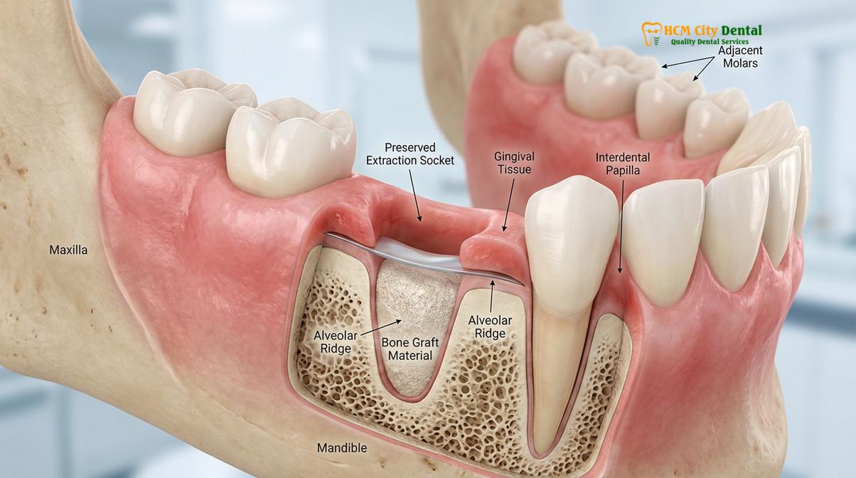
A socket preservation bone graft is an elegant, interceptive surgical procedure designed to counteract the natural resorption process described above. By filling the empty socket with a biocompatible material immediately after the tooth is removed, the clinician provides a physical scaffold that maintains the space and guides the body’s natural bone-healing mechanisms. This socket grafting technique is a cornerstone of modern implant dentistry, ensuring that the site remains viable for future restoration.
The Step-by-Step Surgical Protocol
The success of a socket preservation procedure relies heavily on meticulous surgical execution. The process begins with an atraumatic extraction. Unlike traditional extractions, which may involve significant force and expansion of the bone, an atraumatic approach utilizes specialized micro-instruments, such as periotomes, to gently sever the periodontal ligament. In cases involving multi-rooted teeth, the roots are carefully sectioned and removed individually. The primary goal is to preserve the delicate bony walls of the socket, particularly the fragile buccal plate, as these walls will contain the graft material.
Once the tooth is removed, the socket is thoroughly debrided. The clinician uses surgical curettes to meticulously remove any granulation tissue, cysts, or remnants of infection. This step is critical; leaving pathological tissue behind can severely compromise the integration of the bone graft. The socket is then irrigated with sterile saline or an antimicrobial solution to ensure a pristine environment for regeneration.
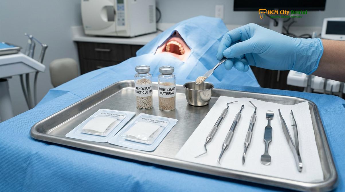
Following debridement, the chosen bone graft material is carefully packed into the socket. The material is compacted just enough to eliminate dead space but not so tightly that it impedes the ingrowth of new blood vessels (angiogenesis), which are essential for healing. To prevent the fast-growing soft gum tissue from invading the socket and displacing the slower-growing bone cells, a barrier membrane is placed over the graft. This principle, known as Guided Bone Regeneration (GBR), ensures that only osteogenic (bone-forming) cells populate the graft site[2]. Finally, the site is secured with sutures, which may be resorbable or require removal after a few weeks.
Understanding Bone Graft Materials
The choice of graft material is tailored to the patient’s specific clinical needs, the size of the defect, and the planned timeline for future implant placement. The materials generally fall into four main categories:
| Graft Type | Source Material | Mechanism of Action | Clinical Application |
|---|---|---|---|
| Autograft | The patient’s own bone (often from the jaw or hip). | Osteogenic, osteoinductive, and osteoconductive. Contains living cells. | Gold standard for large defects, but requires a secondary surgical site. Rarely used for simple socket preservation. |
| Allograft | Human donor bone from a certified tissue bank. | Osteoinductive and osteoconductive. Stimulates the patient’s cells to form new bone. | Highly common in socket preservation. Remodels into natural bone relatively quickly (4-6 months). |
| Xenograft | Animal source (typically bovine or porcine), thoroughly sterilized. | Osteoconductive. Acts as a long-lasting physical scaffold. | Excellent for maintaining ridge volume over a longer period. Very slow resorption rate. |
| Alloplast | Synthetic materials (e.g., calcium phosphate, hydroxyapatite). | Osteoconductive. Provides a synthetic matrix for bone ingrowth. | Used when patients prefer non-biologic materials. Resorption rates vary based on chemical composition. |
In many clinical scenarios, a combination of materials may be used to leverage the rapid remodeling properties of an allograft with the long-term volume maintenance of a xenograft. The barrier membranes used to cover the graft can also be either resorbable (made of collagen, which dissolves naturally over several weeks) or non-resorbable (made of dense polytetrafluoroethylene, or dPTFE, which must be removed later but provides superior protection in certain complex cases).
Preventing Ridge Collapse and Maintaining Gum Tissue Volume
Beyond preserving the underlying bone, this procedure supports the overlying gingival tissues, preventing aesthetic deformities and maintaining the natural gum line.
While the primary focus of a socket preservation bone graft is often on the hard tissue (the bone), its impact on the soft tissue (the gums) is equally profound and clinically significant. The architecture of the gingiva is directly dictated by the underlying bone. If the bone collapses, the gums will inevitably follow, leading to a sunken, concave appearance in the jawline. This alveolar ridge volume conservation is critical not just for structural integrity, but for achieving a natural, aesthetically pleasing final result.
The Importance of Soft Tissue Biotypes
In clinical dentistry, patients present with different gingival biotypes, generally categorized as either thick-flat or thin-scalloped. Patients with a thin-scalloped biotype are at a significantly higher risk for severe aesthetic complications following a tooth extraction. Their delicate gum tissue is highly susceptible to recession, and the underlying buccal bone plate is usually paper-thin. If a tooth is extracted in a patient with a thin biotype without immediate socket preservation, the resulting tissue collapse can be dramatic.
This collapse is particularly devastating in the aesthetic zone—the front teeth that are visible when a person smiles. The loss of bone volume leads to the loss of the interdental papillae (the small, pink triangles of gum tissue between the teeth). Once these papillae are lost, they are notoriously difficult, if not impossible, to surgically reconstruct. The result is the appearance of “black triangles” between the teeth or a future implant crown that looks unnaturally long and disproportionate compared to the adjacent natural teeth.
Clinical Case Example: Aesthetic Zone Preservation
A 34-year-old patient presented to HCMC Dental Clinic in Ho Chi Minh City with a fractured upper central incisor that required extraction. The patient had a high smile line and a thin gingival biotype. Dr. Cuong performed an atraumatic extraction followed immediately by a socket preservation bone graft using a slow-resorbing xenograft and a collagen membrane. Six months later, the site exhibited perfect preservation of both the buccal bone contour and the interdental papillae. The subsequent implant placement was straightforward, and the final ceramic crown emerged from the gums looking completely natural, indistinguishable from the adjacent teeth.
By filling the socket with graft material, the clinician provides a stable foundation that supports the overlying soft tissue during the healing phase. The graft acts like a tent pole, keeping the “tent” of the gums elevated and maintaining the natural convex contour of the ridge. This meticulous attention to soft tissue management is what separates an acceptable functional outcome from a truly exceptional aesthetic restoration.
Furthermore, maintaining the width of the keratinized gingiva (the tough, pink tissue immediately surrounding the teeth) is vital for the long-term health of a future implant. Implants surrounded by adequate keratinized tissue are more resistant to mechanical trauma from brushing and chewing, and they exhibit lower rates of peri-implantitis (inflammation and bone loss around an implant)[3]. Socket preservation plays a key role in ensuring this protective band of tissue is not lost during the post-extraction remodeling phase.
How Socket Preservation Simplifies Future Implant Placement
By maintaining optimal bone dimensions, socket grafting ensures that future dental implants can be placed in the ideal anatomical position without requiring additional invasive surgeries.
The ultimate goal of a socket preservation bone graft is usually future implant site preparation. In modern implantology, the paradigm has shifted from “bone-driven” implant placement to “prosthetically-driven” implant placement. In the past, surgeons would place the implant wherever there was available bone, which often resulted in implants emerging at awkward angles, making it difficult for the restorative dentist to create a functional and aesthetic crown. Today, the ideal position of the final crown dictates exactly where the implant must be placed in the bone.
Achieving Primary Stability and Ideal 3D Positioning
For an implant to be successful, it must be placed in the precise three-dimensional position (mesio-distal, bucco-lingual, and apico-coronal). If the ridge has collapsed due to a lack of socket preservation, the surgeon may be forced to place the implant off-axis or use a narrower, shorter implant that may not adequately support the biting forces. By preserving the original dimensions of the socket, the surgeon has a robust, wide foundation that allows for the placement of an appropriately sized implant in the exact ideal trajectory.
Moreover, preserved bone volume is crucial for achieving primary stability. Primary stability refers to the mechanical friction and tightness of the implant within the bone at the exact moment of surgical placement. It is measured by insertion torque and Implant Stability Quotient (ISQ) values. High primary stability is a prerequisite for successful osseointegration (the biological fusion of bone to the titanium implant surface). A preserved socket provides dense, regenerated bone that firmly grips the implant threads, significantly reducing the risk of early implant failure.
“Prosthetically driven implantology demands an uncompromised foundation. Socket preservation is the most reliable method to ensure that when the time comes for implant placement, the bone architecture perfectly aligns with the restorative goals.”
Avoiding Complex Secondary Surgeries
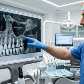
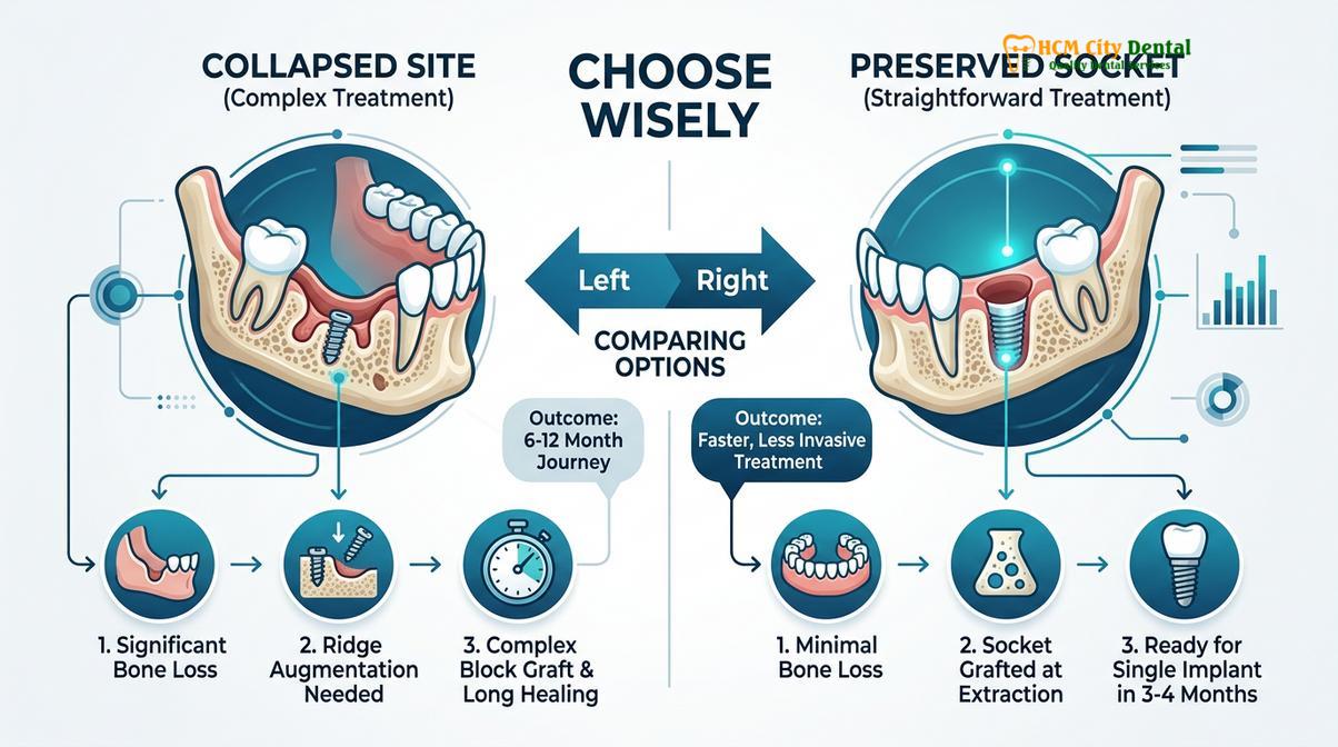
Perhaps the most significant advantage of socket preservation is its ability to eliminate the need for complex, secondary bone grafting procedures in the future. If a patient loses a tooth in the posterior maxilla (upper back jaw) and the bone resorbs, the floor of the maxillary sinus often drops down into the space previously occupied by the tooth roots (a process called sinus pneumatization). If this patient later desires an implant, they will likely require a sinus lift—a delicate and costly procedure to elevate the sinus membrane and pack bone graft underneath it to create enough vertical height for an implant.
Similarly, severe horizontal bone loss in the lower jaw may necessitate a block bone graft, where a solid block of bone is harvested from the patient’s ramus (the back of the jaw) or chin, screwed into the deficient area, and allowed to heal for several months before an implant can even be considered. These advanced procedures carry higher risks of morbidity, increased post-operative pain, longer overall treatment times, and significantly higher costs. A simple socket preservation performed at the time of Tooth Extraction can often completely circumvent the need for these invasive interventions.
For patients exploring comprehensive tooth replacement options, understanding the foundation is key. Whether considering single implants or evaluating Dental Implants vs Dentures: Which Is the Better Investment?, the presence of adequate bone volume is the determining factor for long-term stability and comfort.
Cost Analysis: Upfront Preservation vs. Future Complex Bone Grafts
While adding a minor upfront cost to an extraction, socket preservation prevents the need for significantly more expensive and painful reconstructive surgeries down the line.
When discussing treatment plans, patients frequently ask about the financial implications of adding a socket preservation bone graft to a standard extraction. It is true that the grafting material, the barrier membrane, and the additional surgical time increase the immediate cost of the procedure. However, when viewed through the lens of long-term dental health and future restorative needs, socket preservation is overwhelmingly the more economical choice.
The Financial and Biological Economics
To understand the true value, one must compare the cost of interceptive preservation against the cost of delayed reconstruction. A standard socket preservation is a relatively straightforward, minimally invasive procedure performed during the same appointment as the extraction. The patient undergoes one surgical event, one period of anesthesia, and one recovery phase.
Conversely, if a patient declines preservation, allows the bone to atrophy, and returns two years later for an implant, the clinical scenario is vastly different. The surgeon will likely need to perform a Guided Bone Regeneration (GBR) procedure, a ridge split, or a block graft to rebuild the lost volume. These advanced surgeries require significantly more expensive materials (larger volumes of graft, titanium tacks, specialized membranes), advanced surgical expertise, and often a secondary surgical site if autogenous bone is harvested[4].
Clinical Warning: The Cost of Delay
Opting out of socket preservation to save money in the short term frequently results in overall treatment costs that are 2 to 3 times higher when the patient eventually decides to pursue dental implants. Furthermore, the biological cost—measured in additional surgeries, increased pain, and extended healing timelines—is substantially greater.
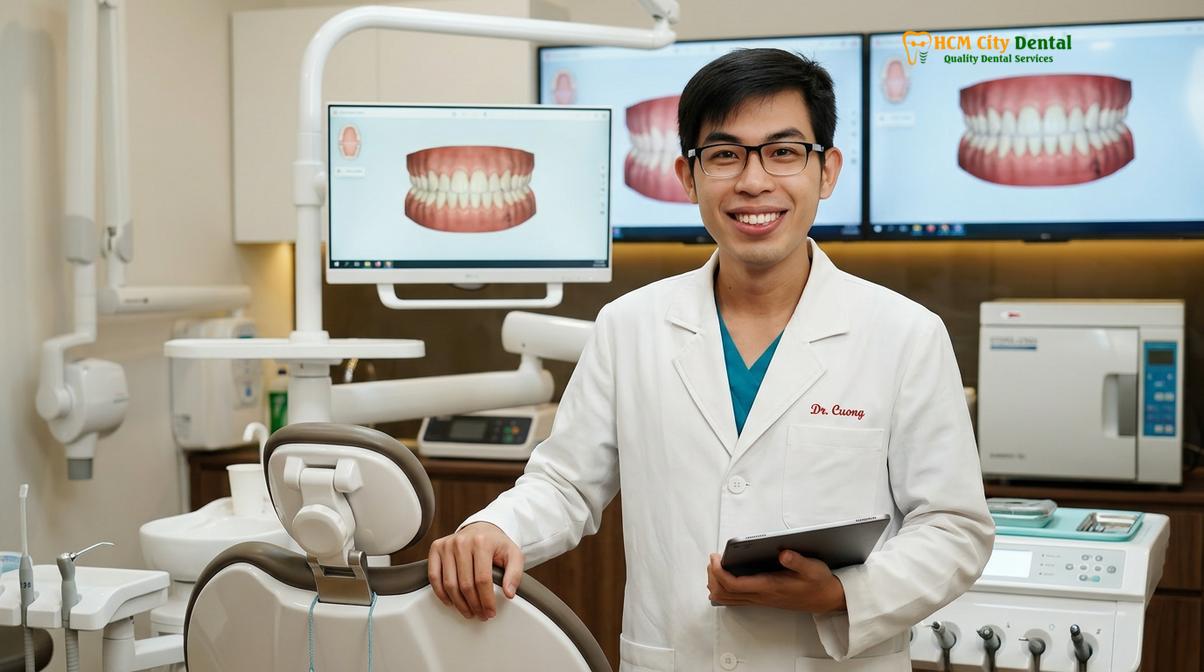
Dr. Cuong advises patients to consider the total timeline of their treatment. A preserved socket typically heals and is ready for an implant in 3 to 6 months. If complex ridge augmentation is required later, the patient must undergo the grafting surgery, wait 6 to 9 months for that graft to consolidate, undergo a second surgery to place the implant, and wait another 3 to 6 months for osseointegration. The total time from the start of treatment to receiving the final crown can easily exceed a year and a half.
Patients researching the How Much Do Dental Implants Cost in Vietnam? (Comprehensive Guide) will find that the most cost-effective implant journeys are those where the foundational bone was preserved early. Furthermore, the choice of premium implant systems, as discussed in Straumann vs Nobel Biocare Implants: What Is the Difference?, relies heavily on having adequate bone volume to support their advanced thread designs and surface technologies.
When to See a Doctor (Important Clinical Considerations)
Consulting a qualified dental specialist is crucial to determine if socket preservation is appropriate for your specific clinical situation and to manage any post-operative healing.
While a socket preservation bone graft is highly beneficial for the majority of patients undergoing tooth extraction, it is a medical procedure that requires careful diagnostic evaluation. Not every extraction site is an immediate candidate for grafting, and specific clinical conditions must be assessed by an experienced oral surgeon or periodontist.
Indications and Contraindications
You should specifically request a consultation for socket preservation if:
- You are having a tooth extracted and plan to replace it with a dental implant, either immediately or in the future.
- The tooth being removed is in the aesthetic zone (the front of the mouth), where preserving the gum line is critical for your smile.
- You are having a tooth extracted near the maxillary sinus, and the surgeon wants to prevent the sinus floor from dropping.
- You are an older adult considering future restorations; as detailed in Dental Implants for Seniors: Am I Too Old for Implants?, preserving bone early is vital for successful outcomes later in life.
However, there are specific contraindications where immediate grafting may be delayed or avoided. If the tooth being extracted is surrounded by an acute, active, and severe purulent infection (an abscess with significant pus), placing a bone graft immediately into that infected site can lead to graft failure. The bacteria can colonize the graft material, turning it into a source of chronic infection rather than a scaffold for new bone. In such cases, the surgeon may opt to extract the tooth, thoroughly clean the socket, prescribe antibiotics, and allow the soft tissue to heal for 4 to 8 weeks before re-entering the site to perform the bone graft (a delayed grafting approach)[5].
Monitoring Your Healing
After undergoing a socket preservation procedure, it is vital to monitor the site closely and attend all scheduled follow-up appointments. You should contact your dental provider immediately if you experience any of the following signs of potential complications:
- Severe, throbbing pain that worsens after the first few days, which may indicate an infection or a dry socket.
- Excessive swelling that extends to the neck or eye, or swelling that continues to increase after 48 hours.
- A persistent fever or a foul taste/odor coming from the surgical site.
- Significant exposure of the bone graft material or the barrier membrane, especially if the sutures have come undone prematurely.
For patients in Ho Chi Minh City, seeking a comprehensive evaluation at a specialized facility like HCMC Dental Clinic ensures that your extraction and subsequent preservation are handled with the highest standards of surgical care and sterile protocol. To explore more about the transformative power of implant dentistry once your bone is preserved, review Dental Implants: Transform Your Smile Today! [Expert Tips] | Dr. Cuong and Are Dental Implants Right For You? Pros & Cons Guide | Dr. Cuong.
Frequently Asked Questions
Is socket preservation always necessary after a tooth extraction?
While not mandatory for every extraction, socket preservation is highly recommended if you plan to receive a dental implant in the future. It is especially critical in the aesthetic zone (front teeth) or when the surrounding bone is already thin, as it prevents severe structural collapse. If you are extracting a third molar (wisdom tooth) with no intention of replacing it, preservation is usually unnecessary.
How long does socket preservation bone heal?
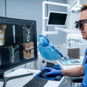
The initial soft tissue healing takes about two weeks, but the underlying bone graft requires three to six months to fully integrate and mature. This extended period allows the graft material to act as a scaffold while your body generates new, solid jawbone. Your dentist will use X-rays or a CBCT scan to confirm when the bone is dense enough for implant placement.
Can I get an implant directly in a preserved socket?
Yes, once the preservation graft has fully healed and integrated into your natural jawbone, the site is typically ideal for implant placement. The preserved bone volume ensures the implant can be anchored securely without the need for further complex grafting procedures. In some highly specific cases, an implant can even be placed at the exact same time as the extraction and graft, known as immediate implant placement.
Does the bone graft material stay in my jaw forever?
No, most bone graft materials used in socket preservation act as a temporary biological scaffold. Over several months, your body’s natural bone-building cells gradually resorb the graft particles and replace them with your own living, healthy bone tissue. The rate of resorption depends on the type of material used; allografts remodel faster, while xenografts resorb very slowly, maintaining volume over a longer period.
Is the socket preservation procedure painful?
The procedure is performed under local anesthesia and is generally no more uncomfortable than the tooth extraction itself. Post-operative discomfort is typically mild and can be effectively managed with standard over-the-counter pain relievers and proper adherence to aftercare instructions. Because the graft fills the empty socket, it can actually reduce the risk of developing a painful “dry socket” compared to leaving the extraction site completely open.
References
- Journal of Periodontology. Alveolar ridge preservation following tooth extraction: A systematic review and meta-analysis. (2019).
- International Journal of Oral and Maxillofacial Implants. Dimensional changes of the alveolar ridge contour after socket preservation. (2020).
- Clinical Oral Implants Research. Healing of extraction sockets and surgically produced defects. (2018).
- Journal of Clinical Periodontology. Efficacy of different bone substitute materials in alveolar ridge preservation. (2021).
- American Association of Oral and Maxillofacial Surgeons. Parameters of Care: Guidelines for extraction site management. (2022).