A well-maintained dental crown typically lasts between 10 to 15 years, though high-quality materials like zirconia or gold can endure for decades. The exact lifespan depends heavily on the material chosen, the precision of the clinical fit, and the patient’s daily oral hygiene habits.
Clinical Summary:
The longevity of a dental restoration is a multifactorial clinical outcome. While the average lifespan ranges from a decade to 15 years, advancements in monolithic zirconia and lithium disilicate have significantly extended this timeline. Success relies on precise margin adaptation during preparation, optimal occlusal load distribution, and rigorous periodontal maintenance. Understanding the biomechanical properties of different materials helps clinicians and patients set realistic expectations for structural durability and aesthetic retention over time.
Key Takeaways:
- Average clinical lifespan ranges from 10 to 15 years, with potential for 20+ years under optimal conditions.
- Material selection (zirconia, E-max, PFM, gold) dictates fracture resistance and aesthetic longevity.
- Bruxism and heavy occlusal forces significantly reduce the survival rate of ceramic restorations.
- Recurrent decay at the crown margin is the leading cause of premature failure.
- Routine professional cleanings and radiographic monitoring are essential for early detection of microleakage.
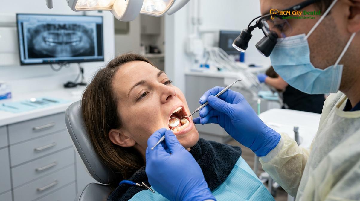
The Clinical Lifespan of Dental Crowns: Material Breakdown
The durability of a crown is intrinsically linked to its composition, with gold offering maximum longevity, while modern ceramics like zirconia provide an optimal balance of strength and aesthetics lasting 10 to 15 years.
When patients ask what is a dental crown made of, the answer involves a complex array of biomaterials designed to mimic the structural integrity and optical properties of natural enamel and dentin. The choice of material is the foundational variable in determining how long the restoration will survive in the hostile environment of the oral cavity. The mouth subjects restorations to extreme temperature fluctuations, acidic pH challenges, and immense biting forces. Therefore, understanding exactly what is tooth crown made of helps clinicians and patients make evidence-based decisions tailored to specific functional and aesthetic needs.
Zirconia: The Modern Standard for Strength
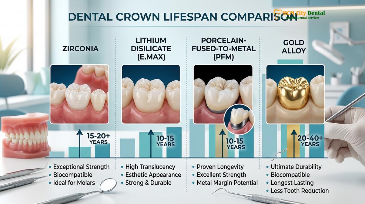
Zirconia (zirconium dioxide) has revolutionized restorative dentistry. Categorized as a polycrystalline ceramic, it possesses exceptional flexural strength, often exceeding 1000 MPa. Monolithic zirconia crowns—milled from a single block of material—are highly resistant to chipping and fracture. According to clinical literature, zirconia restorations exhibit excellent long-term survival rates, often lasting 15 to 20 years or more with proper care [1].
One of the unique biomechanical properties of zirconia is “phase transformation toughening.” When a micro-crack begins to propagate through the material, the crystalline structure locally transforms from a tetragonal phase to a monoclinic phase. This transformation is accompanied by a slight volume expansion, which effectively compresses the crack and stops it from spreading [2]. This makes zirconia an ideal choice for posterior teeth (molars) that endure heavy masticatory (chewing) forces.
Lithium Disilicate (E-max): The Aesthetic Champion
Lithium disilicate, commonly known by the brand name E-max, is a glass-ceramic material celebrated for its unparalleled optical properties. It mimics the natural translucency and light-scattering characteristics of human enamel better than almost any other material. While its flexural strength (around 400-500 MPa) is lower than zirconia, it is still significantly stronger than traditional feldspathic porcelain.
E-max crowns are typically bonded to the tooth structure using adhesive resin cements, which creates a monoblock effect, reinforcing both the crown and the underlying tooth. With meticulous clinical execution, lithium disilicate crowns generally last 10 to 15 years. They are the premier choice for a cosmetic dental crown on anterior (front) teeth where aesthetics are paramount.
Porcelain-Fused-to-Metal (PFM): The Traditional Workhorse
For over half a century, Porcelain-Fused-to-Metal (PFM) crowns were the gold standard for combining strength and aesthetics. These restorations consist of a cast or milled metal substructure (coping) overlaid with layered feldspathic porcelain. The metal provides structural rigidity, while the porcelain provides a tooth-like appearance.
PFM crowns typically last 10 to 15 years. However, they have distinct clinical drawbacks. Over time, the layered porcelain is susceptible to chipping or delamination from the metal core. Furthermore, as patients age and natural gingival recession occurs, the dark metal margin of the crown often becomes visible at the gumline, compromising the aesthetic outcome. Despite these limitations, PFMs remain a viable option for long-span restorative dental procedures and complex full-mouth rehabilitations.
Gold and Noble Metal Alloys: Unmatched Longevity
From a purely biomechanical standpoint, cast gold alloys remain the ultimate restorative material. Gold is highly biocompatible, non-abrasive to opposing natural teeth, and possesses a coefficient of thermal expansion very similar to natural tooth structure. Because gold is ductile, it can be burnished at the margins, creating a nearly microscopic seal that is highly resistant to bacterial microleakage.
It is not uncommon to see gold crowns functioning perfectly after 30 or even 40 years in the mouth. However, due to the obvious aesthetic limitations of a metallic appearance, gold is almost exclusively reserved for second molars in patients with severe bruxism (teeth grinding) or those who prioritize longevity above all else.
| Material Type | Average Lifespan | Flexural Strength | Primary Clinical Indication |
|---|---|---|---|
| Monolithic Zirconia | 15 – 20+ Years | Very High (>1000 MPa) | Posterior molars, heavy biters, bruxers |
| Lithium Disilicate (E-max) | 10 – 15 Years | Moderate (400-500 MPa) | Anterior teeth, high aesthetic demands |
| Porcelain-Fused-to-Metal (PFM) | 10 – 15 Years | High (Metal Core) | Bridges, traditional posterior restorations |
| Cast Gold Alloy | 20 – 40+ Years | High (Ductile) | Non-visible molars, severe bruxism |
Biomechanical Factors Influencing Crown Longevity
A crown’s survival depends not just on the material, but on the patient’s bite forces, periodontal health, and the structural integrity of the underlying natural tooth.
The lifespan of a dental restoration is never determined in a vacuum. Even the strongest monolithic zirconia crown will fail prematurely if placed in a biomechanically compromised environment. The oral cavity is a dynamic system, and the longevity of any prosthesis is dictated by the interplay between the restorative material, the supporting periodontium (gums and bone), and the patient’s occlusal (bite) dynamics.
The Importance of the Ferrule Effect
One of the most critical concepts in restorative dentistry is the “ferrule effect.” A ferrule is a band of solid, healthy tooth structure (dentin) that extends above the gumline, which the crown encircles and grasps. Clinical guidelines dictate that a minimum of 1.5 to 2.0 millimeters of vertical tooth structure must be present circumferentially to provide adequate resistance and retention form.
If a tooth is severely broken down due to extensive decay or trauma, and a crown is placed without an adequate ferrule, the lateral forces of chewing will eventually cause the crown to debond or the root to fracture. In cases where insufficient tooth structure remains, a core buildup or a post-and-core procedure is required to create a stable foundation before the permanent tooth cap can be fabricated.
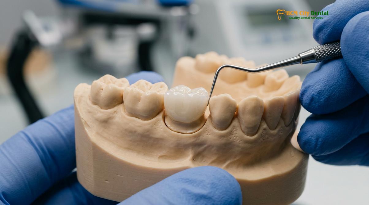
Occlusal Forces: Bruxism and Clenching
Parafunctional habits, such as sleep bruxism (grinding) and clenching, exert catastrophic forces on dental restorations. While normal chewing generates approximately 50 to 70 pounds per square inch (psi) of force, a severe bruxer can generate forces exceeding 250 psi. Over time, this immense pressure leads to material fatigue.
In patients with heavy occlusal loads, layered ceramics (like PFM or layered zirconia) are highly prone to cohesive fractures (chipping of the porcelain). For these patients, clinicians strongly recommend monolithic materials and the fabrication of a custom acrylic occlusal guard (night guard) to protect the restorations during sleep [4]. Failure to manage occlusal forces is a primary reason why crowns fail before their expected lifespan.
Margin Adaptation and Biologic Width
The “margin” is the junction where the edge of the dental crown meets the natural tooth structure. The precision of this fit is arguably the most critical factor in preventing secondary caries (recurrent decay). If the margin is open or bulky, it creates a microscopic ledge where plaque and cariogenic bacteria accumulate, shielded from the patient’s toothbrush bristles.
“The longevity of a crown is won or lost at the margin. A restoration that fits seamlessly against the prepared tooth structure virtually eliminates the micro-environments where decay-causing bacteria thrive, ensuring the underlying dentin remains protected for decades.”
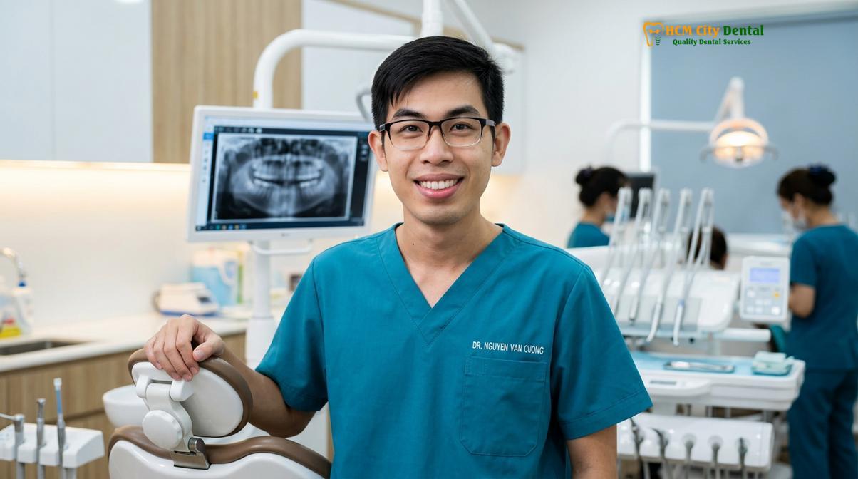
— Dr. Nguyen Van Cuong, Lead Clinician
Furthermore, the placement of the margin relative to the crest of the alveolar bone is crucial. If a crown margin is placed too deep beneath the gumline, it violates the “biologic width”—the natural attachment apparatus of the gingiva. This violation triggers chronic localized inflammation, bleeding, and eventual bone loss around the restored tooth [3]. Meticulous tissue management during the preparation phase is essential for long-term periodontal health.
The Treatment Workflow: From Preparation to Final Cementation
Achieving a long-lasting restoration requires a meticulous multi-step clinical protocol, transitioning from precise tooth reduction and temporary protection to the final bonding of the permanent prosthesis.
The clinical workflow for fabricating a dental crown is a highly technique-sensitive process. Any error during the preparation, impression, or provisionalization phases will compound, ultimately compromising the fit and longevity of the final restoration. Understanding this workflow highlights why quality dentistry requires time, precision, and advanced technology.
Tooth Preparation and Tissue Management
The procedure begins with the administration of local anesthesia to ensure profound numbness. The clinician then uses specialized diamond burs to reduce the anatomical contours of the tooth. The amount of reduction is strictly dictated by the chosen restorative material; for example, a gold crown requires minimal reduction (0.5 – 1.0 mm), while a PFM or E-max crown requires more aggressive reduction (1.5 – 2.0 mm) to allow adequate thickness for the ceramic.
Once the tooth is prepared, tissue management is performed. The dentist carefully packs a retraction cord—a tiny thread often soaked in a hemostatic agent—into the gingival sulcus (the space between the tooth and the gum). This temporarily pushes the gum tissue away from the margin, ensuring that the subsequent impression captures the entire preparation clearly.
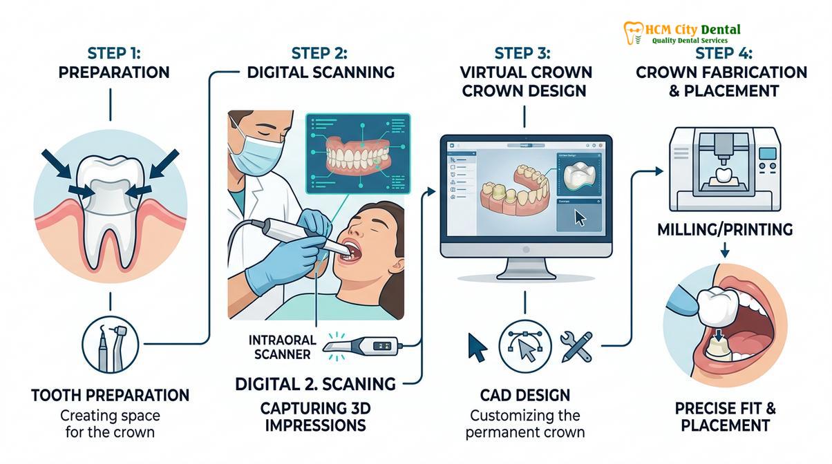
Digital Impressions vs. Conventional Methods
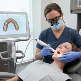
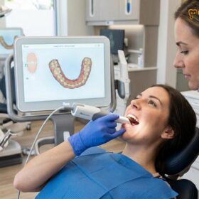
Historically, impressions were taken using polyvinyl siloxane (PVS) putty, which was messy and uncomfortable for the patient. Today, modern clinics utilize digital intraoral scanners. These devices capture thousands of optical images per second, stitching them together to create a highly accurate 3D model of the prepared tooth and the opposing arch. Digital impressions eliminate the risk of material distortion, air bubbles, and temperature-related dimensional changes, resulting in a superior marginal fit for the final restoration.
The Critical Role of the Provisional Restoration
While the dental laboratory fabricates the final prosthesis, the prepared tooth must be protected. The clinician will fabricate a temporary cap on tooth using a bis-acryl composite resin. This provisional restoration serves several vital functions: it prevents thermal sensitivity, maintains the position of the tooth (preventing it from drifting), and guides the healing of the gingival tissues.
Clinical Warning: Never leave a temporary crown in place longer than prescribed. Provisional cements wash out over time, allowing bacteria to infiltrate and decay the underlying dentin rapidly. If your temporary crown dislodges, contact your dentist immediately for recementation.
Patients must exercise caution during the provisional phase. For detailed guidance on managing provisional restorations, especially if you are away from home, refer to our comprehensive guide on Temporary Dental Crown Care While Traveling. Once the laboratory completes the fabrication, the patient returns for the delivery appointment. The temporary is removed, the tooth is thoroughly decontaminated, and the permanent tooth cap is bonded or cemented into place using specialized luting agents. This precise workflow applies to single units as well as larger dental caps and bridges.
Financial Considerations and Long-Term Value
While initial expenses vary based on material and location, investing in high-quality fabrication and precise clinical execution ultimately reduces the lifetime cost of dental restorations.
During initial consultations, patients frequently inquire about the overall crown in tooth cost. Understanding exactly how much standard dental crown cost variations depend on the chosen material, the complexity of the case, and the expertise of the dental laboratory is crucial for long-term financial planning. Dentistry is an investment in your systemic health and daily quality of life.
Breaking Down the Cost Factors
When evaluating the cap for teeth cost, it is important to look beyond the initial crown for teeth price and consider the longevity of the restoration. A cheaper crown that suffers from poor marginal adaptation may fail within three to five years, necessitating a replacement and potentially a root canal due to secondary decay. Conversely, a premium restoration placed with meticulous isolation protocols may last two decades, offering a significantly better return on investment.
The crown on front tooth cost is typically higher than posterior restorations. Anterior teeth have intense aesthetic demands. The dental ceramist must painstakingly layer different opacities and translucencies of porcelain to perfectly match the adjacent natural dentition. This custom shade matching and artistic characterization require immense skill and time. Patients considering anterior restorations often explore Whitening Before Dental Crowns to ensure the final prosthetics match a brighter, rejuvenated smile.
Clinical Case Study: Long-Term Aesthetic Rehabilitation
A 45-year-old patient visited HCMC Dental Clinic in Ho Chi Minh City presenting with failing, discolored composite veneers and severe occlusal wear. After a comprehensive digital smile design, Dr. Cuong recommended a combination of E-max crowns and Porcelain Veneers. By utilizing high-strength lithium disilicate and establishing a protective canine-guided occlusion, the clinic delivered a highly aesthetic result designed to withstand functional forces for 15+ years, maximizing the patient’s investment.
For international patients, the cost-to-quality ratio is a major driving factor. Many individuals travel abroad to receive premium materials (like authentic Ivoclar E-max or Vita Zirconia) at a fraction of the cost in their home countries. For a detailed breakdown of how to plan such a trip, review our guide on Dental Tourism Vietnam Crowns. Ultimately, whether you are choosing a crown or evaluating a Dental Crown vs Veneer, the focus should always remain on clinical precision and material durability.
Signs of a Failing Crown and When to See a Doctor
Immediate clinical evaluation is required if you experience localized pain, thermal sensitivity, visible margin gaps, or a sensation of mobility in the restored tooth.
Even with the highest quality materials and exceptional clinical execution, dental crowns do not last forever. The oral environment is constantly changing, and the underlying tooth structure remains susceptible to disease. Recognizing the early warning signs of a failing restoration can mean the difference between replacing a crown and losing the entire tooth to extraction.
Secondary Caries (Recurrent Decay)
The most common reason for crown failure is secondary caries. Because the crown itself cannot decay, bacteria target the vulnerable junction where the crown meets the root. If plaque is not meticulously removed from the gumline, acid-producing bacteria will dissolve the cement seal and infiltrate beneath the restoration [5].
“Patients often assume that a crowned tooth is invincible. In reality, the margin is highly susceptible to microleakage. Without rigorous daily flossing and biannual professional cleanings, recurrent decay can silently hollow out the tooth beneath the crown, often without causing pain until the nerve is irreversibly damaged.”
— Dr. Nguyen Van Cuong, HCMC Dental Clinic
If you notice a foul odor or taste coming from a specific tooth, or if the dental floss consistently frays or catches on the margin, it is a strong indicator of an open margin or active decay. Early detection via digital bitewing radiographs is essential.
Structural Fractures and Cement Washout
While materials like zirconia are incredibly strong, layered porcelain is prone to cohesive fractures. If you bite down on a hard object (like an olive pit or ice) and feel a sharp edge on your crown with your tongue, the ceramic has likely chipped. While minor chips can sometimes be polished, significant fractures compromise the structural integrity and require complete replacement.
Additionally, if a crown feels loose, mobile, or makes a subtle “squishing” sound when you chew, the underlying luting cement has likely washed out. A loose crown is a dental emergency because the underlying dentin is completely exposed to bacterial invasion. Do not attempt to glue the crown back yourself using household adhesives; this is highly toxic and will destroy the tooth.
When to Seek Immediate Clinical Care
You should schedule an immediate evaluation if you experience any of the following symptoms:
- Spontaneous Pain: A constant, throbbing ache that wakes you up at night indicates irreversible pulpitis (nerve infection) beneath the crown.
- Prolonged Thermal Sensitivity: If the tooth aches for more than 10 seconds after consuming hot or cold liquids, the nerve is highly inflamed. Read more about Tooth Crown Pain Chewing Sensitivity.
- Gingival Swelling: A localized pimple (fistula) or swelling on the gums near the crowned tooth indicates an active abscess.
- Visible Darkening: While a dark line at the gum is normal for older PFM crowns, sudden darkening of the tooth structure beneath a ceramic crown suggests severe decay or nerve necrosis.
If you suspect your restoration is compromised, review the Failed Dental Crown Symptoms and contact a qualified prosthodontist or restorative dentist for a comprehensive radiographic and clinical examination.
Frequently Asked Questions
Can a dental crown last a lifetime?
While some gold or zirconia crowns can last over 30 years, it is clinically inaccurate to guarantee a lifetime lifespan. Longevity depends on meticulous oral hygiene, the absence of heavy grinding forces, and routine professional monitoring to prevent underlying decay. The underlying tooth root and surrounding bone are biological tissues that remain susceptible to periodontal disease and caries, regardless of the crown’s material strength.
Why would a crown need to be replaced after only 5 years?
Premature crown failure is typically caused by recurrent decay at the margin, severe bruxism fracturing the ceramic, or poor initial clinical fit. A compromised cement seal allows bacterial microleakage, necessitating early replacement to save the underlying tooth structure. Additionally, if the tooth was prepared with an inadequate ferrule, the crown may repeatedly debond under normal chewing forces.
Does a root canal treated tooth with a crown last as long as a vital tooth?
A root canal treated tooth can last just as long, provided there is sufficient healthy dentin remaining (the ferrule effect) to support the crown. However, non-vital teeth are more brittle, making the structural integrity of the core buildup critical for long-term success. Endodontically treated teeth lose their proprioception (pressure sensing), meaning patients may unknowingly bite down too hard, increasing the risk of root fracture.
How can I extend the lifespan of my dental crown?
You can maximize your crown’s lifespan by brushing twice daily, flossing meticulously around the margins, wearing a custom night guard if you grind your teeth, and attending biannual professional cleanings to monitor the restoration’s integrity. Avoiding highly acidic diets and refraining from using your teeth as tools (e.g., opening packages or biting nails) will also significantly reduce the risk of ceramic chipping and cement degradation.
Is it normal for an old crown to show a dark line at the gum?
A dark line is very common with older porcelain-fused-to-metal (PFM) crowns as the gums naturally recede over time, exposing the metal base. While aesthetically displeasing, it does not always indicate clinical failure unless accompanied by decay or an open margin. If the dark line is an aesthetic concern, the crown can be replaced with a modern all-ceramic material like monolithic zirconia or lithium disilicate, which contain no metal substructure.
References
- Journal of Prosthetic Dentistry. Long-term survival rates of ceramic and metal-ceramic crowns. (2021).
- Dental Materials. Fracture resistance and phase transformation of monolithic zirconia restorations. (2022).
- Journal of Clinical Periodontology. Periodontal responses to subgingival restorative margins and biologic width. (2020).
- International Journal of Prosthodontics. The impact of sleep bruxism on the survival of dental restorations. (2019).
- Journal of the American Dental Association. Pathophysiology of secondary caries at crown margins. (2023).