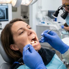
Failed dental crown symptoms include persistent pain, sensitivity to temperature, a loose or wobbly sensation, visible cracks, and gum inflammation around the restoration. Recognizing these signs early is crucial to prevent underlying tooth decay, infection, and the need for more complex restorative procedures.
Clinical Summary:
Dental crowns are highly durable restorations designed to protect compromised teeth, but they can fail over time due to recurrent caries, structural fractures, or cement degradation. Clinical indicators of failure include localized pain, thermal sensitivity, marginal discoloration, and gingival inflammation. Advanced diagnostic protocols, including digital intraoral scanning, are essential for evaluating the integrity of the underlying tooth structure and the marginal seal. When a crown fails, timely intervention is required to remove the defective prosthesis, eradicate any underlying pathology, and fabricate a precise replacement using modern CAD/CAM materials like Zirconia or Lithium Disilicate. Early detection significantly improves the prognosis for preserving the natural tooth.
Key Takeaways:
- Persistent pain or sensitivity often indicates a compromised marginal seal or underlying nerve inflammation.
- Recurrent decay is a primary cause of crown failure, requiring immediate clinical evaluation.
- Modern replacements utilize digital scanning for enhanced precision and patient comfort.
- High-strength materials like Zirconia HT offer superior resistance to structural fractures.
- Addressing a failing crown promptly can often save the underlying natural tooth structure.
How to Recognize a Failed Dental Crown
Identifying a compromised restoration early involves monitoring for localized pain, bite discrepancies, and marginal discoloration, which indicate the protective seal has been breached.
A dental crown is engineered to function seamlessly within the oral cavity, mimicking the biomechanics and aesthetics of a natural tooth. However, when the structural integrity of the crown or its bond to the underlying tooth is compromised, a cascade of clinical symptoms typically follows. The most immediate and noticeable symptom is persistent pain or discomfort. This pain may manifest as a sharp, localized ache when biting down, which often suggests that the crown is sitting too high, creating occlusal trauma, or that the underlying tooth structure has fractured [1]. Alternatively, a dull, throbbing ache that radiates through the jaw may indicate that the dental pulp (the nerve center of the tooth) has become inflamed or infected due to bacterial penetration.
Thermal sensitivity is another hallmark sign of a failing restoration. If you experience a sharp, lingering pain when consuming hot or cold foods and beverages, it is highly probable that the dentin beneath the crown has been exposed. This exposure usually occurs when the dental cement washes out over time, creating a microscopic gap between the crown margin and the natural tooth. According to the hydrodynamic theory of tooth sensitivity, fluid movement within the exposed dentinal tubules stimulates the nerve endings, resulting in acute pain [2].
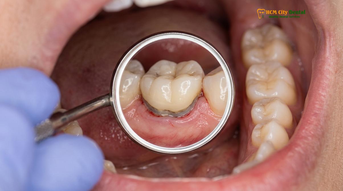
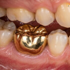
Physical changes in the restoration itself are also critical indicators. A crown that feels loose, wobbly, or shifts slightly when you chew is a clear sign of cement failure or underlying tooth decay. In some cases, patients may notice a distinct clicking sound when they bite. Furthermore, visual inspection may reveal chips, cracks, or a dark line at the gumline. While a dark line can sometimes simply be the metal base of an older Porcelain-Fused-to-Metal (PFM) crown showing through receding gums, it can also signify active decay. If a crown sustains significant physical damage, a broken dental crown replacement becomes a clinical necessity to restore function and prevent sharp edges from lacerating the tongue or buccal mucosa.
Finally, olfactory and gustatory changes should not be ignored. A persistent bad odor (halitosis) or an unpleasant, metallic, or sour taste originating from a specific tooth strongly suggests that food particles and bacteria are trapped beneath a poorly fitting crown. This stagnant environment is a breeding ground for anaerobic bacteria, which produce volatile sulfur compounds as they metabolize debris, leading to localized gingival inflammation and further degradation of the tooth structure.
Decay Underneath the Crown (Recurrent Caries)
Recurrent decay occurs when bacteria penetrate compromised crown margins, silently eroding the underlying dentin and threatening the tooth’s structural integrity.
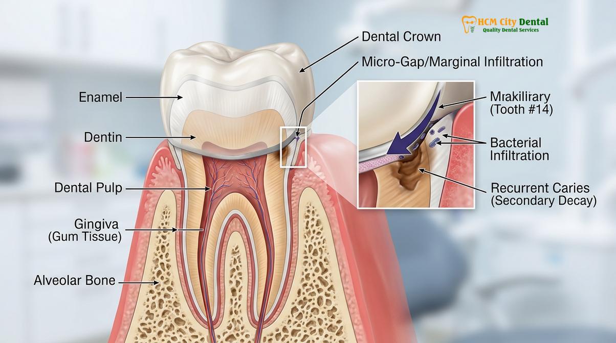
One of the most pervasive misconceptions in restorative dentistry is that a crowned tooth is immune to cavities. While the ceramic or metallic material of the crown itself cannot decay, the natural tooth structure supporting it remains highly vulnerable. Recurrent caries, or decay that forms underneath an existing restoration, is a leading cause of crown failure. This process typically begins at the crown margin—the delicate junction where the restorative material meets the natural tooth structure [3].
Microleakage is the primary mechanism driving recurrent decay. Over years of functional use, the dental cement that seals the crown to the tooth can slowly dissolve or degrade due to the acidic environment of the mouth and the mechanical stresses of chewing. Once the cement seal is breached, microscopic gaps form. Saliva, laden with cariogenic bacteria and fermentable carbohydrates, seeps into these gaps. Because this area is completely inaccessible to a toothbrush or dental floss, the bacteria proliferate unchecked, producing acids that demineralize the dentin.
“The insidious nature of recurrent caries beneath a crown lies in its ability to progress silently; because the crown obscures the underlying tooth from visual inspection and routine X-rays, the decay often reaches advanced stages before clinical symptoms manifest.”
When recurrent caries under crown margins is left untreated, the decay can rapidly progress toward the dental pulp. If the pulp becomes irreversibly inflamed or necrotic, a root canal treatment will be required to eradicate the infection. In cases where the tooth has already undergone a root canal, the patient may not feel any pain as the decay advances, making regular clinical and radiographic examinations absolutely critical. The decay can hollow out the tooth from the inside, eventually leaving insufficient healthy tooth structure to support the crown, leading to a catastrophic fracture of the abutment tooth.
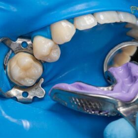
The clinical approach to saving decayed tooth under crown involves a meticulous workflow. The prosthodontist must carefully section and remove the failing crown to gain direct access to the lesion. All decayed and softened dentin is excavated using specialized burs and caries-detecting dyes. Once healthy, sound tooth structure is reached, the clinician must evaluate whether enough tooth remains to support a new crown. If a significant amount of structure has been lost, a core buildup—often reinforced with a fiber post placed into the root canal space—is performed to provide a stable foundation for the new restoration. This meticulous process ensures that the new crown has a solid, infection-free base, maximizing its long-term prognosis.
Fractures and Structural Failures
Structural failures range from minor porcelain chipping to catastrophic framework fractures, often driven by excessive occlusal forces, bruxism, or material fatigue.
While modern dental materials are engineered for exceptional durability, they are not impervious to the immense biomechanical forces generated within the human mouth. The average human bite can exert forces exceeding 200 pounds of pressure per square inch, and these forces are exponentially magnified in patients who suffer from bruxism (chronic teeth grinding and clenching). Over time, this relentless mechanical stress can lead to material fatigue and eventual structural failure of the dental crown.
The type of structural failure often depends on the material composition of the crown. Traditional Porcelain-Fused-to-Metal (PFM) crowns are susceptible to a specific type of failure known as delamination or cohesive fracture, where the aesthetic porcelain layer chips or shears off from the underlying metal substructure. This not only compromises the aesthetics of the smile, exposing the dark metal underneath, but also alters the patient’s bite and creates rough edges that can irritate oral tissues. In contrast, all-ceramic crowns, while highly aesthetic, can suffer from bulk fractures if they are subjected to excessive point-loading or if the underlying tooth preparation did not provide adequate thickness for the material.
To mitigate the risk of structural failures, contemporary prosthodontics heavily favors advanced monolithic materials. Zirconia HT (High Translucency) is a prime example. Milled from a single block of zirconium dioxide using CAD/CAM technology, monolithic zirconia exhibits extraordinary flexural strength—often exceeding 1000 MPa—making it highly resistant to chipping and fracturing, even in the posterior regions of the mouth where chewing forces are highest [4]. For anterior teeth, where aesthetics are paramount, Lithium Disilicate (E.max) offers a superior balance of lifelike translucency and robust strength.
At HCMC Dental Clinic, we emphasize the importance of selecting the appropriate material based on the patient’s specific functional requirements and aesthetic goals. For patients with a history of bruxism, we routinely recommend the fabrication of a custom-fitted occlusal nightguard. Worn during sleep, this appliance acts as a protective shock absorber, dissipating grinding forces and significantly extending the lifespan of both natural teeth and ceramic restorations.
Understanding the financial parameters of Dental Crowns & Bridges is an important aspect of treatment planning. We offer a transparent pricing structure, utilizing only authentic, certified materials from Germany and Liechtenstein. For international patients and dental tourists, we provide an exclusive 40% pre-arrival discount for bookings made via WhatsApp, allowing for premium care at highly accessible rates.
| Material / Treatment Type | Standard Walk-in Price | WhatsApp Pre-booking (-40%) |
|---|---|---|
| PFM Crown (Porcelain-Fused-to-Metal) | ~$140 (3.5M VND) | From $84 (2.1M VND) |
| Zirconia Crown (Cercon, Germany) | ~$200 (5.0M VND) | From $120 (3.0M VND) |
| Zirconia HT Crown (Cercon HT, Germany) | ~$260 (6.5M VND) | From $156 (3.9M VND) |
| E.max Crown (Ivoclar, Liechtenstein) | ~$280 (7.0M VND) | From $168 (4.2M VND) |
| 3-Unit Bridge (Zirconia HT) | ~$480 (12.0M VND) | From $288 (7.2M VND) |
| 4-Unit Bridge (Zirconia HT) | ~$640 (16.0M VND) | From $384 (9.6M VND) |
| Inlay / Onlay (Sứ Lab) | ~$180 (4.5M VND) | From $108 (2.7M VND) |
Gum Recession and Margins
Gum recession exposes the vulnerable tooth root and crown margin, leading to aesthetic concerns, heightened sensitivity, and increased susceptibility to plaque accumulation.
The relationship between a dental crown and the surrounding gingival (gum) tissue is a critical determinant of long-term success. A healthy periodontium provides a protective seal around the tooth, preventing bacterial ingress. However, when a crown fails to harmonize with the surrounding biology, it can trigger gingival inflammation and subsequent gum recession. Failed crown margins gum recession is a common clinical presentation that compromises both the health and the appearance of the restoration.
One of the primary causes of gum recession around a crown is a violation of the biologic width. The biologic width is the natural dimension of soft tissue attached to the portion of the tooth above the alveolar bone. If a crown margin is placed too deeply beneath the gumline (subgingival margin) in an attempt to hide the junction, it can impinge on this delicate attachment apparatus. The body’s natural response to this chronic irritation is chronic inflammation, bone loss, and eventual apical migration of the gingival tissue—resulting in recession.
Clinical Warning: Persistent redness, swelling, or bleeding of the gums specifically around a crowned tooth is a strong indicator of marginal discrepancy, cement residue, or biologic width violation. Ignoring these signs can lead to localized periodontal disease and the eventual loss of the supporting alveolar bone.
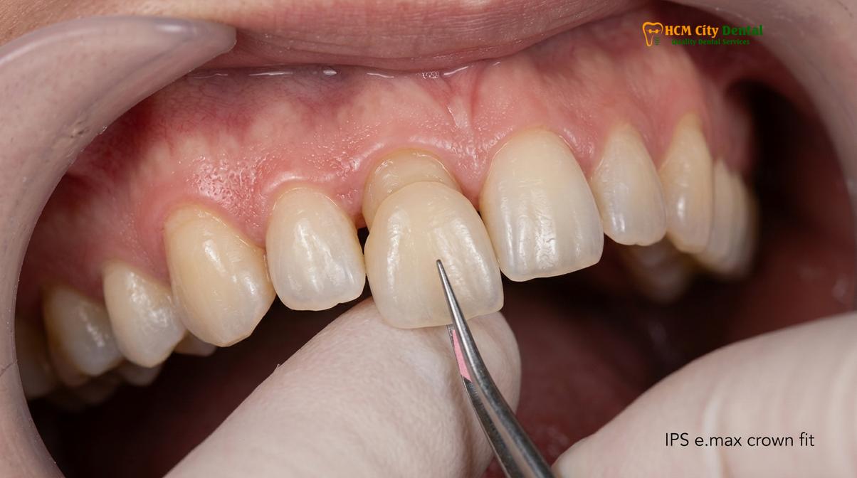
The design of the tooth preparation also plays a vital role in marginal integrity. Prosthodontists utilize specific margin designs, such as a chamfer or a shoulder preparation, depending on the restorative material chosen. A precise preparation ensures that the crown transitions smoothly into the natural root surface without any bulky overhangs or deficient ledges. Overhanging margins act as plaque traps, making effective oral hygiene impossible and accelerating periodontal breakdown.
When recession occurs, the root surface of the tooth becomes exposed. Unlike the enamel-covered crown, the root is covered by cementum, which is significantly softer and more prone to rapid decay and toothbrush abrasion. Aesthetically, recession often reveals the dark junction between the crown and the root, which is particularly problematic in the aesthetic zone (the front teeth). In cases where the biologic width has been severely compromised, a minor surgical procedure known as functional crown lengthening may be required before a new crown can be placed. This procedure gently recontours the gum and bone levels to re-establish a healthy biologic width, ensuring that the new restoration will be surrounded by stable, healthy tissue.
Replacement Process for a Failed Crown
Replacing a compromised crown involves careful removal, treating underlying pathology, and utilizing digital scanning for a precise, custom-milled permanent restoration.
When clinical diagnostics confirm that a dental crown has failed, a systematic replacement protocol is initiated to restore the tooth’s health, function, and aesthetics. The process begins with the careful removal of the defective prosthesis. Using specialized diamond burs, the prosthodontist sections the old crown, allowing it to be gently wedged apart and removed without applying excessive torque to the underlying tooth structure. Once the tooth is exposed, a thorough clinical assessment is performed to evaluate the extent of any recurrent decay or structural damage.
Dr. Nguyen Van Cuong, a leading specialist at HCMC Dental Clinic, emphasizes the importance of meticulous tooth preparation during this phase. “The success of the new restoration is entirely dependent on the foundation we build,” notes Dr. Cuong. “We must ensure that all carious tissue is completely eradicated and that the remaining tooth structure is optimally shaped to provide maximum retention and resistance form for the new CAD/CAM crown.” If the tooth requires a core buildup or a post-and-core procedure following a root canal, this is completed to reinforce the abutment.
Clinical Case Study: A 45-year-old patient presented to HCMC Dental Clinic in Ho Chi Minh City with severe thermal sensitivity and a loose sensation in a lower right molar. Clinical examination by Dr. Nguyen Van Cuong revealed a failing 10-year-old PFM crown with significant recurrent decay at the distal margin. The old crown was sectioned and removed, the decay was meticulously excavated, and a composite core buildup was performed. Using the Medit i700 scanner, a highly precise digital impression was captured. A new monolithic Zirconia HT crown was milled and cemented within 4 days, completely resolving the patient’s symptoms and restoring optimal occlusal function.
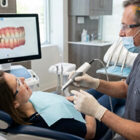
Following the preparation, the traditional, uncomfortable silicone impression materials are entirely bypassed. Instead, state-of-the-art digital scanning technology, such as the iTero or Medit i700 intraoral scanners, is utilized. These devices capture highly accurate 3D digital models of the prepared tooth, the adjacent teeth, and the patient’s bite registration in a matter of minutes. This digital workflow not only maximizes patient comfort but also eliminates the dimensional distortions inherent in traditional impression materials, ensuring a virtually seamless marginal fit for the final restoration [5].
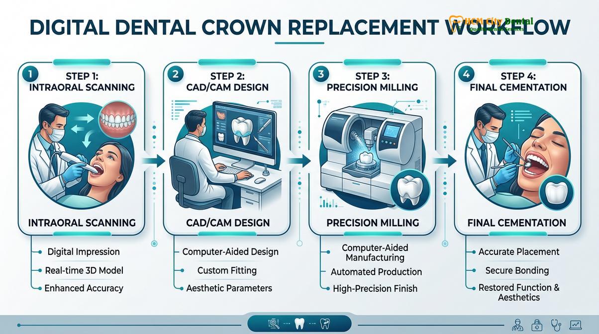
The digital data is then transmitted instantly to our direct lab partnership facility located right here in Ho Chi Minh City. This close collaboration between the clinical team and the master ceramists allows for unparalleled quality control and precise shade matching. For international patients and expats, this streamlined workflow enables an express 3-5 day turnaround time. During this brief interim, the patient is fitted with a custom-fabricated temporary crown to protect the prepared tooth and maintain aesthetic appearance. Upon completion, the final Zirconia or E.max crown is rigorously evaluated for marginal adaptation, proximal contacts, and occlusal harmony before being permanently bonded into place using advanced resin cements.
When to See a Doctor
While some signs of crown wear are gradual, certain symptoms require immediate clinical attention to prevent irreversible damage to the tooth or surrounding tissues. You should schedule an urgent dental evaluation if you experience severe, throbbing pain that disrupts your sleep or is not relieved by over-the-counter analgesics. This level of pain is a strong indicator of acute pulpitis or a developing periapical abscess, which requires prompt endodontic intervention.
Visible swelling of the gums, face, or jaw, particularly if accompanied by a fever or a foul-tasting discharge (pus) around the crowned tooth, constitutes a dental emergency. These are signs of an active, spreading bacterial infection that can compromise your systemic health if left untreated.
“If a dental crown becomes completely dislodged, do not attempt to reattach it using household glues or over-the-counter adhesives, as these are toxic and can permanently damage the tooth. Keep the crown safe and seek professional care immediately for proper evaluation and recementation.”
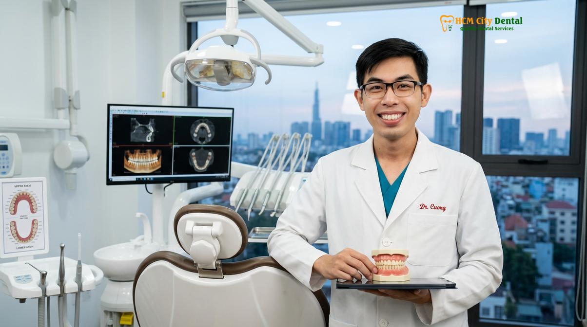
If your crown feels significantly loose or falls out entirely, it is imperative to see a dentist as soon as possible. An exposed prepared tooth is highly sensitive to temperature and is extremely vulnerable to rapid decay and structural fracture. Furthermore, adjacent teeth can begin to shift into the empty space within a matter of days, which may prevent the old crown from fitting properly even if it is structurally intact. At HCMC Dental Clinic, we offer emergency loose crown recementation services and provide a comprehensive global warranty policy with remote follow-up support via WhatsApp, ensuring our patients receive continuous care and guidance regardless of their geographic location.
Frequently Asked Questions
What are the symptoms of a failed dental crown?
The primary symptoms of a failed dental crown include persistent pain, thermal sensitivity, a loose sensation, visible cracks, and swollen gums. If you experience a bad taste or odor around the restored tooth, it may indicate bacterial leakage beneath the crown margin. Early detection of these signs is critical to prevent the decay from reaching the tooth’s nerve.
Can a decayed tooth under a crown be saved?
Yes, a decayed tooth under a crown can often be saved if the decay is detected and treated early. The clinical process involves removing the old crown, excavating the recurrent caries, and potentially performing a root canal or core buildup before placing a new restoration. However, if the decay has destroyed too much of the root structure, extraction and implant placement may be necessary.
How do you replace a failed dental crown?
Replacing a failed dental crown involves carefully sectioning and removing the old prosthesis, treating any underlying decay or infection, and reshaping the tooth. A digital intraoral scan is then taken to design and mill a precise new crown using CAD/CAM technology. The new crown is then permanently cemented, restoring the tooth’s function and aesthetics.
Is it normal for a new dental crown to hurt when chewing?
Mild sensitivity is normal for a few days after placement, but sharp pain when chewing usually indicates that the crown’s bite is too high. A simple occlusal adjustment by your prosthodontist can quickly resolve this issue and prevent trauma to the underlying tooth. If the pain persists, it may indicate underlying nerve inflammation requiring further evaluation.
How long do zirconia and E.max crowns typically last?
With proper oral hygiene and regular clinical examinations, high-quality Zirconia and E.max crowns can last 10 to 15 years, or even longer. Their longevity depends heavily on the precision of the marginal fit, the patient’s bite forces, and the absence of bruxism. Wearing a custom nightguard can significantly extend the lifespan of these ceramic restorations.
References
- Journal of Prosthodontic Research. Flexural strength and clinical performance of monolithic zirconia crowns. (2021).
- Journal of Esthetic and Restorative Dentistry. Translucency and marginal adaptation of lithium disilicate restorations. (2020).
- International Journal of Prosthodontics. Microleakage and recurrent caries beneath fixed dental prostheses. (2019).
- Journal of Dentistry. Accuracy of CAD/CAM milling and digital intraoral scanners in prosthodontics. (2022).
- American Dental Association. Clinical guidelines for the management of defective dental restorations. (2018).