When patients invest in advanced restorative dentistry, understanding zirconia crown longevity years becomes a primary focus during the treatment planning phase. Zirconia crowns typically last between 10 to 15 years, with a significant number of clinical cases demonstrating functional and aesthetic longevity exceeding two decades. Their exceptional durability is not merely a product of the material itself, but rather a combination of precise tooth preparation, high-quality digital fabrication, and consistent daily oral hygiene practices designed to prevent underlying tooth decay or periodontal disease.
The evolution of dental prosthetics has seen a massive shift away from traditional metal-based restorations toward high-strength, metal-free ceramics. Zirconium dioxide, commonly referred to as zirconia, has revolutionized this landscape by offering a biocompatible solution that mimics the natural translucency of enamel while providing structural resilience that rivals, and often surpasses, cast metal. However, achieving the maximum potential lifespan of these restorations requires a comprehensive understanding of biomechanical forces, proper cementation protocols, and the patient’s commitment to long-term maintenance.
Clinical Summary:
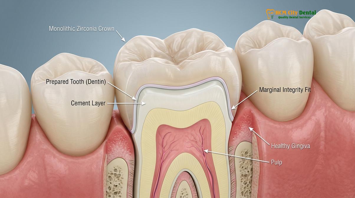
Zirconia crowns represent a premier choice in modern restorative dentistry due to their outstanding biocompatibility, superior flexural strength, and excellent aesthetic properties. While extensive clinical studies indicate an average functional lifespan of 10 to 15 years, meticulous digital fabrication, advanced adhesive cementation protocols, and diligent patient maintenance can significantly extend this duration. Advanced digital workflows, including high-resolution intraoral scanning and precision CAD/CAM milling, ensure optimal marginal adaptation, which is critical for long-term success and the prevention of secondary caries. Understanding the unique biomechanical properties of yttria-stabilized zirconia helps both clinicians and patients optimize treatment outcomes for complex functional and aesthetic rehabilitations.
Key Takeaways:
- Zirconia crowns offer exceptional fracture resistance, making them highly suitable for both anterior aesthetic zones and posterior load-bearing areas.
- Clinical longevity typically ranges from 10 to 15 years, though this is heavily influenced by patient oral hygiene, diet, and occlusal forces.
- Advanced digital scanning and CAD/CAM milling ensure precise marginal fit, drastically reducing the risk of bacterial microleakage and secondary decay.
- Parafunctional habits like bruxism can impact crown lifespan, often necessitating the use of a custom protective occlusal nightguard.
- Routine professional evaluations and radiographic assessments are essential to monitor cement integrity and surrounding gingival health over time.
Zirconia Strength: Flexural Resistance and Durability
Zirconia exhibits unparalleled flexural resistance, often exceeding 1000 MPa, making it highly resistant to fracture and wear under heavy occlusal forces. This structural integrity ensures reliable, long-lasting dental restorations even in the posterior segments of the mouth.
The remarkable durability of zirconia in dental applications stems from its unique crystalline structure and advanced material science. Zirconium dioxide is a polymorphic material that exists in distinct crystallographic phases depending on the ambient temperature: monoclinic, tetragonal, and cubic. In modern dentistry, yttria-stabilized tetragonal zirconia polycrystal (Y-TZP) is predominantly utilized. The addition of yttria stabilizes the tetragonal phase at room temperature, which is crucial for the material’s mechanical properties. When a microscopic crack begins to propagate through the ceramic material due to extreme occlusal stress or trauma, the localized stress at the tip of the crack triggers a phase transformation from the tetragonal phase to the monoclinic phase.
This specific phase transformation is accompanied by a localized volumetric expansion of approximately 3% to 4%. This expansion effectively places the crack tip under severe compressive stress, actively pinching the crack shut and halting its progression through the material[1]. This remarkable phenomenon, known in materials science as transformation toughening, is the primary reason why the flexural strength profile of zirconia is vastly superior to traditional feldspathic porcelains and lithium disilicate glass-ceramics[2]. It allows monolithic zirconia to withstand the immense biting forces generated in the molar regions without catastrophic failure.
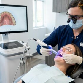
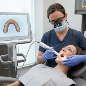
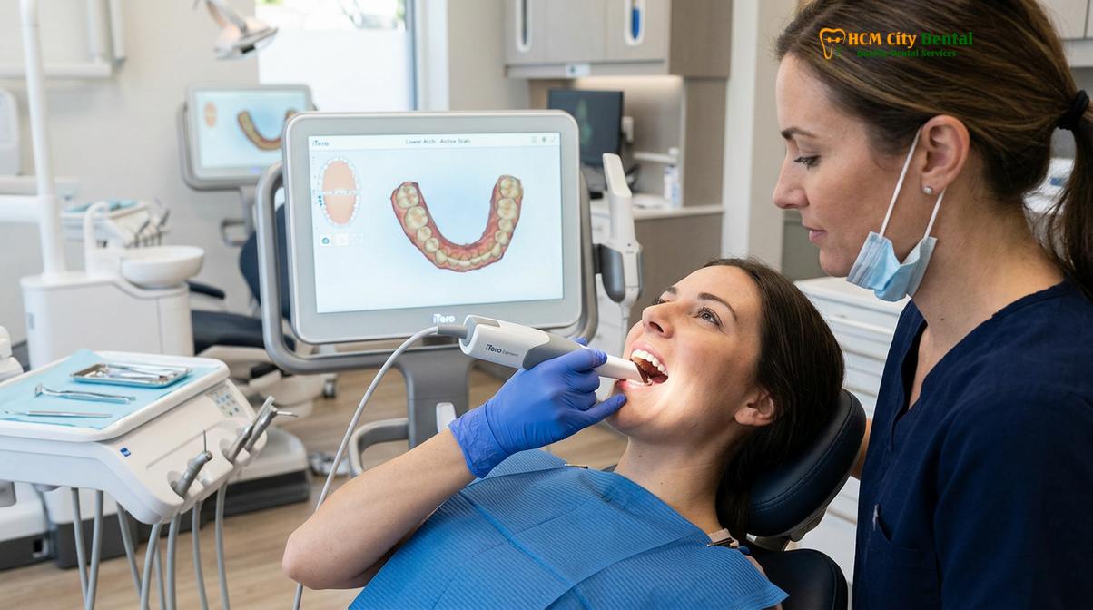
To fully harness this immense strength, modern prosthodontics relies heavily on advanced digital workflows. The traditional method of taking messy, uncomfortable silicone impressions has been largely superseded by state-of-the-art digital scanning technology. Clinicians can now capture highly accurate, three-dimensional topographical maps of the prepared tooth and the surrounding dentition in real-time. This digital impression provides maximum comfort for the patient and eliminates the dimensional distortions, tearing, and shrinkage inherent in elastomeric impression materials. The digital file is then exported to a specialized dental laboratory where the crown is meticulously designed using CAD (Computer-Aided Design) software and milled from a solid, homogeneous block of pre-sintered zirconia using multi-axis CAM (Computer-Aided Manufacturing) technology. The extreme precision of CAD/CAM milling ensures an intimate, gap-free marginal fit, which is paramount for the long-term biological success of the restoration.
Average Lifespan in Clinical Studies
Extensive clinical research demonstrates that monolithic zirconia crowns maintain a survival rate of over 90% after ten years of function. With optimal clinical protocols and dedicated patient care, their lifespan frequently extends well beyond fifteen years.
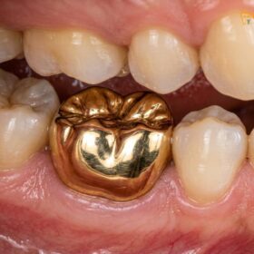
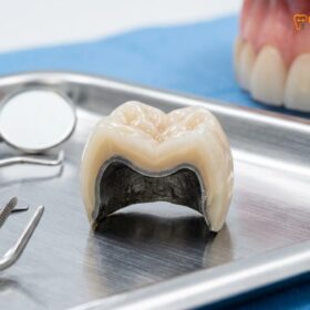
When evaluating zirconia crown longevity years, evidence-based dentistry provides a clear, highly optimistic picture. Numerous longitudinal clinical studies and systematic reviews have tracked the performance of zirconia-based restorations over extended periods across diverse patient populations. The data consistently indicates that monolithic zirconia crowns exhibit exceptional survival rates, often outperforming traditional porcelain-fused-to-metal (PFM) alternatives in terms of structural integrity. Failures, when they do occur, are exceedingly rarely due to the catastrophic fracture of the zirconia core itself. Instead, the most common complications leading to crown failure are biological in nature, such as secondary caries developing at the margin where the crown meets the natural tooth, or endodontic complications requiring a root canal therapy beneath the existing restoration.
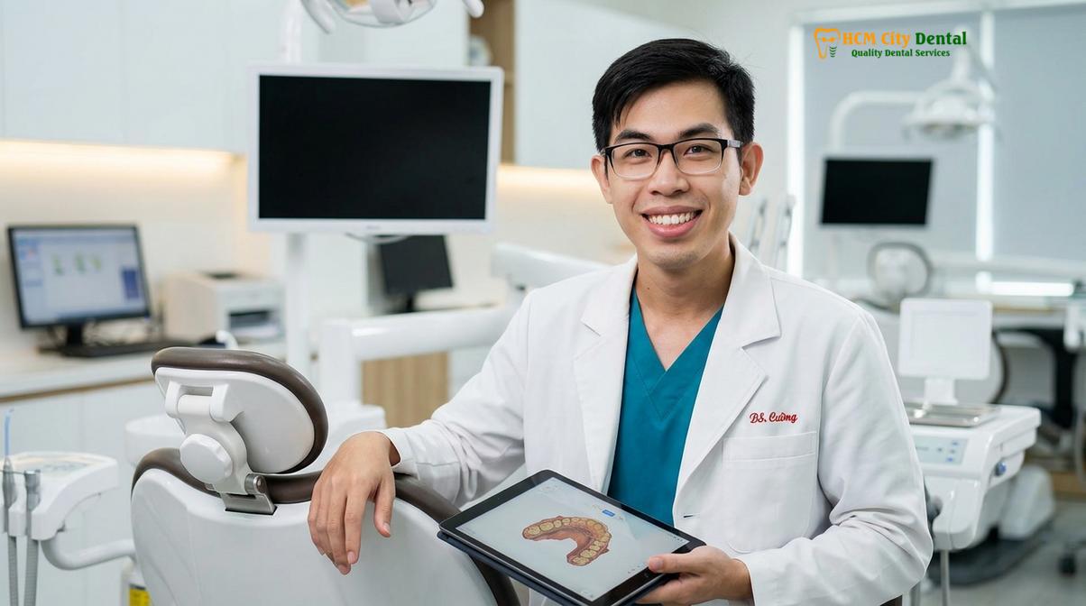
The clinical perspective on cosmetic material selection and meticulous tooth preparation is vital for maximizing this lifespan. Dr. Nguyen Van Cuong, a highly regarded restorative expert, emphasizes that a precise preparation design is essential to provide adequate thickness for the ceramic material while ensuring a smooth, continuous finish line. At HCMC Dental Clinic, Dr. Cuong and the clinical team utilize advanced digital workflows to ensure every restoration meets rigorous international standards. His approach focuses on preserving healthy dentin while achieving the precise marginal adaptation required for long-term clinical success. Respecting the biologic width during preparation and impression taking prevents chronic gingival inflammation, which can lead to localized bone loss and the eventual failure of the abutment tooth.
Understanding the financial investment is an important aspect of comprehensive treatment planning. We offer a transparent pricing structure that provides significant value, especially for international patients utilizing our streamlined pre-booking system.
| Material / Treatment Type | Standard Walk-in Price | WhatsApp Pre-booking (-40%) |
|---|---|---|
| PFM Crown (Standard) | ~$140 (3.5M VND) | From $84 (2.1M VND) |
| Zirconia Crown (Cercon) | ~$200 (5.0M VND) | From $120 (3.0M VND) |
| Zirconia HT Crown (Cercon HT) | ~$260 (6.5M VND) | From $156 (3.9M VND) |
| E.max Crown (Ivoclar) | ~$280 (7.0M VND) | From $168 (4.2M VND) |
| 3-Unit Bridge (Zirconia HT) | ~$480 (12.0M VND) | From $288 (7.2M VND) |
Factors Influencing Zirconia Crown Longevity Years
The ultimate lifespan of a zirconia restoration is dictated by a combination of precise clinical execution, including optimal tooth preparation and advanced adhesive cementation, alongside the patient’s unique oral environment.
While the material properties of zirconia are inherently robust, the clinical longevity of the final restoration is heavily dependent on several critical factors executed during the treatment phase. The first and arguably most important factor is the quality of the initial tooth preparation. A tooth must be prepared with a specific geometry to provide adequate retention and resistance form. This ensures that the crown remains securely in place under the dynamic forces of chewing and speaking. Furthermore, the clinician must establish a clear, well-defined margin—typically a heavy chamfer or a rounded shoulder—which allows the milling machine to create a crown with a perfectly adapted edge. If the margin is poorly defined, the resulting crown may have microscopic gaps, leading to cement dissolution and bacterial infiltration.
Another crucial element is the presence of the ferrule effect. When a tooth has been severely compromised by decay or previous root canal therapy, it is essential that the crown grasps a sufficient band of healthy, solid tooth structure (the ferrule) above the gumline. This ferrule acts like the metal band around a wooden barrel, preventing the underlying tooth root from fracturing under stress. Without an adequate ferrule, even the strongest zirconia crown is likely to fail due to the catastrophic fracture of the underlying natural tooth.
Finally, the cementation protocol plays a pivotal role in longevity. Unlike traditional metal crowns that can be cemented with conventional luting agents, modern zirconia often benefits from advanced adhesive resin cements, particularly when retention is compromised. To achieve a strong chemical bond, the internal surface of the zirconia crown must be properly treated, typically by sandblasting with aluminum oxide particles to increase surface area, followed by the application of a specialized primer containing MDP (10-methacryloyloxydecyl dihydrogen phosphate) monomers. This meticulous chemical bonding process ensures a durable, long-lasting seal that protects the underlying dentin from bacterial microleakage.
Daily Maintenance and Cleaning Practices
Preserving a zirconia crown requires rigorous daily oral hygiene, including brushing with non-abrasive toothpaste and meticulous interdental cleaning, to prevent plaque accumulation at the crown margin and ensure long-term gingival health.
While the high-density ceramic material of a monolithic zirconia crown is completely impervious to bacterial decay, the underlying natural tooth structure and the surrounding periodontal tissues remain highly susceptible to oral diseases. Therefore, maintaining the longevity of these advanced restorations is fundamentally tied to the patient’s daily oral hygiene regimen. The most critical anatomical area to maintain is the crown margin—the microscopic junction where the restorative material meets the natural tooth structure. If dental plaque biofilm is allowed to accumulate and mature at this interface, the acid-producing bacteria can slowly dissolve the cement seal and initiate secondary caries deep beneath the crown[3]. Furthermore, chronic plaque accumulation inevitably leads to gingivitis and periodontitis, which can cause the gums to recede, exposing the vulnerable root surface and compromising both the aesthetic and structural integrity of the restoration.
“The longevity of any advanced dental restoration is a partnership between the clinician’s precision in the operatory and the patient’s dedication at the bathroom sink. Even the most perfectly milled zirconia crown relies on healthy, inflammation-free gingival tissues for long-term survival.”
Patients must adopt a meticulous and consistent cleaning routine to protect their investment. Brushing at least twice daily with a soft-bristled toothbrush is mandatory. It is crucial to use a non-abrasive toothpaste; highly abrasive whitening toothpastes containing harsh silica particles can scratch the highly polished surface of the zirconia over time. While zirconia is incredibly hard, maintaining its smooth, glass-like surface polish is essential to prevent it from becoming abrasive to the opposing natural teeth. Flossing daily is equally important to remove interproximal plaque that a standard toothbrush cannot reach. For patients with dental bridges, splinted crowns, or larger embrasure spaces, the use of interdental brushes or water flossers is highly recommended to effectively flush out food debris and bacteria from beneath the pontic and around the critical abutment margins.
The Impact of Bruxism (Teeth Grinding)
Chronic teeth grinding exerts excessive, destructive forces that can compromise both the restoration and opposing natural teeth. Wearing a custom-fitted occlusal nightguard is highly recommended to mitigate these risks.
Bruxism, the involuntary grinding or clenching of teeth, is a highly prevalent parafunctional habit that poses a significant threat to all dental restorations, including high-strength ceramics. While monolithic zirconia is exceptionally resistant to fracture, the immense occlusal forces generated during sleep bruxism—which can be exponentially higher than normal, conscious chewing forces—can have detrimental effects on the entire stomatognathic system. The primary clinical concern is not necessarily the fracture of the zirconia itself, but rather the severe wear inflicted on the opposing dentition. If the zirconia crown is not perfectly polished after occlusal adjustments, or if the patient grinds heavily, the microscopic roughness of the ceramic can act as an abrasive file, causing rapid and severe wear of the opposing natural enamel[4].
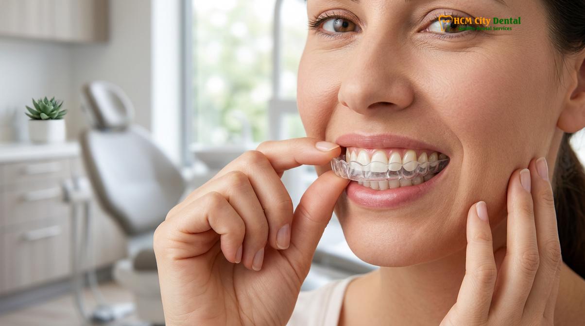
Clinical Warning: Unmanaged bruxism can lead to catastrophic failure of both natural teeth and dental restorations. The excessive forces can cause widening of the periodontal ligament, tooth mobility, and vertical root fractures. Patients who wake up with jaw soreness, tension headaches, or notice flattening of their teeth should seek immediate evaluation for a protective occlusal splint.
Furthermore, the excessive forces transmitted through the rigid crown can severely affect the underlying tooth structure and the supporting periodontal ligament. This continuous trauma can lead to a phenomenon known as abfraction (notching at the gumline), widening of the periodontal ligament space, increased tooth mobility, or even vertical root fractures, which inevitably result in the complete loss of the tooth. To manage these significant risks, precise bite adjustment during the crown delivery appointment is critical. The clinician must ensure that the occlusal contacts are perfectly even and that there are no premature interferences during excursive movements of the jaw. Following delivery, the fabrication of a custom, hard-acrylic nightguard is often considered mandatory for patients with a known history of bruxism to protect their investment and preserve their remaining natural dentition.
Signs That Your Zirconia Crown Needs Replacement
Indicators for crown replacement include visible marginal gaps, localized gingival inflammation, persistent thermal sensitivity, or structural damage. Prompt clinical evaluation is necessary when these symptoms arise to prevent further complications.
Despite their impressive longevity and robust material properties, zirconia crowns are not permanent fixtures and may eventually require replacement due to biological changes or mechanical wear. Recognizing the early signs of failure can prevent more severe complications, such as extensive tooth decay, pulpal infection, or the need for a complex dental extraction. One of the most common and critical indicators of failure is a compromised marginal seal. Over time, the dental cement securing the crown can slowly wash out or degrade due to continuous exposure to oral fluids, temperature fluctuations, and bacterial acids. This degradation creates a microscopic gap, allowing cariogenic bacteria to infiltrate the underlying vulnerable dentin. According to the clinical guidelines established by the Vietnam Odonto-Stomatology Association (VOSA), patients should seek immediate professional evaluation if they notice a dark line developing at the gumline, localized gingival bleeding around a specific crowned tooth when brushing, or a persistent foul odor emanating from the area[5].
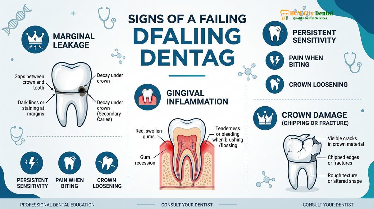
Another significant sign of impending failure is the onset of pain or sensitivity. A properly sealed, well-integrated crown should feel and function exactly like a healthy natural tooth. If a patient begins to experience persistent sensitivity to hot or cold temperatures, pain upon biting down, or a dull, throbbing ache, it often indicates that the nerve of the tooth is inflamed or infected. This pulpal distress is frequently caused by recurrent decay that has progressed beneath the crown, reaching the inner pulp chamber. In such cases, the existing crown must be carefully sectioned and removed to access the underlying tooth structure, allowing the clinician to perform necessary endodontic therapy (root canal) before fabricating a new restoration.
“Early detection of marginal leakage or cement failure can often save the underlying abutment tooth. Delaying a clinical evaluation when symptoms arise significantly increases the risk of requiring complex endodontic therapy or facing complete tooth loss.”
Aesthetic degradation can also be a primary reason for replacement, particularly in the anterior (front) region of the mouth. While the zirconia material itself will not discolor, the surrounding gums may recede over time due to aggressive brushing, periodontal disease, or natural aging. This gingival recession can expose the junction between the crown and the tooth root, which often appears darker and less aesthetically pleasing. If this aesthetic compromise affects the patient’s smile and confidence, replacing the crown to establish a new, harmonious margin with the current gumline is a highly effective solution.
Patient Case Study: Restoring Function in Ho Chi Minh City
A clinical case demonstrates how replacing failing traditional restorations with precision-milled monolithic zirconia can restore both biomechanical function and gingival health for patients experiencing occlusal distress.
Clinical Case Study: Comprehensive Arch Rehabilitation in Ho Chi Minh City
To illustrate the profound clinical impact of utilizing high-quality materials and advanced digital workflows, consider the case of a 45-year-old expatriate patient who recently visited HCMC Dental Clinic in Ho Chi Minh City. The patient presented with severe discomfort and aesthetic concerns regarding an older porcelain-fused-to-metal (PFM) crown on a lower first molar. The existing restoration exhibited significant porcelain chipping due to years of unmanaged nocturnal bruxism. Furthermore, the surrounding gingival tissue was highly inflamed, bleeding easily upon probing, due to a bulky, over-contoured metal margin that consistently trapped plaque and hindered proper oral hygiene.
Dr. Nguyen Van Cuong conducted a comprehensive clinical and radiographic evaluation. Utilizing high-resolution digital intraoral scanning, the team assessed the occlusal clearance, the extent of the marginal discrepancies, and the patient’s dynamic bite. The treatment plan involved carefully sectioning and removing the failing PFM crown, meticulously refining the underlying tooth preparation to ensure a smooth, precise chamfer margin, and allowing the inflamed gingival tissues to heal with a well-adapted, highly polished temporary crown.
Once optimal periodontal health was stabilized, a monolithic zirconia crown was digitally designed and milled to exact specifications. The final restoration provided exceptional fracture resistance against the patient’s bruxism while establishing a precise, hygienic margin that completely resolved the localized gingival inflammation. This case highlights the critical importance of combining superior materials with meticulous clinical execution at HCMC Dental Clinic to achieve long-lasting, predictable oral health outcomes.
References
- Journal of Prosthetic Dentistry. Long-term survival rates of zirconia-based restorations. (2021).
- International Journal of Prosthodontics. Flexural strength and phase transformation of yttria-stabilized zirconia. (2020).
- Journal of Clinical Periodontology. Marginal adaptation and periodontal health around CAD/CAM crowns. (2019).
- Dental Materials. Wear behavior of monolithic zirconia against natural enamel. (2022).
- Vietnam Odonto-Stomatology Association (VOSA). Clinical guidelines for the maintenance of all-ceramic restorations in Vietnamese populations. (2023).
If you are experiencing issues with your current restorations, noticing signs of marginal leakage, or wish to explore your options for advanced Dental Crowns & Bridges, contact HCMC Dental Clinic, Ho Chi Minh City today for a comprehensive evaluation and a personalized treatment plan tailored to your unique clinical needs.