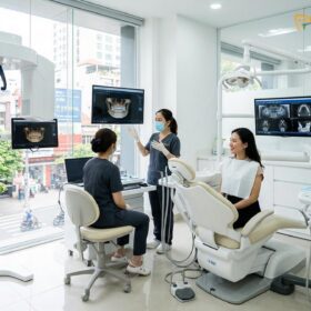
A dental crown cap is a custom-fabricated, full-coverage prosthetic restoration designed to encase a damaged, severely decayed, or structurally compromised tooth. By restoring the tooth’s original anatomical shape, size, and strength, this clinical intervention preserves natural dentition and ensures optimal biomechanical function within the oral cavity.
Clinical Summary:
A dental crown—frequently referred to by patients as a “cap”—is an essential restorative treatment utilized in modern dentistry to salvage teeth that cannot be repaired with standard direct fillings. Fabricated from advanced materials such as monolithic zirconia, lithium disilicate, or porcelain-fused-to-metal, these prosthetics provide a durable, aesthetic shield over the natural tooth structure. The clinical workflow involves precise tooth preparation, digital or physical impressions, and meticulous cementation to ensure marginal integrity and long-term occlusal stability. Proper diagnostic evaluation by a qualified prosthodontist or general dentist is critical to determining the appropriate material and technique for each unique clinical scenario, ultimately preventing tooth loss and restoring full masticatory function.
Key Takeaways:
- A dental crown and a dental cap refer to the exact same full-coverage restorative prosthetic.
- Crowns protect structurally compromised teeth following severe decay, trauma, or root canal therapy.
- Modern material options include high-strength zirconia, aesthetic glass-ceramics, and durable metal alloys.
- The clinical procedure typically requires two visits, though CAD/CAM technology enables same-day restorations in specific cases.
- Longevity depends heavily on marginal fit, occlusal alignment, and the patient’s daily oral hygiene.
Anatomical and Clinical Definition of a Dental Crown Cap
A dental crown cap is a fixed prosthetic device cemented onto existing teeth or implants to restore structural integrity, masticatory function, and natural aesthetics.
In the realm of restorative dentistry, the terminology used to describe full-coverage prosthetics can sometimes cause confusion among patients. The terms “dental crown” and “dental cap” are entirely synonymous. From a clinical perspective, the anatomical “crown” of a tooth refers to the portion of the tooth that naturally sits above the gingival margin (gum line) and is covered by highly mineralized enamel. When this natural anatomical crown is severely compromised by bacterial decay, mechanical trauma, or extensive wear, a synthetic replacement must be fabricated. Because this prosthetic is designed to fit over the remaining prepared tooth structure much like a protective helmet, the lay term “cap” became widely adopted in popular vernacular. However, in professional dental literature and clinical practice, the procedure is universally referred to as crown placement.
The primary objective of a dental crown cap is to provide circumferential reinforcement to a weakened tooth. Unlike direct restorations (such as composite resin fillings or dental amalgams) which are packed into a cavity preparation and rely on the remaining tooth walls for support, a crown encases the entire visible portion of the tooth. This full-coverage design fundamentally alters the biomechanics of the tooth. When masticatory (chewing) forces are applied, a heavily filled tooth is prone to flexure, which can lead to catastrophic vertical root fractures or the shearing off of cusps. A crown, conversely, acts as a rigid ferrule—a band that binds the remaining tooth structure together, distributing occlusal loads evenly down the long axis of the tooth root and into the supporting alveolar bone. [1]
According to Dr. Nguyen Van Cuong, a leading restorative specialist, the decision to utilize a full-coverage crown rather than a partial restoration (such as an inlay or onlay) hinges heavily on the amount of remaining healthy enamel and the specific occlusal load the tooth must bear. If the structural loss exceeds fifty percent of the coronal volume, or if the remaining walls are undermined and thin, a crown is the most predictable and conservative long-term option to prevent eventual tooth extraction.
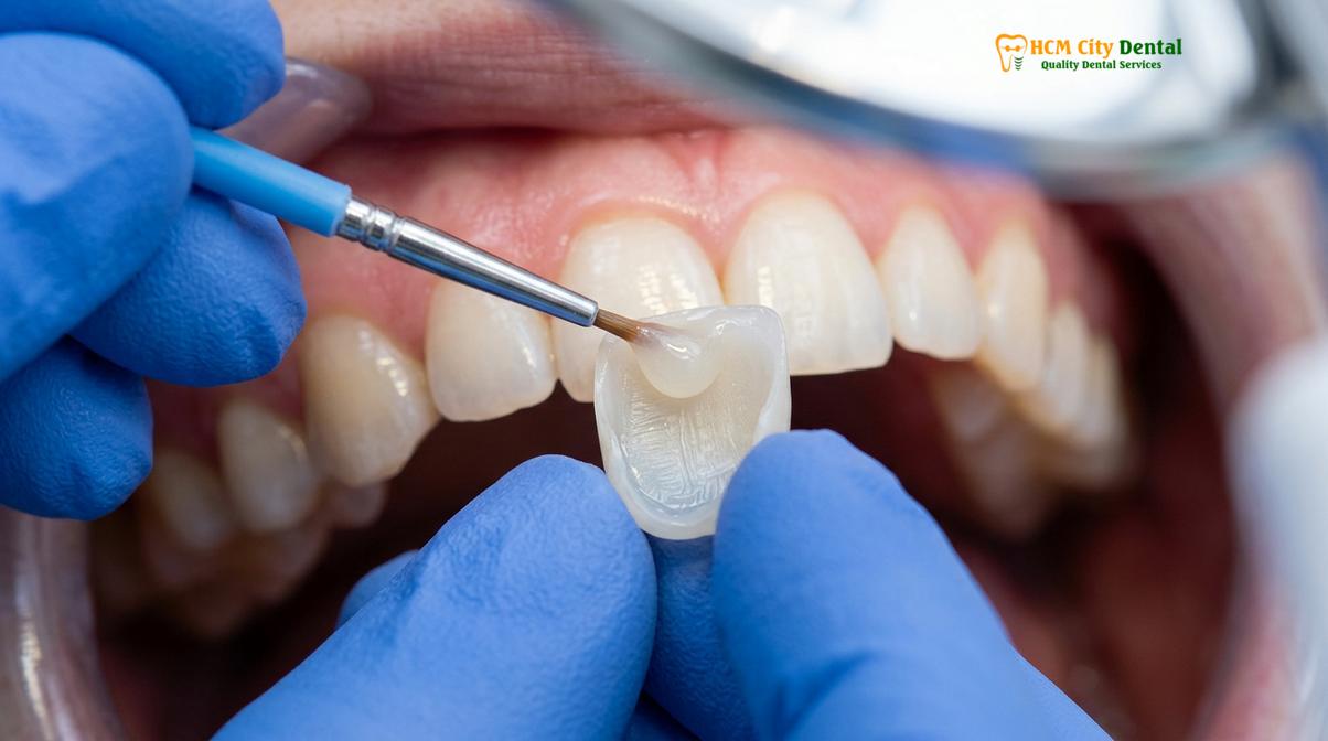
Material Science: Types of Dental Crown Caps
Dental crowns are fabricated from various advanced materials, including high-strength monolithic zirconia, highly aesthetic glass-ceramics, traditional porcelain-fused-to-metal, and durable gold alloys.
The evolution of dental biomaterials has dramatically expanded the options available for fabricating a dental crown cap. The selection of the appropriate material is a critical clinical decision that balances the need for mechanical strength, aesthetic integration, biocompatibility, and the preservation of opposing dentition. No single material is universally superior; rather, the choice must be tailored to the specific functional and cosmetic demands of the patient’s oral environment.
Zirconia: The Pinnacle of Strength
Zirconia (specifically yttria-stabilized tetragonal zirconia polycrystal, or Y-TZP) has revolutionized modern prosthodontics. Zirconia is a crystalline dioxide of zirconium, a metal with properties similar to titanium. However, in its oxidized dental form, it presents as a highly opaque, tooth-colored ceramic. The defining characteristic of zirconia is its extraordinary flexural strength, often exceeding 1000 MPa, making it virtually indestructible under normal masticatory forces. Furthermore, zirconia exhibits a unique property known as “phase transformation toughening.” When a micro-crack begins to propagate through the material, the crystal structure at the tip of the crack undergoes a localized volumetric expansion, effectively squeezing the crack shut and preventing catastrophic failure. [2] Monolithic zirconia crowns are milled from a single solid block using CAD/CAM technology, eliminating the risk of porcelain chipping that plagues layered restorations. They are the premier choice for posterior molars and patients with severe bruxism (teeth grinding).
Lithium Disilicate (Glass-Ceramics)
For anterior (front) teeth where aesthetic demands are paramount, lithium disilicate (commonly known by the brand name IPS e.max) is often the material of choice. This glass-ceramic offers unparalleled optical properties, mimicking the natural translucency, opalescence, and fluorescence of human enamel. Similar to the advanced ceramics utilized in premium Porcelain Veneers, lithium disilicate can be either pressed or milled. While its flexural strength (approximately 400-500 MPa) is lower than that of zirconia, it is more than sufficient for the anterior segment and premolars. Additionally, lithium disilicate can be adhesively bonded to the underlying tooth structure, creating a monobloc effect that significantly enhances the overall fracture resistance of the restored tooth.
Porcelain-Fused-to-Metal (PFM)
For decades, the porcelain-fused-to-metal (PFM) crown was the gold standard in restorative dentistry. A PFM crown consists of a cast or milled metal substructure (coping) that provides high tensile strength, over which layers of feldspathic porcelain are baked in a high-heat vacuum furnace to provide a tooth-like appearance. While highly durable and clinically proven, PFM crowns have distinct aesthetic limitations. The underlying metal is dark, requiring an opaque masking layer of porcelain that can make the final crown look somewhat flat or lifeless compared to all-ceramic options. Furthermore, as patients age and natural gingival recession occurs, the metal margin of the PFM crown often becomes visible as a distinct “dark line” at the gum line, compromising the cosmetic outcome.
Gold Alloys and Base Metals
Despite the surge in aesthetic ceramics, cast gold alloys remain an exceptional restorative material. Gold is highly biocompatible, non-toxic, and possesses a coefficient of thermal expansion very similar to natural tooth structure. Most importantly, the wear characteristics of gold are nearly identical to human enamel, meaning a gold crown will not cause excessive wear on the opposing natural teeth. Gold is also highly malleable, allowing the dental laboratory to achieve an incredibly precise marginal seal, which minimizes the risk of recurrent decay. While their metallic appearance restricts their use primarily to out-of-sight second molars, gold crowns boast the longest clinical track record of survival in dentistry.
| Material Type | Flexural Strength | Aesthetic Quality | Primary Clinical Indication |
|---|---|---|---|
| Monolithic Zirconia | Very High (>1000 MPa) | Moderate to High | Posterior molars, heavy bruxers, implant crowns |
| Lithium Disilicate (e.max) | Moderate (400-500 MPa) | Exceptional | Anterior teeth, premolars, high aesthetic zones |
| Porcelain-Fused-to-Metal (PFM) | High (Metal Core) | Moderate | Long-span bridges, posterior restorations |
| Cast Gold Alloy | High (Malleable) | Low (Metallic) | Out-of-sight molars, patients with severe wear |
“The selection of crown material is not merely an aesthetic choice; it is a complex biomechanical decision dictated by the patient’s parafunctional habits, opposing dentition, and the specific location within the dental arch.”
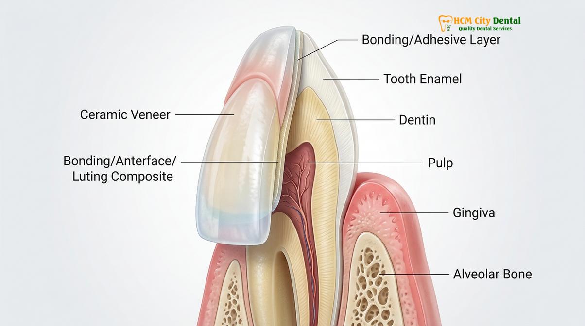
The Clinical Workflow: From Preparation to Final Cementation
The crown placement process involves meticulous diagnostic evaluation, precise tooth preparation, accurate impression taking, provisionalization, and the final adhesive cementation of the permanent prosthetic.
The fabrication and placement of a dental crown cap is a highly precise, multi-step clinical procedure that demands rigorous attention to detail. The success of the final restoration is directly proportional to the accuracy achieved during each phase of the workflow. For patients seeking comprehensive care, understanding this general dental guide to the crown procedure helps alleviate anxiety and sets appropriate clinical expectations.
Diagnostic Evaluation and Core Build-Up
Prior to initiating any irreversible tooth preparation, a thorough diagnostic assessment is mandatory. This includes periapical radiographs to evaluate the health of the tooth roots and surrounding alveolar bone, as well as vitality testing to ensure the dental pulp is healthy. If the tooth exhibits profound decay that has compromised the pulp, endodontic therapy (a root canal) must be completed first. Once the tooth is deemed biologically sound, the dentist must assess the remaining structural volume. If significant portions of the tooth are missing due to fracture or previous large fillings, a “core build-up” is performed. This involves using a highly filled composite resin to rebuild the missing internal structure, providing a solid, geometric foundation upon which the final crown can be securely anchored.
Principles of Tooth Preparation
Tooth preparation is a subtractive process where the dentist utilizes specialized high-speed diamond burs to reduce the circumference and height of the tooth. The goal is to create sufficient restorative space for the chosen crown material while preserving as much healthy tooth structure as possible. The preparation must possess specific geometric features: a slight convergence angle (taper) of the axial walls to allow the crown to seat fully, and adequate height to provide “resistance and retention form,” which prevents the crown from dislodging under lateral chewing forces. At the gingival margin, the dentist creates a precise finish line—typically a chamfer or a modified shoulder—which dictates exactly where the crown will end and seamlessly transition into the natural tooth root.
Tissue Management and Impression Techniques
Capturing an exact replica of the prepared tooth is arguably the most critical step in the workflow. To ensure the dental laboratory can identify the exact finish line, the dentist must manage the surrounding gingival tissue. This is often achieved using a retraction cord—a tiny braided thread soaked in a hemostatic agent (like aluminum chloride)—which is gently packed into the gingival sulcus to temporarily push the gums away from the tooth margin. Following tissue retraction, an impression is taken. While traditional polyvinyl siloxane (PVS) putty impressions are still widely used, modern clinics increasingly rely on optical technology. At premium facilities like HCMC Dental Clinic, digital intraoral scanners have largely replaced traditional putty, utilizing confocal microscopy to capture highly accurate, real-time 3D models of the patient’s dentition. This digital data is then transmitted instantly to the dental laboratory for CAD/CAM design.
Provisionalization (The Temporary Crown)
Because the fabrication of a custom permanent crown typically takes one to two weeks, the prepared tooth must be protected in the interim. A provisional, or temporary, crown is fabricated chairside using bis-acryl composite or polymethyl methacrylate (PMMA) resin. The temporary crown serves several vital biological functions: it seals the dentinal tubules to prevent severe thermal sensitivity, it maintains the position of the tooth within the arch (preventing it from drifting or super-erupting), and it guides the healing of the gingival tissues around the newly established margins.
Cementation Protocols and Adhesion Chemistry
The final cementation of a dental crown requires strict isolation and specific chemical bonding protocols tailored to the exact restorative material used.
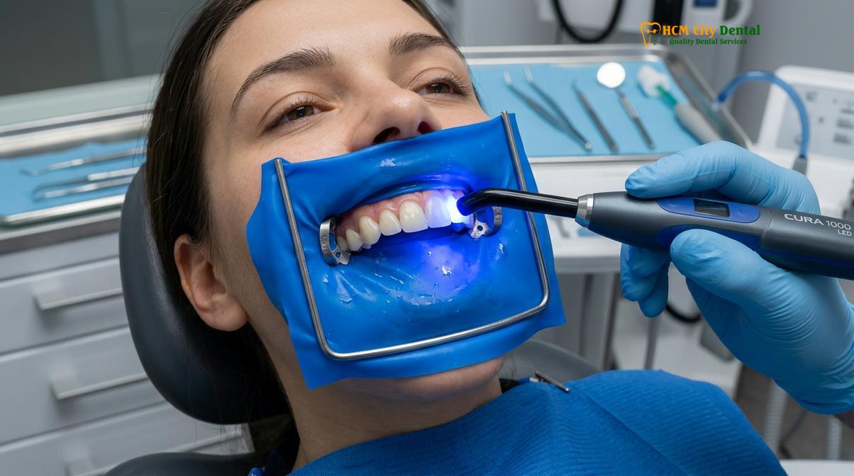
The final phase of the crown workflow is the delivery and cementation of the permanent prosthetic. This appointment begins with the careful removal of the temporary crown and the thorough cleaning of the prepared tooth surface to remove any residual temporary cement. The permanent crown is then “tried in” to verify several critical parameters. The dentist checks the interproximal contacts (how tightly the crown touches the adjacent teeth) using dental floss, ensuring food will not become impacted between the teeth. The marginal fit is evaluated using a sharp dental explorer and often confirmed with a bitewing radiograph to ensure there are no microscopic gaps or overhangs. Finally, the occlusion (the patient’s bite) is checked using articulating paper to ensure the new crown hits simultaneously and harmoniously with the rest of the dentition.
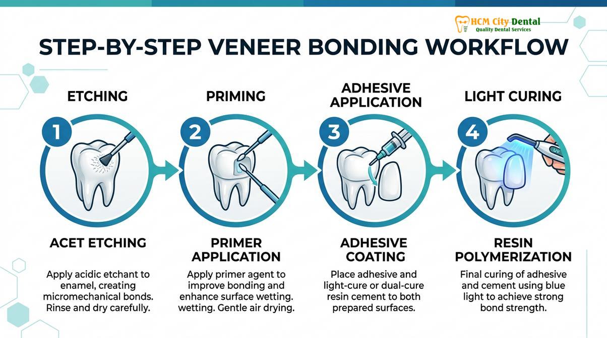
Once the fit is verified, the cementation protocol begins. The choice of cement is dictated by the crown material. For traditional PFM or cast gold crowns, conventional luting agents such as resin-modified glass ionomer (RMGI) cements are typically utilized. These cements rely primarily on the mechanical retention provided by the geometry of the tooth preparation. However, for highly aesthetic glass-ceramics like lithium disilicate, an adhesive resin bonding protocol is required. [5] This involves a complex chemical process: the internal surface of the ceramic crown is etched with hydrofluoric acid to create micro-porosities, and then treated with a silane coupling agent. Simultaneously, the natural tooth structure is etched with phosphoric acid and coated with a dentin bonding agent. When the resin cement is applied and cured with a specialized dental curing light, a profound micromechanical and chemical bond is formed, effectively fusing the crown to the tooth.
Biomechanical Considerations and Long-Term Longevity
The long-term success of a dental crown depends on precise marginal adaptation, harmonious occlusal dynamics, and the maintenance of surrounding periodontal health.
A well-executed dental crown cap is designed to be a long-term, highly predictable restoration. However, its survival in the hostile environment of the oral cavity depends on several critical biomechanical factors. The most significant of these is marginal integrity. The junction where the restorative material meets the natural tooth structure is the most vulnerable point of the entire system. If the gap at this margin exceeds acceptable clinical limits (typically 50 to 100 microns), the cement can slowly wash out over time, allowing cariogenic bacteria to infiltrate beneath the crown. [3] Because the crown itself cannot decay, this recurrent caries attacks the underlying dentin, often progressing silently until the tooth structure is completely undermined and the crown simply falls off.
Occlusal dynamics also play a paramount role in crown longevity. The human masticatory system can generate immense forces, particularly during parafunctional activities like nocturnal bruxism. If a crown is left even a fraction of a millimeter too high, it will bear a disproportionate amount of the occlusal load. This premature contact can lead to several adverse outcomes: the ceramic material may fracture or chip, the cement seal may break under the stress, or the periodontal ligament surrounding the tooth root may become acutely inflamed, leading to severe pain upon biting. In some cases, malocclusion may contribute to premature crown wear, necessitating a multidisciplinary approach that includes orthodontic evaluation, as detailed in guides concerning Braces in Vietnam Price and alignment strategies.
Furthermore, the relationship between the crown margin and the surrounding gingival tissue is critical. The margin must respect the “biologic width”—the natural dimension of the soft tissue attachment to the tooth. If a crown margin is placed too deeply beneath the gums, it violates this space, triggering a chronic inflammatory response. The gums will remain perpetually red, swollen, and prone to bleeding, eventually leading to localized periodontal bone loss.
A 45-year-old patient presented to HCMC Dental Clinic in Ho Chi Minh City with severe occlusal wear, loss of vertical dimension, and multiple fractured amalgam restorations. Dr. Nguyen Van Cuong conducted a comprehensive diagnostic workup, utilizing a combination of monolithic zirconia crowns for the posterior molars to withstand heavy grinding forces, and highly aesthetic lithium disilicate crowns for the anterior segment. By meticulously restoring the vertical dimension of occlusion and ensuring precise marginal fit across all restorations, the patient achieved both functional stability and a highly aesthetic outcome, demonstrating the profound efficacy of material-specific treatment planning.
Advanced Applications: Crowns in Comprehensive Treatment Plans
Beyond single-tooth restoration, dental crowns are integral components in complex prosthodontic rehabilitations, including implantology and fixed partial dentures.
While the single-tooth dental crown cap is a staple of restorative dentistry, the principles of full-coverage restorations extend into much more complex prosthodontic applications. When a tooth is deemed entirely non-restorable and must be extracted, implant placement becomes necessary. Patients often research international options, comparing the clinical protocols of implant-supported crowns across different regions, such as evaluating Dental Implants Vietnam vs Thailand. An implant crown differs from a natural tooth crown in its foundation; rather than being cemented onto a prepared dentin core, it is attached to a titanium abutment that is screwed directly into the osseointegrated implant fixture. These implant crowns can be either cement-retained or screw-retained, with the latter offering the distinct advantage of easy retrievability for maintenance.
Crowns also serve as the foundational retainers for fixed partial dentures, commonly known as dental bridges. In this scenario, the teeth adjacent to an edentulous space (the missing tooth gap) are prepared for full-coverage crowns. These retainer crowns are then fused to a pontic (a false tooth) to create a single, continuous prosthetic unit that bridges the gap. The biomechanical demands on bridge retainers are exceptionally high, as they must support not only their own occlusal load but also the forces applied to the suspended pontic.
Additionally, the relationship between endodontics and prosthodontics is deeply intertwined. Following a root canal procedure, a tooth loses its internal blood supply and hydration, rendering the remaining dentin significantly more brittle and prone to fracture. Furthermore, the access cavity required to perform the root canal removes a substantial portion of the structural roof of the tooth. Therefore, it is a standard clinical protocol to place a full-coverage crown on almost all endodontically treated posterior teeth to protect them from catastrophic splitting. [4]
“Endodontically treated posterior teeth exhibit a significantly higher survival rate when protected by a full-coverage coronal restoration, as the crown distributes occlusal forces and prevents catastrophic vertical root fractures.”
For international patients planning their treatment schedule, understanding the fabrication timeline for custom ceramics is crucial. Whether undergoing a single crown procedure or a more extensive cosmetic makeover, reviewing resources like the Dental Veneers Timeline Vietnam Stay can provide valuable insights into the necessary laboratory turnaround times. Clear communication regarding these complex treatment plans is vital, which is why consulting an experienced, fluent practitioner—such as an English Speaking Dentist Ho Chi Minh City—is highly recommended for expatriates and dental tourists to ensure all clinical nuances are thoroughly understood.
When to See a Doctor for Crown Evaluation
While a dental crown cap is designed for long-term durability, it is not immune to complications. Patients must be vigilant and recognize the clinical signs that indicate a crown may be failing or that the underlying tooth is in distress. Prompt professional evaluation is critical to preventing the complete loss of the tooth.
You should schedule an immediate clinical evaluation if you experience any of the following symptoms:
- Pain Upon Mastication: If biting down causes a sharp, localized pain, it may indicate that the crown is sitting too high (hyperocclusion), that the underlying tooth has developed a vertical root fracture, or that the periodontal ligament is inflamed.
- Thermal Sensitivity: If the crowned tooth remains vital (has not had a root canal) and suddenly develops severe, lingering sensitivity to hot or cold stimuli, it may be a sign that the dental pulp is dying or that the cement seal has washed out, exposing the sensitive dentin.
- Gingival Inflammation and Bleeding: Persistent redness, swelling, or bleeding of the gums immediately surrounding the crown margin can indicate a violation of the biologic width, an allergic reaction to a base metal alloy, or the accumulation of plaque due to an overhanging margin.
- Foul Odor or Taste: A persistent bad taste or odor emanating from a specific crowned tooth is a strong indicator of microleakage. This suggests that bacteria have breached the cement seal and are actively decaying the underlying tooth structure.
- Mobility or Dislodgement: If the crown feels loose, shifts when you chew, or completely falls off, it must be evaluated immediately. Do not attempt to re-cement the crown yourself with household adhesives, as this can permanently damage the tooth and alter your occlusion.
Regular radiographic monitoring during routine dental check-ups is the most effective way to ensure the ongoing health of a crowned tooth, allowing the dentist to detect microscopic issues long before they become symptomatic.
Frequently Asked Questions
What is the difference between a dental crown and a dental cap?
There is no clinical difference between a dental crown and a dental cap; they are two terms for the exact same full-coverage restoration. Dentists typically use the anatomical term “crown,” while patients historically refer to the prosthetic as a “cap” because it caps or covers the remaining tooth structure. Both terms describe a prosthetic device designed to restore the shape, size, strength, and appearance of a damaged tooth.
How long does a permanent dental crown typically last?
A permanent dental crown typically lasts between 10 and 15 years, though many can last a lifetime with excellent oral hygiene and regular professional maintenance. The longevity of the restoration depends heavily on the material chosen, the precision of the marginal fit achieved by the dentist, and the patient’s occlusal habits. Patients who suffer from bruxism (teeth grinding) may experience premature wear or fracture of the crown material if they do not wear a protective occlusal night guard.
Is the procedure for getting a tooth cap painful?
The procedure for getting a tooth cap is not painful, as it is performed under profound local anesthesia to ensure complete patient comfort. The dentist will numb the tooth and surrounding tissues before any preparation begins. Post-operative sensitivity may occur for a few days as the dentin and pulpal tissues recover from the vibration and preparation process, but this is generally mild and transient, easily managed with over-the-counter anti-inflammatory medications.
Can a decayed tooth be crowned without a root canal?
Yes, a decayed tooth can be crowned without a root canal provided the decay has not irreversibly damaged the inner dental pulp. If the bacterial infection has not breached the pulp chamber and the tooth remains vital and asymptomatic, the dentist can remove the decay, place a protective core build-up, and cement a crown directly over the vital tooth. A root canal is only necessary if the pulp is infected, necrotic, or irreversibly inflamed.
How should I care for my new dental crown?
You should care for your new dental crown by brushing twice daily with a non-abrasive fluoride toothpaste, flossing meticulously around the gingival margins, and attending regular professional cleanings. While the ceramic or metal crown itself cannot decay, the natural tooth structure exposed at the gum line remains highly susceptible to cavities. Maintaining the health of the surrounding gum tissue is critical to preventing recurrent decay beneath the edges of the crown.
References
- Journal of Prosthetic Dentistry. Biomechanical principles of tooth preparation for full-coverage restorations. (2021).
- Dental Materials. Phase transformation toughening in yttria-stabilized tetragonal zirconia polycrystals. (2020).
- International Journal of Periodontics & Restorative Dentistry. Marginal adaptation and biologic width considerations in fixed prosthodontics. (2019).
- Journal of Endodontics. Survival rates of endodontically treated posterior teeth with and without full-coverage crowns. (2022).
- Clinical Oral Investigations. Adhesive cementation protocols for lithium disilicate and zirconia ceramics. (2018).