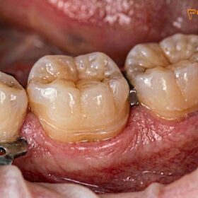
When comparing an inlay vs veneer for front teeth, the clinical decision hinges on the location and extent of the structural damage. Veneers are thin cosmetic shells covering the facial surface to improve aesthetics, while inlays are structural restorations used to repair deep internal decay while preserving visible enamel.
Clinical Summary:
The debate between an inlay vs veneer for front teeth centers on the primary goal of the treatment: structural repair versus cosmetic enhancement. Porcelain veneers are ultra-thin ceramic layers bonded to the anterior (front) surface of the teeth to correct discoloration, minor misalignment, or surface chips. They require minimal facial enamel reduction. Conversely, anterior inlays are utilized when a front tooth suffers from significant interproximal (between the teeth) or lingual (back side) decay. Instead of shaving the entire tooth for a full crown, an inlay precisely fills the cavity, preserving the intact facial enamel. Both treatments rely on advanced CAD/CAM technology and high-strength materials like lithium disilicate to ensure a precise marginal seal and lifelike translucency. A thorough clinical evaluation is necessary to determine which conservative restoration best serves the patient’s long-term oral health and aesthetic goals.
Key Takeaways:
- Primary Function: Veneers enhance facial aesthetics, while inlays restore internal structural integrity.
- Preservation Philosophy: Both are conservative alternatives to full crowns, preserving maximum natural tooth structure.
- Advanced Materials: High-end ceramics provide exceptional durability and natural light transmission.
- Digital Precision: Intraoral scanners eliminate messy impressions, ensuring highly accurate CAD/CAM marginal fits.
- Clinical Assessment: The choice depends entirely on whether the damage is on the visible front surface or hidden between/behind the teeth.
Restoring Anterior Teeth: Aesthetic and Functional Demands
Anterior teeth require a delicate balance between flawless aesthetics and the mechanical strength necessary to withstand shearing forces during biting and incising.
The anterior teeth, comprising the incisors and canines, play a dual role in the human dentition. Aesthetically, they form the foundation of the smile zone, dictating facial harmony, lip support, and phonetic articulation. Functionally, they are responsible for the initial incision of food and provide anterior guidance, which protects the posterior teeth during jaw excursions. Because of these critical functions, restoring anterior teeth demands a meticulous approach that addresses both visual perfection and biomechanical resilience[1].
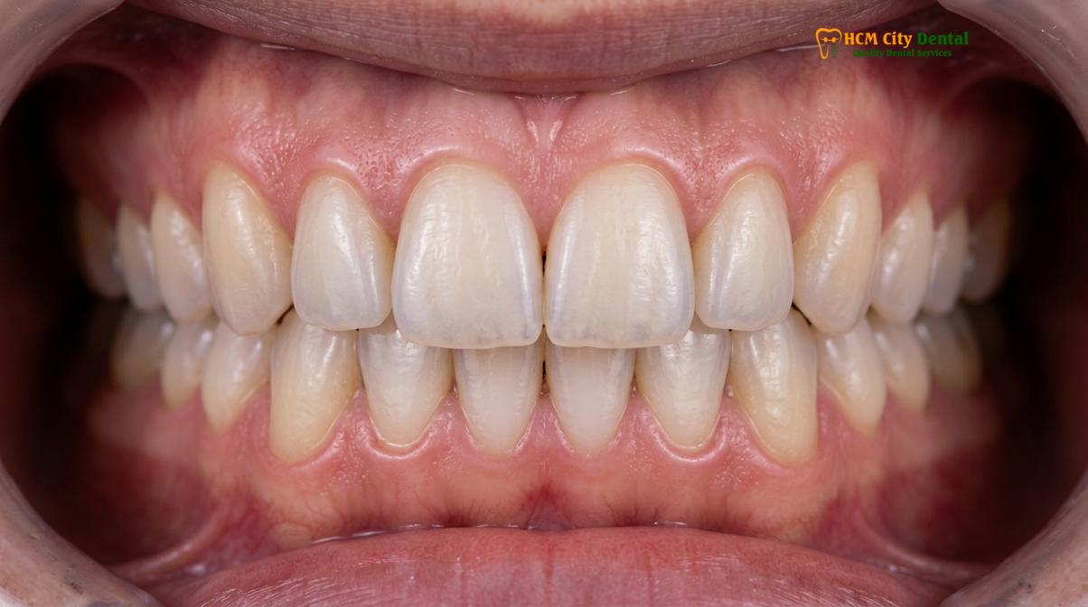
When an anterior tooth is compromised by decay, trauma, or wear, the traditional approach often involved aggressive reduction for a full-coverage crown. However, modern dentistry has shifted toward a strict preservation philosophy. Dr. Nguyen Van Cuong, a leading specialist at HCMC Dental Clinic in Ho Chi Minh City, emphasizes that preserving the natural enamel—especially the cingulum and the facial surface—is paramount for maintaining the tooth’s long-term vitality and flexibility. His approach to Inlays & Onlays tại HCMC Dental Clinic focuses on maximizing healthy tooth structure while ensuring highly predictable aesthetic results.
The choice of restoration depends heavily on the type of mechanical stress the tooth will endure. Incisors are subjected to significant shearing forces. If a restoration lacks adequate bonding strength or is poorly designed, these forces can lead to micro-leakage, debonding, or structural failure. Therefore, when evaluating cosmetic restorations for anterior teeth, clinicians must analyze the patient’s bite alignment, the presence of parafunctional habits, and the remaining volume of healthy dentin and enamel.
“The ultimate goal in anterior restorative dentistry is to mimic nature seamlessly while sacrificing as little healthy tooth structure as possible. Conservative restorations not only look better but behave more like natural teeth under functional loading.”
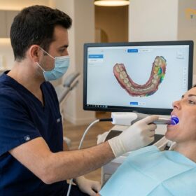
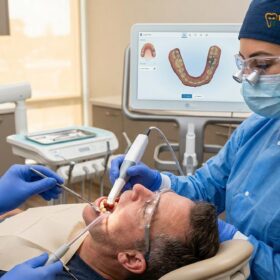
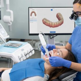
To achieve this, modern clinics utilize advanced diagnostic tools. Digital intraoral scanners capture the exact topography of the teeth. This digital workflow allows the dental laboratory to design restorations that integrate perfectly with the patient’s unique anatomy, ensuring that whether a veneer or an inlay is chosen, the final result meets the highest aesthetic and functional demands.
What is a Veneer and When is it Used?
A dental veneer is a highly aesthetic, ultra-thin ceramic layer bonded to the facial surface of an anterior tooth to correct color, shape, or alignment issues.
Porcelain veneers are the cornerstone of modern cosmetic smile design. They are custom-made, wafer-thin shells of ceramic that are chemically bonded to the front (facial) surface of the teeth. Veneers are primarily indicated for teeth that are structurally sound but suffer from aesthetic deficits. These deficits may include severe intrinsic staining that does not respond to traditional whitening, minor chips, enamel hypoplasia, or slight misalignments and gaps[2].
The success of a veneer relies heavily on the tooth preparation and the enamel bonding protocols. Typically, a clinician will remove approximately 0.3 to 0.5 millimeters of facial enamel. This minimal reduction provides the necessary space for the ceramic material, ensuring the final tooth does not appear bulky or over-contoured. The preparation usually ends at the gingival margin with a light chamfer, which allows for a smooth transition between the restoration and the natural root, preventing gum line irritation and promoting periodontal health.
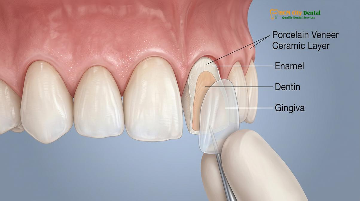
Material selection is critical for anterior veneers. High-end materials like lithium disilicate are favored because they offer an excellent combination of flexural strength and lifelike translucency. Unlike older metal-based restorations, these metal-free, biocompatible ceramics allow light to penetrate and scatter just as it does in natural enamel. This optical property is what makes a well-crafted veneer virtually indistinguishable from a natural tooth.
The cementation process for veneers is highly technique-sensitive. It involves isolating the tooth, etching the enamel with phosphoric acid to create micro-porosities, and applying a silane coupling agent to the internal surface of the etched ceramic. A light-cure resin-adhesive cement is then used to bond the two surfaces together. When bonded entirely to enamel, the bonding strength of a porcelain veneer is exceptionally high, creating a unified complex that can withstand normal biting forces for many years.
For patients exploring options like a partial crown vs full crown, it is crucial to understand that veneers are not designed to repair deep cavities or structural fractures. If a tooth has significant decay, a different restorative approach is required.
Inlays/Onlays on Front Teeth: Restoring Structural Bite Damage
While less common than posterior applications, anterior inlays and onlays are utilized to repair significant lingual or interproximal decay without sacrificing the intact facial enamel.
When discussing advanced ceramic restorations, most patients immediately think of the posterior (back) teeth, where these are routinely used to replace large amalgam fillings or repair fractured cusps. However, dental inlays and dental onlays also have highly specific and valuable applications in the anterior region. They are the treatment of choice when a front tooth has sustained structural damage—such as deep interproximal decay (cavities between the teeth) or significant lingual erosion (damage on the back side of the tooth)—but the facial enamel remains healthy and aesthetically pleasing.
In the past, a dentist faced with a large anterior cavity might have opted for a massive composite restoration or a full dental crown. While composite resin is excellent for small to moderate cavities, large composite fillings are susceptible to polymerization shrinkage. As the resin cures and hardens, it shrinks slightly, which can create micro-gaps at the margins. Over time, these gaps can harbor bacteria, leading to secondary decay and staining. Furthermore, large composites may lack the mechanical strength to withstand the shearing forces of the incisors.
Alternatively, a full crown requires 360-degree shaving of the tooth, removing perfectly healthy facial enamel just to access and repair the decay on the back or sides. This violates the preservation philosophy. An anterior inlay solves this dilemma. The clinician meticulously removes only the decayed tissue, creating a precise cavity outline form. The healthy facial enamel is left completely untouched.
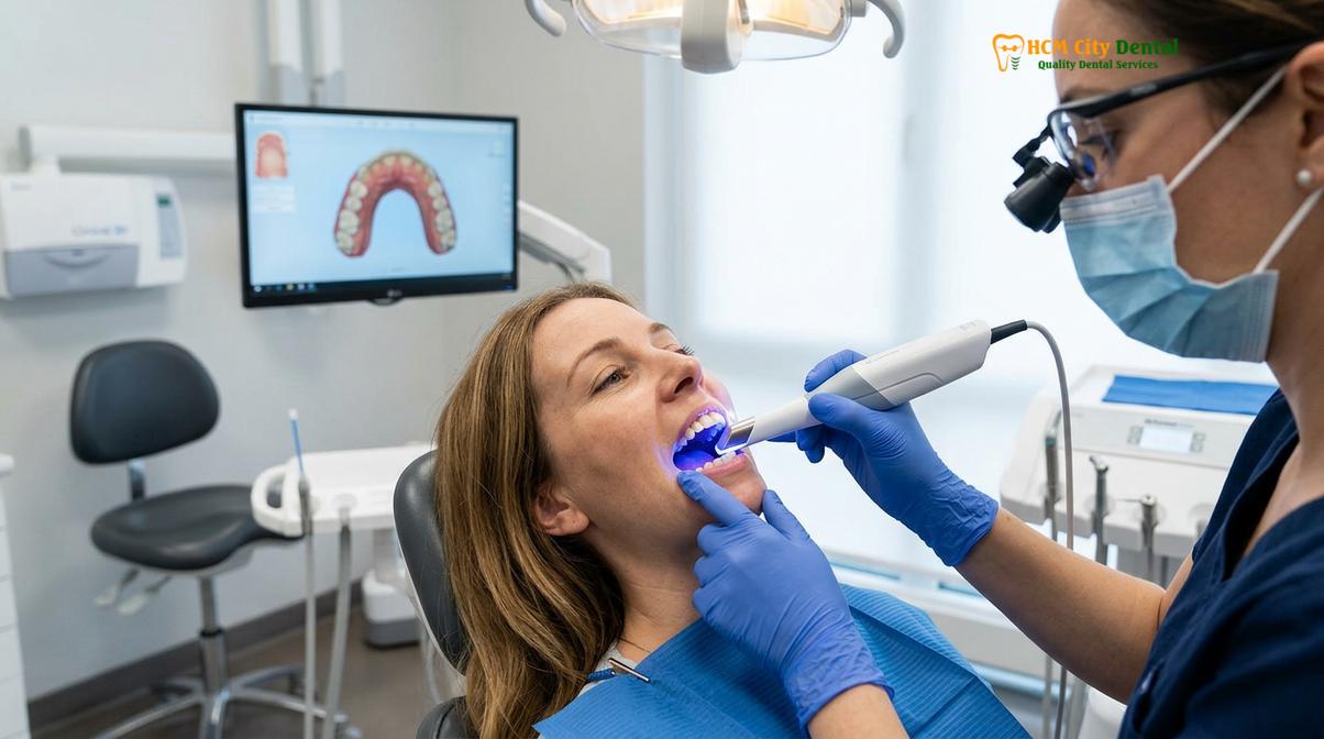
Because of the complex geometry of anterior inlays, digital CAD/CAM milling is essential. The digital impression is transmitted to the milling center, where the inlay is milled from a solid block of high-strength ceramic, ensuring an exact marginal fit. This precise margin sealing helps prevent bacterial infiltration and secondary decay[3].
For teeth that have undergone endodontic therapy, an anterior onlay or overlay after root canal might be necessary to cover and protect the access hole and weakened lingual structure, providing structural reinforcement while maintaining the natural smile.
Comparing Tooth Preparation: Surface Veneer vs. Interproximal Inlay
Veneer preparation focuses on minimal facial enamel reduction, whereas inlay preparation requires precise internal cavity design to manage mechanical stress and ensure a perfect marginal seal.
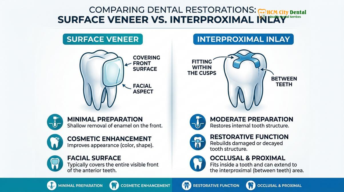
Understanding the clinical differences between a porcelain veneer vs inlay front teeth requires a detailed look at how the tooth is prepared for each restoration. The preparation design is dictated by the primary goal: aesthetics for the veneer, and structural repair for the inlay.
For a veneer, the preparation is largely confined to the facial (front) surface and the incisal (biting) edge. The dentist uses specialized diamond burs to remove a uniform layer of enamel, typically 0.3mm to 0.5mm deep. The goal is to stay entirely within the enamel layer, as bonding to enamel provides a significantly higher and more predictable bond strength than bonding to the underlying dentin. The margins are usually placed at or slightly above the gum line using a light chamfer finish line. This ensures the ceramic transitions smoothly into the natural tooth, preventing plaque accumulation[4].
In contrast, preparing an anterior tooth for an inlay is a more complex geometric exercise. The preparation is driven by the location of the tooth decay or old filling replacement. The clinician must remove all compromised tissue while ensuring the remaining cavity walls are smooth and divergent, allowing the rigid ceramic inlay to slide into place without binding. The internal line angles must be rounded to prevent stress concentration, which could lead to fracture of either the ceramic or the natural tooth under mechanical stress.
The following table summarizes the key clinical differences between the two preparations:
| Clinical Parameter | Porcelain Veneer | Anterior Inlay |
|---|---|---|
| Primary Indication | Cosmetic enhancement (color, shape, minor alignment) | Structural repair (deep decay, fractured lingual surface) |
| Preparation Location | Facial (front) surface and incisal edge | Internal cavity, interproximal (sides), or lingual (back) |
| Preparation Depth | 0.3mm – 0.5mm (strictly within enamel) | Variable, dictated by the extent of decay (often into dentin) |
| Bonding Surface | Primarily Enamel | Mixed (Enamel margins, Dentin internal walls) |
| Restorative Goal | Aesthetic transformation | Functional reinforcement |
Regardless of the preparation type, the use of digital intraoral scanners has revolutionized the workflow. The scanner captures the preparation margins with micron-level accuracy, eliminating the distortion and patient discomfort associated with traditional silicone impressions. This digital data is seamlessly integrated into the CAD/CAM software, allowing the technician to design a restoration with perfect contacts and occlusion.
Combining Veneers and Inlays/Onlays for Smile Makeovers
Comprehensive smile rehabilitations often combine facial veneers for aesthetics with posterior or lingual inlays/onlays to restore vertical dimension and overall bite stability.
In many complex clinical scenarios, a patient presents with a combination of aesthetic dissatisfaction and structural dental breakdown. This is particularly common in patients with a history of severe acid reflux, eating disorders, or chronic teeth grinding (bruxism). In these cases, a single restorative modality is rarely sufficient. Instead, a multidisciplinary approach combining veneers and inlays/onlays is required to achieve a comprehensive smile makeover.
For example, a patient may have severe wear on the biting surfaces of their posterior teeth, leading to a collapsed bite. This loss of vertical dimension of occlusion places excessive stress on the anterior teeth, causing them to chip, thin out, and appear shorter. To correct this, the clinician cannot simply place veneers on the front teeth; the underlying bite alignment must be corrected first, or the new veneers will quickly fracture under the unbalanced mechanical stress[5].
The treatment protocol often begins by utilizing night grinding onlay protection to restore the posterior teeth. Using high-strength materials, the dentist can rebuild the lost cusps and elevate the bite back to its natural, healthy vertical dimension. Once the posterior support is re-established and the bite is stabilized, the anterior teeth can be safely restored.
If the anterior teeth have suffered lingual erosion but the facial enamel is intact, anterior inlays or palatal veneers can be bonded to the back of the teeth to restore their thickness and strength. Finally, if the facial aesthetics are compromised, ultra-thin porcelain veneers are bonded to the front. This comprehensive approach ensures that the cosmetic smile design is built upon a foundation of biomechanical stability.
“A beautiful smile must be a functional smile. By combining the aesthetic brilliance of veneers with the structural reinforcement of inlays and onlays, we can rehabilitate the entire mouth conservatively, ensuring results that last a lifetime.”
This hybrid approach requires meticulous planning, often involving digital smile design software and 3D-printed mock-ups, allowing the patient to preview their new smile and bite before any irreversible tooth preparation occurs.
Case Study: Front Teeth Reconstruction
A multidisciplinary approach utilizing both facial veneers and structural inlays can salvage severely compromised anterior teeth while delivering a natural, highly aesthetic outcome.
To illustrate the practical application of these techniques, consider a recent case managed at HCMC Dental Clinic in Ho Chi Minh City. A 34-year-old patient presented with significant aesthetic and structural concerns regarding her upper central incisors. She had previously received large composite restorations to repair interproximal decay, but these fillings had discolored over time and were beginning to leak, causing secondary decay. Additionally, she was unhappy with the overall shape and yellowish hue of her front teeth.
Clinical Case: Comprehensive Anterior Rehabilitation
Patient Profile: 34-year-old female, presenting with failing interproximal composites and aesthetic dissatisfaction.
Clinical Findings: Deep mesial and distal decay on teeth #8 and #9 (upper central incisors). The facial enamel was largely intact but discolored. The existing composite restorations were leaking, compromising the structural integrity of the teeth.
Treatment Plan: A combined approach was selected. To address the deep interproximal decay without sacrificing the remaining healthy facial enamel for a full crown, the clinician opted to replace failing fillings with onlays and inlays designed specifically for the anterior region. Once the structural integrity was restored internally, ultra-thin porcelain veneers were bonded to the facial surfaces to achieve the desired aesthetic transformation.
Outcome: The patient achieved a bright, natural-looking smile. The internal inlays provided the necessary structural support to withstand biting forces, while the facial veneers delivered flawless aesthetics. By avoiding full crowns, the maximum amount of natural tooth structure was preserved.
When to Consult a Dentist for Anterior Restorations
Early intervention is critical when dealing with anterior tooth damage to prevent the need for more invasive procedures like root canals or full crowns.
Patients should seek a professional dental consultation if they notice any signs of structural damage or aesthetic deterioration on their front teeth. Symptoms such as sensitivity to hot or cold, visible dark spots between the teeth, or chipping along the biting edge indicate that the enamel has been compromised. Delaying treatment can allow decay to progress deeper into the dentin, eventually reaching the dental pulp and necessitating endodontic therapy.
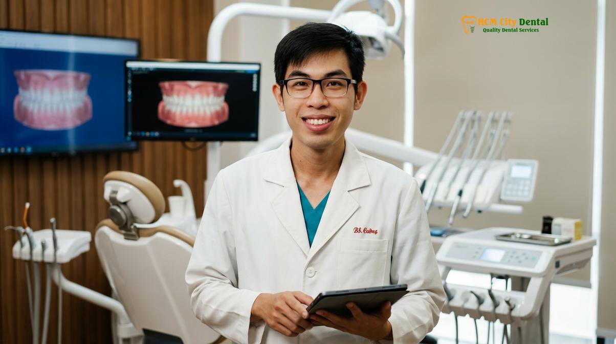
Furthermore, if old composite fillings on the front teeth begin to turn yellow, brown, or appear to have a dark line around the edges, this is a strong indicator of micro-leakage. A dentist can evaluate whether these failing restorations should be replaced with new composites, structural inlays, or cosmetic veneers. According to the Vietnam Odonto-Stomatology Association (VOSA) guidelines on conservative aesthetic restorations, preserving natural tooth structure through timely, minimally invasive interventions yields the best long-term prognosis for anterior teeth[6].
If you are experiencing any of these issues, or if you simply wish to enhance the appearance of your smile, a comprehensive evaluation at a qualified dental clinic is the first step toward determining the most appropriate and conservative treatment plan.
Frequently Asked Questions
Can you get an inlay on a front tooth?
Yes, an inlay can be placed on a front tooth to restore deep interproximal (between teeth) or lingual (back surface) decay while preserving the visible facial enamel. This conservative approach maintains the natural aesthetic front of the tooth while providing structural support.
What is the difference between a veneer and an inlay?
A veneer is an ultra-thin ceramic shell bonded to the front (facial) surface of a tooth primarily for cosmetic enhancement, whereas an inlay is a structural restoration fitted into the internal cavity of a tooth to repair decay or damage.
Do veneers cover more tooth than inlays?
Veneers cover the entire visible front surface and sometimes wrap slightly over the incisal edge, whereas inlays only fill the specific internal cavity or damaged area, leaving the rest of the natural tooth structure completely untouched.
Which is more durable for front teeth?
Both offer excellent durability when properly indicated. Inlays provide superior structural reinforcement for teeth with deep cavities, while veneers offer exceptional surface durability against staining and minor chipping, provided the underlying tooth structure is sound.
How do I choose between a composite restoration and a porcelain veneer for front teeth?
A composite restoration is ideal for minor chips or small cavities and can be completed in one visit, while a porcelain veneer is recommended for comprehensive cosmetic changes, offering superior stain resistance, longevity, and a more natural translucency.
References
- Journal of Esthetic and Restorative Dentistry. Clinical performance of porcelain veneers and inlays.
- International Journal of Prosthodontics. Biomechanics of anterior teeth restorations.
- Journal of Dentistry. Marginal adaptation in CAD/CAM lithium disilicate restorations.
- American Dental Association. Guidelines on conservative restorative dentistry.
- Journal of Prosthodontic Research. Bonding protocols for anterior ceramic restorations.
- Vietnam Odonto-Stomatology Association (VOSA). National guidelines on conservative aesthetic restorations.
Are you unsure whether your front teeth require cosmetic enhancement or structural repair? The expert team at HCMC Dental Clinic can help you determine the most conservative and effective treatment plan. To learn more about your options, including onlay vs crown clinical differences, schedule a consultation with us today. Visit HCMC Dental Clinic in Ho Chi Minh City for world-class restorative dentistry.