The choice between an onlay vs crown depends entirely on the extent of structural tooth damage and the clinical goal of preservation. An onlay restores only the damaged cusps while preserving healthy enamel, whereas a crown requires reshaping the entire tooth. Both utilize durable ceramics to restore function, but onlays offer a significantly more conservative, minimally invasive approach.
Clinical Summary:
Dental onlays and crowns are both highly effective indirect restorations, but they serve distinctly different clinical needs. Onlays (often referred to as partial crowns) are minimally invasive, preserving up to 70% of natural tooth structure by only replacing the compromised cusps and chewing surfaces. Conversely, full coverage crowns require 360-degree tooth shaving but provide maximum structural reinforcement for severely compromised, fractured, or root-canal-treated teeth. Advanced CAD/CAM milling and digital intraoral scanning ensure precise marginal sealing for both options, preventing secondary decay, optimizing stress distribution, and restoring optimal bite alignment with biocompatible ceramics.
Key Takeaways:
- Onlays preserve significantly more natural tooth structure compared to the aggressive reduction required for full crowns.
- Digital intraoral scanners (like the iTero and Medit i700) eliminate the need for uncomfortable traditional silicone impressions.
- High-strength materials such as IPS e.max and Cercon HT Zirconia provide exceptional durability and lifelike aesthetics.
- Proper enamel bonding protocols ensure a seamless, leak-proof margin for conservative partial restorations.
- Full crowns remain the definitive standard of care for teeth with extensive decay, deep fractures, or post-endodontic fragility.
The Concept of Minimally Invasive Dentistry
Minimally invasive dentistry prioritizes the preservation of healthy tooth structure, utilizing advanced bonding techniques to restore function without the unnecessary removal of natural tissue.
For decades, the prevailing philosophy in restorative dentistry was “extension for prevention,” a concept that often required the removal of healthy tooth structure to create mechanical retention for traditional amalgam fillings or conventional crowns. However, the evolution of adhesive dentistry has fundamentally shifted this paradigm toward a minimally invasive approach. Today, the primary objective is to retain as much natural, healthy tissue as possible. This philosophy is the cornerstone of modern restorative treatments, particularly when evaluating the clinical indications for custom ceramic restorations.
One of the most critical aspects of this conservative approach is the respect for the biological width—the natural dimension of soft tissue attached to the portion of the tooth above the alveolar bone [1]. Traditional full crowns often require subgingival margins (edges that sit below the gum line) to achieve adequate retention and aesthetic blending. Unfortunately, encroaching on the biological width can trigger chronic localized gingival inflammation, recession, and unpredictable bone loss. In contrast, dental inlays and dental onlays are designed with supragingival margins (edges that sit above the gum line). This placement is inherently kinder to the periodontal tissues, facilitating easier home hygiene and significantly reducing the risk of iatrogenic gum disease.
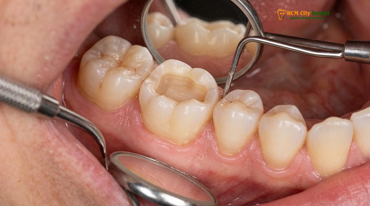
Dr. Nguyen Van Cuong, a leading restorative specialist, frequently emphasizes this preservation philosophy in his clinical practice. According to his clinical perspective on conservative restoration material selection at HCMC Dental Clinic in Ho Chi Minh City, the goal is not merely to fill a hole, but to rebuild the tooth biomimetically. By utilizing 100% metal-free, biocompatible ceramics, the risk of allergic reactions and metallic tattooing of the gums is eliminated. This holistic approach ensures that the restored tooth functions harmoniously within the oral cavity, mimicking the natural flexure and optical properties of virgin enamel.
How Much Tooth Structure is Saved by an Onlay?
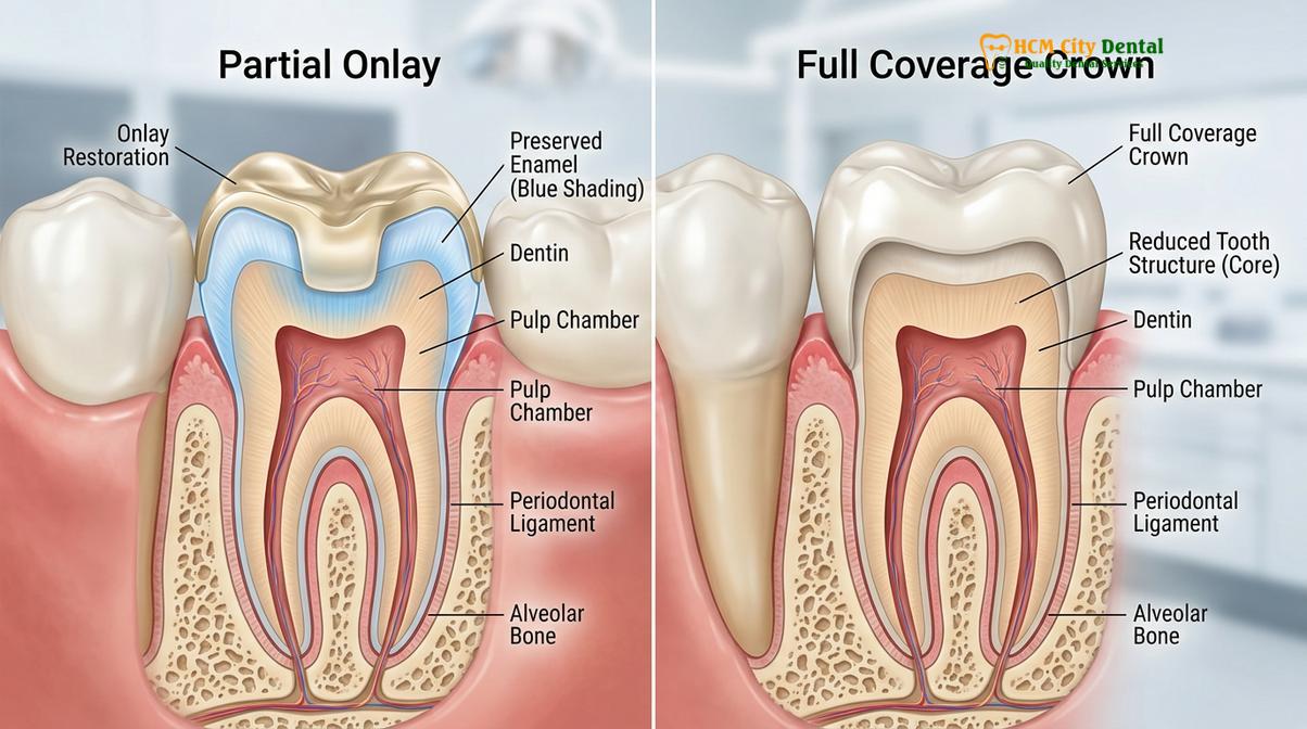
An onlay typically preserves 60% to 80% of the natural tooth volume, whereas a full crown requires the aggressive removal of up to 75% of the clinical crown.
To truly understand the debate of onlay vs full crown, one must visualize the volumetric differences in tooth preparation. A natural molar is a robust structure designed to withstand hundreds of pounds of occlusal (biting) force. The outer layer, the structural enamel, is the hardest substance in the human body and serves as a rigid shell protecting the more flexible dentin underneath. When a tooth suffers moderate decay or a fractured cusp, the traditional response was often to place a full coverage crown. However, preparing a tooth for a full crown requires circumferential reduction—meaning the dentist must shave down all four sides and the top of the tooth to create a “peg” or ferrule onto which the crown is cemented.
This 360-degree tooth shaving process removes a staggering amount of healthy tissue. Studies indicate that a standard crown preparation destroys between 65% and 75% of the coronal tooth volume [2]. Once this structural enamel is milled away, it is gone forever. The tooth is permanently committed to requiring full coverage restorations for the rest of the patient’s life.
“The most durable dental material known to science is intact human enamel. The primary directive of modern restorative dentistry must be to preserve it whenever clinically feasible, utilizing partial coverage restorations to maintain the tooth’s innate biomechanical integrity.”
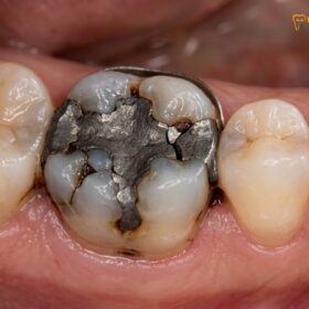
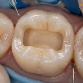
Conversely, an onlay—often categorized as a partial restoration—is designed to replace only the missing or diseased portion of the tooth. If a molar has a large cavity that undermines one or two cusps, the dentist will remove the decay, smooth the sharp edges, and leave the remaining healthy walls completely intact. The laboratory then fabricates a custom porcelain overlay that fits precisely into the prepared defect like a puzzle piece. This targeted approach generally preserves 60% to 80% of the original tooth structure. By choosing a conservative partial crown vs full crown, the patient retains the natural rigidity of the tooth, significantly reducing the risk of future root fractures and endodontic complications.
Biomechanical Advantages of Keeping Natural Enamel
Retaining natural enamel allows for superior resin-adhesive cementation, creating a biomimetic bond that optimizes stress distribution across the tooth during heavy chewing.
The advantages of preserving tooth structure extend far beyond mere volume; they are deeply rooted in biomechanics and adhesive chemistry. The dentino-enamel junction (DEJ) is a complex, naturally occurring interface that seamlessly transfers occlusal loads from the rigid enamel to the shock-absorbing dentin. When a tooth is aggressively prepared for a crown, this natural stress distribution mechanism is severely compromised. The artificial crown must now bear the entirety of the load and transfer it directly to the root, which can sometimes lead to catastrophic vertical root fractures over time.
When an onlay is placed, the remaining natural enamel walls continue to share the occlusal load. Furthermore, the success of modern conservative restorations relies heavily on advanced enamel bonding protocols. Unlike traditional crowns, which are often held in place by mechanical friction and luting cements, onlays are chemically bonded to the tooth. The process involves etching the enamel with phosphoric acid to create microscopic porosities, applying a silane coupling agent to the ceramic, and using a dual-cure resin-adhesive cementation system to fuse the restoration to the tooth [3].
This chemical fusion creates a monoblock effect. The bond strength to preserved enamel is exceptionally high—often exceeding the cohesive strength of the tooth itself. This is particularly crucial when replacing large, failing amalgam restorations. Traditional silver fillings do not bond to the tooth; they are wedged into undercuts. Over decades, the expansion and contraction of the metal can cause micro-fractures in the surrounding enamel. When replacing these fillings, an onlay not only fills the void but actively binds the weakened walls together, preventing the polymerization shrinkage of large fillings that occurs when using direct composite resin for massive cavities.
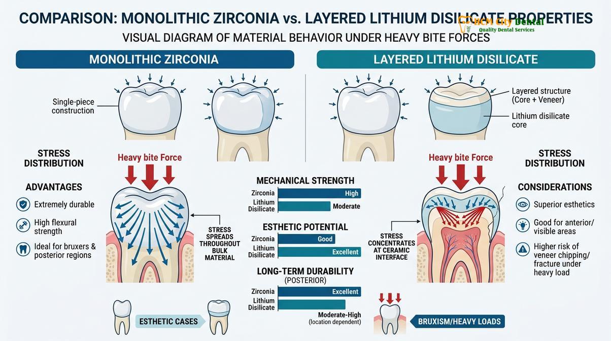
Material selection plays a pivotal role in this biomechanical harmony. For the anterior aesthetic zone and premolars, clinicians often select high-end materials from Ivoclar, specifically IPS e.max lithium disilicate. This glass-ceramic offers unparalleled translucency and bonds exceptionally well to tooth structure. For posterior molars subjected to extreme grinding forces, Dentsply Sirona Cercon HT Zirconia is frequently utilized due to its massive flexural strength and resistance to fracture.
When is a Full Crown Absolutely Necessary?
A full crown becomes clinically necessary when a tooth has suffered extensive structural loss, deep vertical fractures, or requires complete encasement following root canal therapy.
Despite the overwhelming benefits of minimally invasive dentistry, there are specific clinical scenarios where an onlay is contraindicated, and a full crown is the definitive standard of care. Understanding the limitations of partial restorations is crucial for long-term clinical success. The decision matrix for an overlay vs crown hinges on the structural integrity of the remaining tooth walls.
If a tooth has suffered massive decay that undermines all four cusps, leaving only thin, fragile shells of enamel, an onlay cannot provide adequate support. In such cases, the remaining walls are prone to flexing and fracturing under bite pressure. A full crown acts like a protective helmet, encasing the weakened structure and holding it together. Similarly, teeth that exhibit deep vertical fracture lines extending toward the gum line require the circumferential binding effect (the ferrule effect) that only a full crown can provide [4].
Clinical Warning: Ignoring the signs of cracked tooth syndrome—such as sharp pain upon release of biting pressure—can lead to catastrophic tooth splitting. If a crack propagates below the gum line or into the root space, the tooth may become unrestorable and require extraction. Prompt evaluation for full coverage protection is essential.
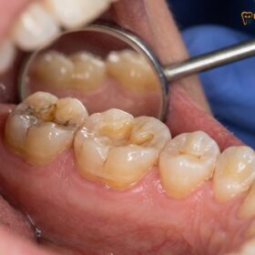
Endodontically treated teeth (teeth that have undergone root canal therapy) present another common indication for full crowns, particularly in the posterior region. Following a root canal, the tooth loses its internal blood supply and hydration, becoming more brittle over time. Furthermore, the access hole drilled to reach the nerve significantly weakens the structural roof of the tooth. While some premolars with minimal access cavities can be successfully restored with an onlay, the vast majority of root-canal-treated molars require a full crown to prevent catastrophic splitting.
Additionally, full crowns are often necessary when performing full-mouth rehabilitations aimed at restoring vertical dimension of occlusion (VDO) for severe bruxism patients. When decades of nighttime grinding have worn the teeth down to flat nubs, crowns are required to rebuild the height, establish proper bite alignment, and protect the exposed dentin from further attrition.
Preparation Differences: 360-Degree Shaving vs. Targeted Milling
Crown preparation involves circumferential shaving to create a uniform ferrule, while onlay preparation uses targeted milling to remove only the compromised tissue and preserve healthy walls.
The physical act of preparing a tooth highlights the stark contrast between these two modalities. For a full crown, the clinician must perform 360-degree tooth shaving. This involves reducing the occlusal (chewing) surface by 1.5 to 2.0 millimeters and shaving down the buccal, lingual, mesial, and distal walls by a similar amount. The goal is to create a miniature, tapered version of the tooth with a distinct finish line—often a heavy chamfer or shoulder margin—at the gum line. This aggressive reduction is necessary to ensure the restorative material has sufficient thickness to resist fracture without making the final tooth look bulky.
In contrast, preparing a tooth for an onlay is an exercise in precision and restraint. The clinician focuses solely on the diseased or structurally compromised areas. The cavity outline form is dictated by the extent of the decay or the old filling, rather than a predetermined geometric shape. The dentist carefully removes the pathology, smooths any sharp internal line angles to reduce stress concentrations, and creates smooth, flowing chamfer margins only where the restoration meets the healthy enamel. The buccal and lingual walls are often left completely untouched.
Clinical Case Example: A 42-year-old patient visited HCMC Dental Clinic in Ho Chi Minh City presenting with a fractured mesio-lingual cusp on a lower first molar due to an old, failing amalgam filling. Instead of aggressively shaving the entire tooth for a crown, Dr. Cuong utilized the Medit i700 scanner to map the defect. A conservative lithium disilicate onlay was milled via CAD/CAM, preserving the remaining three healthy cusps and restoring full function with zero postoperative sensitivity.
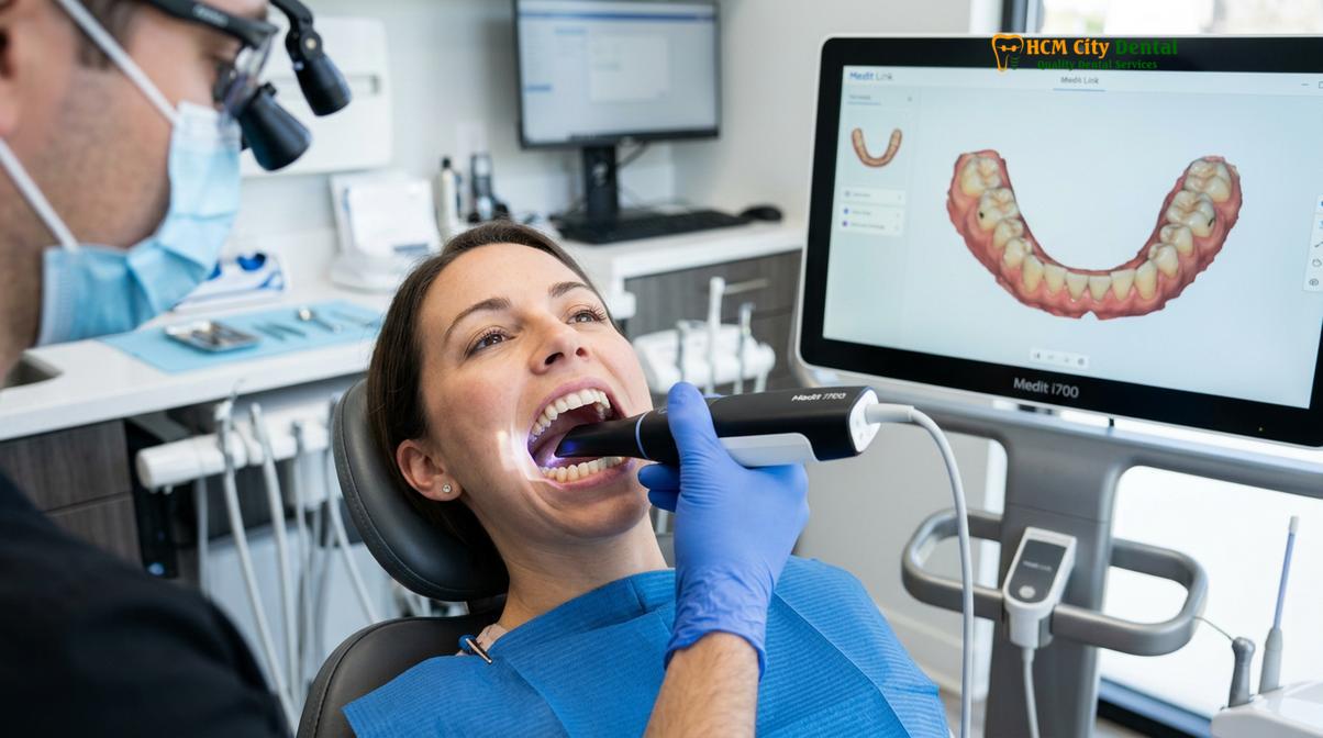
The workflow for both procedures has been revolutionized by digital dentistry. Modern clinics have largely abandoned the use of messy, gag-inducing polyvinyl siloxane (PVS) impression materials. Instead, the prepared tooth is captured using highly advanced iTero + Medit i700 Intraoral Scanners. These devices take thousands of pictures per second, stitching them together to create a flawless 3D digital model of the patient’s mouth.
This digital data is then transmitted instantly to the laboratory. Through a Direct Lab Partnership & CAD/CAM (Computer-Aided Design and Computer-Aided Manufacturing) workflow, the restoration is designed virtually. The software calculates the exact occlusal contacts and proximal contours. A precision milling machine then carves the restoration out of a solid block of ceramic. This digital process ensures an exact marginal fit, which is the most critical factor in margin sealing and the prevention of secondary decay [5].
Summary Comparison Table: Onlay vs. Crown
Evaluating the clinical indications, structural impact, and financial investment helps determine whether an onlay or a crown is the optimal restorative choice for your specific dental needs.
To simplify the decision-making process, the following table outlines the primary differences between these two restorative options across various clinical and practical parameters.
| Feature / Parameter | Dental Onlay (Partial Crown) | Full Coverage Crown |
|---|---|---|
| Tooth Structure Preserved | High (60% – 80% retained) | Low (25% – 35% retained) |
| Preparation Style | Targeted defect removal; healthy walls kept | Aggressive 360-degree circumferential shaving |
| Primary Indication | Moderate decay, fractured cusps, large filling replacement | Severe decay, deep cracks, post-root canal therapy |
| Margin Location | Typically supragingival (above the gums) | Often subgingival (at or below the gums) |
| Risk of Nerve Trauma | Very Low (due to minimal drilling) | Moderate (due to proximity to the pulp chamber) |
| Material Options | IPS e.max (Lithium Disilicate), Composite Resin | Cercon HT Zirconia, IPS e.max, PFM, Gold |
Beyond the clinical parameters, patients must also consider the financial investment. Because onlays require highly precise preparation and advanced adhesive bonding protocols, their cost is often comparable to, or slightly less than, a full crown. However, international patients seeking world-class care can benefit significantly from transparent pricing structures.
According to the latest clinic fee schedule at HCMC Dental Clinic, the pricing structure is highly competitive, especially when utilizing the 40% pre-arrival discount for WhatsApp bookings. An Inlay or Onlay (Lab-made ceramic) typically costs around $385 (10.0M VND) for walk-in patients, but drops to From $231 (6.0M VND) with the pre-booking discount. Similarly, an Overlay or Partial Crown is priced at ~$446 (11.6M VND) for walk-ins, but is available From $269 (7.0M VND) via WhatsApp pre-booking. This investment is protected by complete peace of mind, featuring a global warranty and remote WhatsApp checkup support to ensure long-term success regardless of where the patient resides.
Important Clinical Considerations (When to See a Doctor)
While modern ceramic restorations are incredibly durable, they are not invincible. The underlying tooth structure remains susceptible to bacterial attack if oral hygiene is neglected. Prompt clinical evaluation is essential to ensure the longevity of your restoration and the health of the supporting tooth.
You should schedule an immediate consultation if you experience any of the following symptoms:
- Sharp Pain Upon Biting: This is a classic symptom of a compromised bond, a fractured ceramic, or a propagating crack in the underlying tooth root.
- Prolonged Thermal Sensitivity: While mild sensitivity to cold is normal for a few days following the placement of a new restoration, severe or lingering pain to hot or cold stimuli may indicate irreversible pulpitis (nerve inflammation) requiring endodontic intervention.
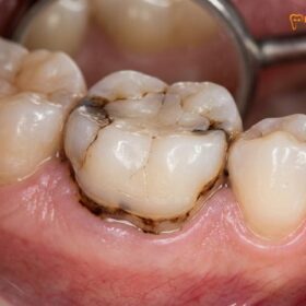
- Visible Margin Discoloration: A dark line or shadowing around the edge of the restoration can be an early indicator of secondary decay detection. If the margin sealing has failed, bacteria can infiltrate beneath the ceramic, causing rapid internal tooth decay.
- Changes in Bite Alignment: If the restoration feels “high” or if you notice a shift in how your teeth come together, occlusal adjustments may be necessary. An improper bite alignment can place excessive stress on the ceramic, leading to premature chipping or failure.
“Timely intervention is the key to preserving dental work. A minor marginal defect detected early during a routine scan can often be repaired conservatively, whereas ignoring the symptoms inevitably leads to catastrophic failure and the need for more invasive procedures.”
Ultimately, the decision between an onlay and a crown should never be made based solely on cost or internet research. It requires a comprehensive clinical and radiographic examination. If you are experiencing symptoms of a failing restoration or have been told you need a crown, seeking a second opinion from a specialist focused on minimally invasive techniques can provide clarity and potentially save valuable tooth structure.
Frequently Asked Questions
Why is an onlay better than a crown?
An onlay is often considered superior for moderate damage because it preserves significantly more natural tooth structure. By only replacing the compromised cusps and leaving healthy walls intact, an onlay maintains the tooth’s innate flexibility and strength while reducing the risk of nerve trauma associated with aggressive preparation. Furthermore, the supragingival margins of an onlay are much kinder to the surrounding gum tissue, reducing the risk of chronic inflammation.
Is an onlay cheaper than a full crown?
Yes, an onlay is generally more cost-effective than a full crown due to the conservative nature of the preparation and reduced material volume. However, the exact price depends on the chosen ceramic material and the complexity of the digital CAD/CAM milling required to achieve a perfect marginal fit. At specialized centers, the cost difference may be minimal because the laboratory expertise required to fabricate a precision partial restoration is exceptionally high.
Can an onlay fail and require a crown later?
While rare with proper bonding protocols, an onlay can fail if the tooth suffers new decay or extreme trauma, potentially necessitating a crown later. The advantage of an onlay is that because so much original tooth structure was preserved, upgrading to a full crown remains a viable clinical option. If a full crown fails, the next step is often a root canal, post and core, or complete extraction and implant placement.
How much tooth is shaved for a crown?
A traditional full crown requires 360-degree tooth shaving, typically removing 1.5 to 2.0 millimeters of enamel and dentin from all sides and the chewing surface. This aggressive reduction equates to losing approximately 65% to 75% of the visible clinical crown volume to create space for the restorative material. This irreversible process permanently alters the biomechanical behavior of the tooth.
How long does a dental onlay last compared to a crown?
Both dental onlays and crowns share a similar lifespan, typically lasting 10 to 15 years or longer with excellent oral hygiene. Longevity depends heavily on the precision of the marginal seal, the strength of the ceramic material used, and the patient’s bite forces and nighttime grinding habits. Regular dental checkups and the use of a protective nightguard for bruxism can significantly extend the life of either restoration.
References
- Journal of Prosthetic Dentistry. Volumetric analysis of tooth structure removal for various restorations. (2020).
- International Journal of Periodontics & Restorative Dentistry. The impact of restorative margins on biological width. (2019).
- Journal of Esthetic and Restorative Dentistry. Clinical performance of CAD/CAM lithium disilicate partial crowns. (2021).
- Dental Materials. Phase transformation toughening in dental zirconia ceramics. (2018).
- American Dental Association. Clinical guidelines for indirect conservative restorations. (2022).