A cracked tooth inlay or onlay is a highly conservative, custom-milled ceramic restoration designed to repair fractured cusps while preserving healthy enamel. Unlike full crowns, these restorations bond directly to the damaged tooth structure, providing immense strength to prevent further splitting under heavy chewing forces.
Clinical Summary:
Treating a cracked tooth requires a delicate balance between removing compromised tissue and preserving healthy enamel. Inlays and onlays represent the gold standard in conservative restorative dentistry for incomplete fractures. By utilizing advanced CAD/CAM technology and high-strength ceramics like lithium disilicate (e.max) or zirconia, clinicians can create a precise marginal seal that binds the weakened cusps together. This approach neutralizes the wedging forces that cause cracks to propagate, effectively halting cracked tooth syndrome without the need for aggressive 360-degree tooth reduction associated with traditional crowns. Early diagnosis and immediate structural reinforcement are critical to preventing irreversible pulpal damage.
Key Takeaways:
- Preservation Philosophy: Onlays preserve maximum natural tooth structure by only covering the damaged cusps, avoiding the aggressive shaving required for full crowns.
- Structural Splinting: Bonded ceramic restorations act as a helmet, holding the tooth together and preventing minor cracks from splitting the root.
- Digital Precision: iTero and Medit i700 intraoral scanners eliminate messy impressions, ensuring a flawless CAD/CAM milled fit.
- Premium Materials: Restorations utilize biocompatible Ivoclar IPS e.max or Dentsply Sirona Cercon HT Zirconia for maximum durability.
- Cost-Effective Care: Pre-booking via WhatsApp offers a 40% discount, making premium lab-made ceramics highly accessible.
Understanding Cracked Tooth Syndrome (CTS)
Cracked Tooth Syndrome involves incomplete fractures of the dentin that cause sharp pain during chewing due to fluid movement within the tooth. Early intervention is critical to prevent the crack from propagating into the dental pulp.
Cracked tooth syndrome is one of the most challenging diagnostic dilemmas in modern dentistry. It occurs when a tooth has an incomplete fracture that originates from the crown and extends apically (toward the root) but has not yet caused the tooth to separate into distinct pieces. The condition is notoriously difficult to pinpoint because the crack is often microscopic, evading detection on standard two-dimensional dental X-rays. Patients typically report a very specific set of symptoms: a sharp, sudden jolt of pain that occurs precisely when releasing biting pressure, accompanied by a heightened, lingering sensitivity to cold temperatures [1].
To understand why this happens, we must look at the microscopic anatomy of the tooth. Beneath the hard outer enamel lies the dentin, a porous layer filled with thousands of microscopic fluid-filled channels called dentinal tubules. These tubules connect directly to the dental pulp, which houses the tooth’s nerve center. When heavy masticatory force is applied to a cracked tooth, the segments of the tooth flex slightly apart. This microscopic movement causes the fluid within the dentinal tubules to shift rapidly. According to Brännström’s hydrodynamic theory, this sudden fluid movement stimulates the A-delta nerve fibers within the pulp, triggering an acute pain response.
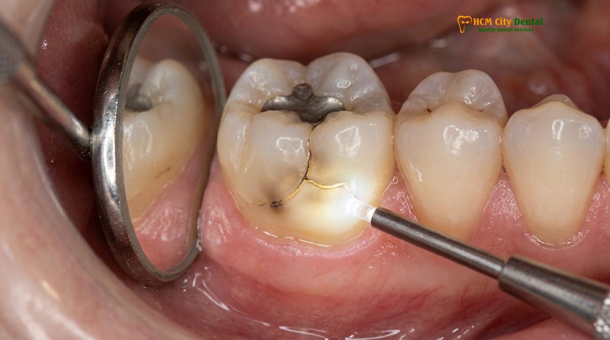
The danger of an untreated crack lies in its progressive nature. Every time you chew, swallow, or grind your teeth, the repetitive stress acts like a wedge, driving the fracture deeper into the dentin. If the crack breaches the pulp chamber, bacteria from the oral cavity will infiltrate the sterile nerve tissue, leading to irreversible pulpitis or a necrotic abscess. At this stage, conservative cracked tooth restoration options are no longer viable, and the patient will require endodontic therapy (a root canal) followed by comprehensive structural reinforcement.
Diagnosing Cusp Fractures vs. Deep Root Cracks
Accurate diagnosis relies on advanced 3D imaging, bite tests, and transillumination to determine if a fracture is confined to the cusp or extends dangerously toward the root. This dictates whether an onlay or extraction is necessary.
The clinical approach to a fractured tooth begins with determining the exact depth, direction, and severity of the crack. A superficial craze line in the enamel is harmless and requires no treatment, whereas a deep vertical root fracture carries a hopeless prognosis and necessitates immediate extraction. The critical middle ground—where the tooth can be saved—is the cusp fracture. A cusp fracture occurs when the crack undermines one of the pointed chewing surfaces of a posterior molar or premolar, often breaking off entirely or remaining loosely attached.
To accurately map the extent of the damage, modern dental clinics employ a multi-faceted diagnostic protocol. First, a specialized instrument called a “Tooth Slooth” is used to isolate individual cusps during a bite test. By having the patient bite down on specific points, the clinician can identify exactly which cusp is flexing and causing pain. Next, fiber-optic transillumination is utilized. A high-intensity light is shined through the tooth structure; healthy enamel will transmit the light completely, whereas a crack will block the light, creating a distinct dark line that reveals the fracture’s exact pathway [2].
Clinical Warning: Ignoring the early signs of a cracked tooth significantly increases the risk of catastrophic tooth splitting. If a crack propagates below the gum line and into the root structure, the tooth becomes non-restorable and must be extracted and replaced with a dental implant.
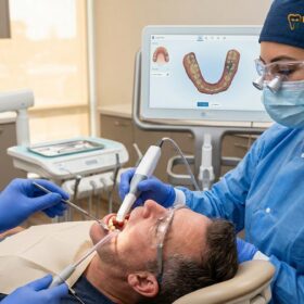
Furthermore, the integration of digital dentistry has revolutionized diagnostic accuracy. Using the iTero or Medit i700 intraoral scanners, clinicians can capture high-resolution, magnified 3D models of the patient’s dentition. These digital scans allow the dentist to evaluate the occlusal wear patterns, identify heavy contact points that may be contributing to the fracture, and assess the structural integrity of the remaining enamel. Differentiating between a restorable cusp fracture and a terminal root crack is the most critical step in determining the appropriate treatment workflow.
How an Onlay/Overlay Binds the Cusp Together
An onlay acts as a structural helmet, utilizing advanced resin-adhesive cementation to bind the weakened cusps together and distribute chewing loads evenly. This prevents the crack from expanding under occlusal stress.
When a tooth is diagnosed with a restorable crack, the primary biomechanical goal is to neutralize the wedging forces that threaten to split the tooth. Traditional silver amalgam or direct composite fillings are often contraindicated for large fractures because they rely on mechanical retention. When a patient bites down on a large filling, the filling acts like a wedge, pushing the remaining walls of the tooth outward. Over time, this outward pressure exacerbates the existing crack. This is where the preservation philosophy of an onlay or overlay becomes clinically invaluable.
An onlay is an indirect restoration, meaning it is custom-fabricated outside the mouth in a dental laboratory. By covering the compromised cusps, the onlay changes the biomechanical dynamics of the tooth from a wedging effect to a splinting effect. It acts like a protective helmet, absorbing the heavy impact of chewing and distributing the binding force evenly across the entire tooth structure. This structural reinforcement is what makes fixing cracked molar onlay procedures so highly successful [3].
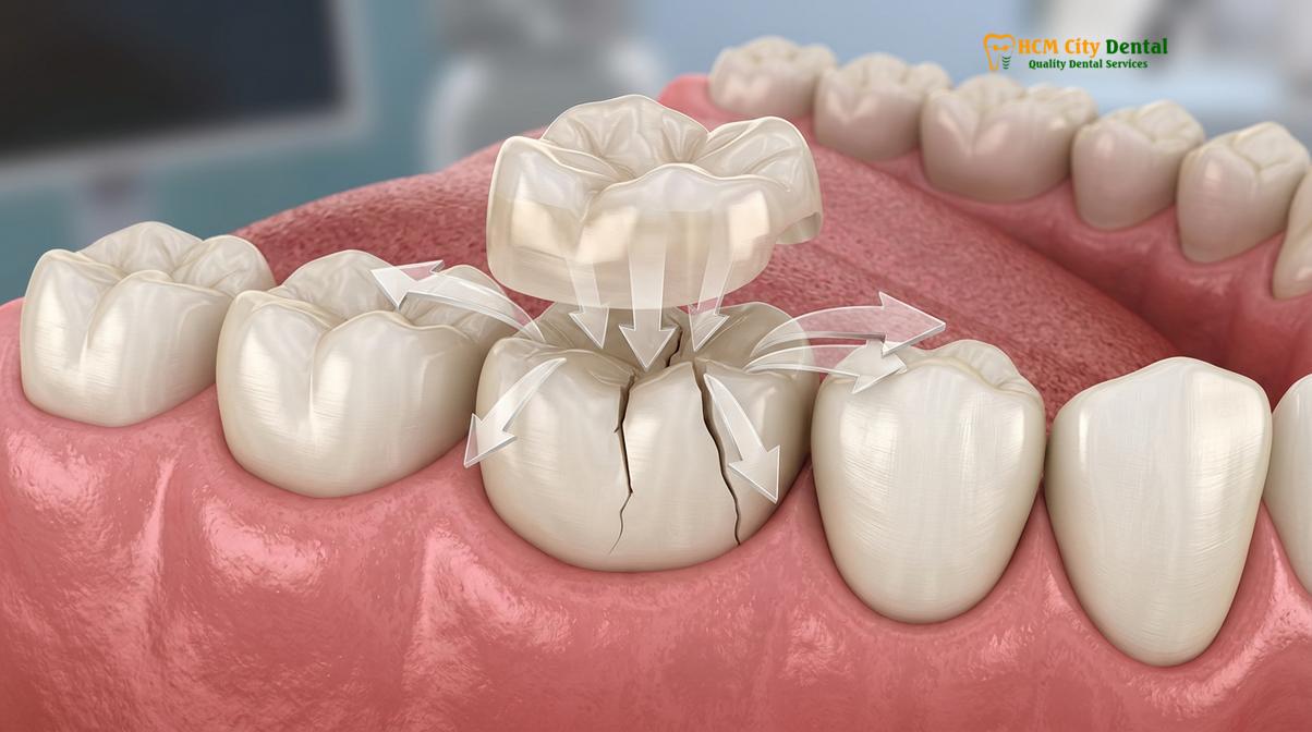
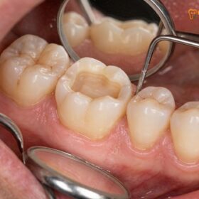
The clinical preparation for an onlay is meticulous. The clinician must design a precise cavity outline form that removes all unsupported enamel and decay while preserving the maximum amount of healthy tooth structure. Smooth, flowing chamfer margins are created to ensure the ceramic material has adequate thickness for strength without creating stress concentrations. Once the tooth is prepared, the success of the restoration relies entirely on the enamel bonding protocols.
“Our primary objective in restorative dentistry is the preservation of natural tooth structure. By utilizing advanced ceramic onlays and precise adhesive protocols, we can structurally reinforce a cracked tooth without the aggressive reduction required for traditional crowns. It is a minimally invasive approach that yields maximum long-term stability.”
— Dr. Nguyen Van Cuong, Lead Clinician
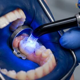
The cementation phase utilizes dual-cure resin-adhesive cements that chemically bond to both the ceramic restoration and the underlying dentin and enamel. The tooth surface is treated with a mild phosphoric acid etchant, followed by a universal bonding agent containing MDP monomers that chemically interact with the calcium in the tooth. Simultaneously, the internal surface of the ceramic onlay is treated with hydrofluoric acid and a silane coupling agent. When these two surfaces are brought together with the resin cement, they form a monolithic, unified structure that effectively seals the crack and restores the tooth to its original strength.
Full Crowns for Severe Fractures: The Ferrule Effect
When a crack compromises more than half the tooth structure, a full crown is required to establish a ferrule effect, ensuring long-term biomechanical stability and preventing catastrophic failure.
While inlays and onlays are the preferred conservative choice, there are clinical scenarios where the damage is simply too extensive, and a full crown restoration becomes mandatory. If a crack extends deep into the tooth, undermines multiple cusps, or if the tooth has previously undergone root canal therapy, the remaining walls may be too thin and fragile to support an onlay. In these cases, the clinician must evaluate the tooth for the ferrule effect.
The ferrule effect is a fundamental principle in prosthodontics. It refers to a band of solid, healthy tooth structure (typically requiring a minimum height of 1.5 to 2.0 mm and a thickness of 1.0 mm) that encircles the base of the prepared tooth, just above the gum line. When a full crown is cemented over this band of enamel and dentin, it creates a “hugging” effect that braces the tooth against lateral forces. This ferrule is what prevents the root from fracturing under the immense leverage of chewing [4].
When discussing fractured cusp repair crown vs onlay, the decision hinges entirely on the amount of remaining healthy tissue. If a robust ferrule cannot be established because the crack extends too far subgingivally (below the gums), the tooth may require a procedure called crown lengthening to expose more solid root structure, or it may be deemed non-restorable.
| Clinical Feature | Ceramic Onlay / Overlay | Full Dental Crown |
|---|---|---|
| Tooth Preservation | High (Preserves up to 70% of natural structure) | Low (Requires 360-degree aggressive reduction) |
| Coverage Area | Only the damaged cusps and central grooves | Encases the entire visible tooth down to the gum line |
| Primary Indication | Moderate cracks, single cusp fractures, large failing fillings | Severe fractures, post-root canal, lack of structural integrity |
| Gum Irritation Risk | Minimal (Margins are usually above the gum line) | Higher (Margins often sit at or below the gingival crest) |
By carefully assessing the biomechanical needs of the tooth, clinicians can avoid over-treating a tooth that only needs an onlay, while ensuring that a severely compromised tooth receives the comprehensive 360-degree protection of a full crown.
Same-Day Temporary Protection Steps
Immediate temporary coverage protects the exposed dentin and prevents bacterial ingress while the permanent ceramic restoration is fabricated, ensuring the patient remains comfortable and the bite remains stable.
The workflow for indirect restorations typically involves two distinct appointments. During the first visit, after the damaged tooth structure is removed and the precise digital impressions are captured, the tooth is left in a vulnerable state. The freshly cut dentin contains open tubules that are highly susceptible to bacterial invasion and thermal sensitivity. Therefore, placing a high-quality temporary restoration is a critical step in the clinical protocol.
The temporary onlay is usually fabricated chairside using a bis-acryl composite material. This material is injected into a preoperative matrix and allowed to set over the prepared tooth. The temporary serves several vital functions: it seals the dentinal tubules to prevent post-operative sensitivity, it maintains the position of the adjacent and opposing teeth to prevent unwanted shifting, and it protects the fragile prepared margins from being damaged during chewing.
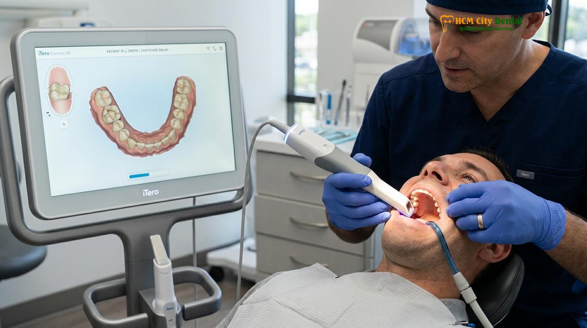
One of the major advantages of indirect restorations over massive direct composite fillings is preventing polymerization shrinkage. When a large composite filling is cured with a dental light, the resin matrix shrinks by 2-3%. In a large cavity, this shrinkage pulls the walls of the tooth inward, creating immense stress that can actually cause new micro-cracks or exacerbate existing ones. Because an onlay is milled from a solid block of ceramic in a laboratory, there is zero shrinkage when it is bonded into the mouth, ensuring a passive, stress-free fit.
Through a Direct Lab Partnership, the digital files captured by the iTero or Medit i700 scanners are instantly transmitted to a premier dental laboratory. There, advanced CAD/CAM milling machines carve the final restoration with micron-level accuracy. This digital workflow eliminates the distortion associated with traditional silicone putty impressions, guaranteeing that the final ceramic onlay will drop perfectly into place with an exact marginal fit.
Expected Success Rates of Restorations on Cracked Teeth
With precise marginal sealing and high-strength ceramics, onlays exhibit exceptional long-term success rates, effectively halting crack progression and restoring full masticatory function.
The long-term prognosis for a cracked tooth restored with a ceramic onlay is highly favorable, provided the crack has not breached the pulp and the patient maintains excellent oral hygiene. Clinical studies consistently show that bonded ceramic onlays have survival rates exceeding 90% over a 10-year period. The success of the restoration is heavily dependent on two factors: material selection and the accuracy of the marginal seal [5].
For patients requiring aesthetic perfection in the premolar regions, Ivoclar IPS e.max (lithium disilicate) is the material of choice. It offers a flexural strength of approximately 500 MPa and mimics the natural translucency of human enamel beautifully. For patients with heavy bruxism (teeth grinding) or for restorations on the second molars where biting forces are most extreme, Dentsply Sirona Cercon HT Zirconia is utilized. Zirconia boasts a flexural strength exceeding 1000 MPa, making it virtually indestructible under normal physiological loads.
Clinical Case Study: A 42-year-old patient presented to HCMC Dental Clinic in Ho Chi Minh City with sharp pain in a lower right molar upon chewing. Transillumination revealed a deep mesial-distal crack undermining the lingual cusp. Dr. Nguyen Van Cuong performed a conservative preparation, preserving the buccal wall, and bonded a custom CAD/CAM milled e.max onlay. The patient reported immediate relief from all symptoms, and the tooth has remained vital and asymptomatic at the 3-year follow-up.
Beyond strength, these modern ceramics offer exceptional biocompatibility. Being 100% metal-free, they eliminate the risk of galvanic toxicity, metallic taste, or the unsightly gray margins often associated with older porcelain-fused-to-metal (PFM) crowns. Furthermore, the smooth, highly polished surface of glazed ceramic resists plaque accumulation, which is vital for secondary decay detection and prevention at the restoration margins.
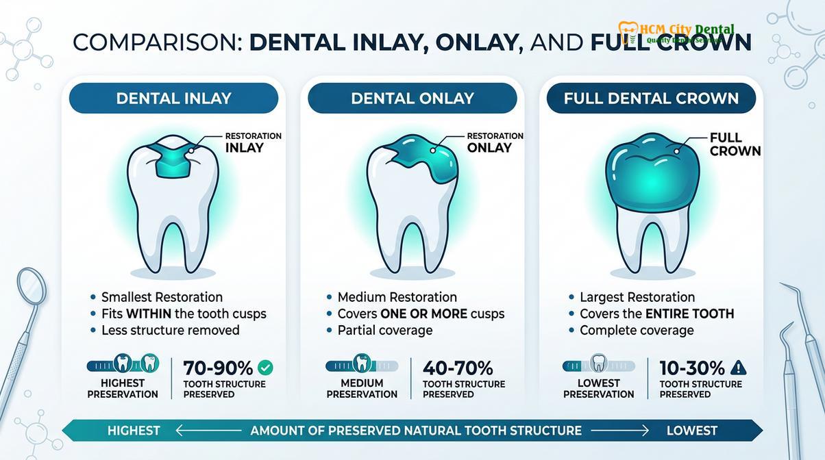
In cases where a patient has suffered widespread enamel wear and multiple cracked teeth due to chronic grinding, onlays and overlays can be used comprehensively to restore the vertical dimension of occlusion (VDO). By rebuilding the lost height of the teeth with conservative ceramic overlays, the clinician can re-establish a harmonious bite, alleviate stress on the temporomandibular joint (TMJ), and protect the remaining dentition from future fractures.
Pricing and Warranty for Inlays and Onlays
Transparent pricing and comprehensive warranties ensure patients receive premium conservative restorations with long-term peace of mind, supported by advanced digital workflows and direct lab partnerships.
Understanding the financial investment required for advanced restorative dentistry is a crucial part of the patient experience. Because inlays and onlays require sophisticated CAD/CAM technology, premium ceramic materials, and highly specialized bonding protocols, they represent a significant upgrade over traditional direct fillings. However, their longevity and ability to prevent the need for costly root canals and full crowns make them a highly cost-effective long-term solution.
At advanced ceramic onlay restorations centers, the pricing structure is designed to be transparent and accessible. According to the latest clinic fee schedule, patients can benefit significantly from planning their treatment in advance:
- Inlay / Onlay (Lab-made ceramic): The standard walk-in rate is approximately $385 (10.0M VND). However, patients who utilize the WhatsApp pre-booking discount can secure this treatment From $231 (6.0M VND), representing a 40% savings.
- Overlay / Partial Crown: For more extensive damage requiring broader coverage, the walk-in rate is approximately $446 (11.6M VND). With the WhatsApp pre-booking discount, the cost is reduced to From $269 (7.0M VND) (-40%).
Beyond the initial cost, patients must consider the value of post-operative security. Reputable clinics provide complete peace of mind through a global warranty on their lab-milled restorations. This ensures that in the rare event of a material failure or debonding, the patient is fully supported. Additionally, remote WhatsApp checkup support allows international or traveling patients to communicate directly with their clinical team, sharing photos and receiving expert guidance without needing to return immediately to the clinic.
When to See a Doctor
A cracked tooth is a progressive condition that will not heal on its own. Because enamel and dentin cannot regenerate, any delay in treatment allows the fracture to deepen under the continuous stress of daily chewing. You should schedule a comprehensive clinical evaluation immediately if you experience any of the following symptoms:
- Rebound Pain: A sharp, sudden jolt of pain that occurs specifically when you release your bite from food.
- Thermal Sensitivity: A lingering, intense ache when the tooth is exposed to hot or cold beverages, which may indicate the crack is nearing the pulp.
- Localized Swelling: A small bump or swelling on the gums near the affected tooth, which is a strong indicator of a developing abscess or root fracture.
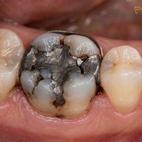
- Visible Damage: You can feel a sharp edge with your tongue, or a piece of an old silver filling has broken away, leaving the surrounding tooth walls unsupported.

Early diagnosis using transillumination and 3D scanning can mean the difference between a conservative, single-visit onlay and a complex, multi-stage root canal and crown procedure. If you suspect you have a cracked tooth, seeking prompt evaluation at a modern facility like HCMC Dental Clinic ensures that the maximum amount of your natural tooth structure can be preserved.
Frequently Asked Questions
Can a cracked tooth be saved with an inlay?
Yes, a cracked tooth can often be saved with an inlay if the fracture is confined to the central chewing surface and does not compromise the outer cusps. The custom ceramic inlay seals the damaged area, restoring structural integrity and preventing bacterial microleakage. However, if the crack extends across a cusp, an onlay is required to provide the necessary structural splinting.
What is cracked tooth syndrome?
Cracked tooth syndrome is a clinical condition characterized by an incomplete fracture in a vital posterior tooth that involves the dentin. It typically causes sharp, localized pain upon releasing biting pressure and heightened sensitivity to extreme temperatures. Because the crack is often microscopic, it requires specialized diagnostic tools like transillumination and bite tests to accurately locate.
When does a cracked tooth need a root canal?
A cracked tooth requires a root canal when the fracture propagates deep enough to irritate or expose the dental pulp. If the nerve tissue becomes irreversibly inflamed or infected, endodontic therapy is mandatory before placing a final restoration. Catching the crack early and placing an onlay can often prevent the need for a root canal entirely.
Does an onlay prevent a cracked tooth from splitting?
Yes, a properly bonded onlay acts as a structural splint that covers the weakened cusps, effectively preventing the tooth from splitting. By distributing masticatory forces evenly across the ceramic material, it neutralizes the wedging effect that causes cracks to expand. This makes it a superior choice over traditional fillings for compromised teeth.
How long does a ceramic onlay last on a cracked tooth?
A high-quality ceramic onlay can last 10 to 15 years or longer when supported by excellent oral hygiene and proper occlusal alignment. Materials like lithium disilicate and zirconia offer exceptional durability against heavy chewing forces. Regular dental check-ups and the use of a night guard for bruxism can further extend the lifespan of the restoration.
References
- Journal of Endodontics. Clinical diagnosis and management of cracked tooth syndrome. (2021).
- International Journal of Prosthodontics. Biomechanical behavior of ceramic onlays on fractured teeth. (2020).
- Journal of Esthetic and Restorative Dentistry. Immediate dentin sealing and bonding protocols. (2022).
- American Dental Association. Guidelines for conservative indirect restorations. (2019).
- Journal of Prosthetic Dentistry. The ferrule effect in severely damaged teeth. (2023).