An overlay after a root canal is a highly effective, conservative restoration that protects the weakened tooth while preserving maximum natural enamel. Unlike traditional full crowns, overlays cover only the damaged cusps, utilizing advanced resin-adhesive cementation to reinforce structural integrity without aggressive 360-degree shaving.
Clinical Summary:
Following endodontic therapy, restoring the biomechanical integrity of the tooth is paramount to prevent catastrophic fractures. While traditional dentistry often defaults to full-coverage crowns, modern biomimetic approaches heavily favor the dental overlay after root canal treatment. This preservation philosophy focuses on retaining the maximum amount of healthy peripheral enamel. By utilizing advanced digital workflows—specifically intraoral scanners—clinicians can capture highly accurate 3D data without uncomfortable silicone impressions. This data is seamlessly transferred to a direct lab partnership for CAD/CAM milling, ensuring an exact marginal fit that prevents secondary decay. High-end, metal-free biocompatible materials like lithium disilicate and zirconia are selected based on occlusal load requirements. Through meticulous enamel bonding protocols and resin-adhesive cementation, an overlay provides robust occlusal protection, effectively mitigating the risk of vertical root fractures while maintaining the tooth’s natural flexural dynamics.
Key Takeaways:
- Overlays are conservative restorations that protect the chewing surface without requiring the aggressive 360-degree enamel removal associated with full crowns.
- Endodontically treated teeth suffer from altered architecture and reduced moisture, making cusp coverage essential to prevent structural failure.
- Digital intraoral scanning and CAD/CAM milling ensure a precise marginal seal, drastically reducing the risk of secondary bacterial microleakage.
- Success relies heavily on adequate residual wall thickness (typically 1.5mm to 2.0mm) and strict isolation during the resin bonding protocol.
- Premium biocompatible materials like lithium disilicate and zirconia offer exceptional durability and aesthetic integration for post-endodontic restorations.
The Biomechanical State of an Endodontically Treated Tooth
A tooth that has undergone endodontic therapy experiences significant architectural changes and moisture loss, requiring specialized restorative techniques to prevent structural failure under chewing forces.
To fully appreciate the necessity of a robust post-endodontic restoration, one must first understand the profound biomechanical changes that occur within the tooth structure during and after a root canal treatment. The primary goal of endodontic therapy is to eradicate infection from the pulp chamber and root canal system. However, achieving this requires the creation of an endodontic access cavity through the occlusal (chewing) surface of the tooth. This access preparation inherently compromises the tooth’s structural integrity.
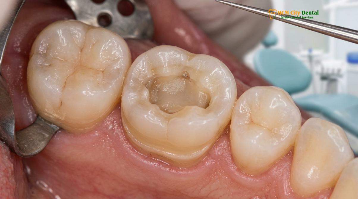
From an architectural standpoint, an intact vital tooth functions much like a closed, rigid box. The roof of the pulp chamber ties the buccal (cheek side) and lingual (tongue side) cusps together, allowing the tooth to distribute heavy occlusal loads evenly down the long axis of the root. When the roof of the pulp chamber is removed to access the infected nerve, this “closed box” is converted into an “open cylinder.” The cusps are no longer tied together, allowing them to flex independently under the immense pressure of mastication. This independent flexing is a primary catalyst for cusp fractures and catastrophic structural failure.
Furthermore, the removal of the vital pulp tissue eliminates the tooth’s internal blood supply and hydration mechanism. Over time, this lack of internal moisture alters the collagen cross-linking within the dentinal tubules, leading to a phenomenon clinically recognized as tooth brittleness[1]. A devitalized tooth is significantly less resilient to shear forces and impact trauma compared to a vital tooth. Additionally, the loss of the pulp means the loss of internal proprioceptive nerve endings. Patients often bite down harder on endodontically treated teeth because the natural feedback mechanism that warns against excessive force is diminished.
Why Traditional Wisdom Mandates a Full Crown
Historically, dentists relied on full-coverage crowns to create a rigid ferrule effect, binding the weakened tooth together, though this requires aggressive removal of healthy enamel.
For decades, the standard of care following endodontic therapy on a posterior tooth was the placement of a full-coverage dental crown. This approach was rooted in the necessity to protect the compromised tooth from the wedge effect of traditional fillings. The clinical rationale was straightforward: by shaving down the entire circumference of the tooth and placing a rigid cap over it, the dentist could create a “ferrule effect”—a 360-degree band of restorative material that tightly hugs the remaining tooth structure, preventing the roots and cusps from splitting apart.
When patients ask if all root canals need full crowns, the historical answer was almost universally yes. The traditional workflow often involved placing a post and core buildup. A metal or fiberglass post was cemented deep into one of the cleaned root canals to provide retention for a core buildup material, which replaced the missing internal dentin. The dentist would then prepare the tooth for a crown by cutting a deep chamfer margin at or below the gumline, effectively removing a significant portion of tooth structure from every single side of the tooth.
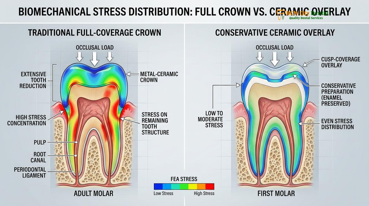
While full crowns are undeniably strong and have a long history of clinical success, the preparation process is highly destructive. Clinical studies comparing survival rates often highlight that while crowns are durable, the aggressive preparation leaves very little natural tooth structure behind[2]. The preservation philosophy of modern biomimetic dentistry questions the wisdom of grinding away perfectly healthy, intact peripheral enamel simply to satisfy the geometric requirements of a traditional crown. Enamel is the strongest substance in the human body, and once it is removed by a dental bur, it is gone forever.
When an Overlay is a Superior Conservative Alternative
An overlay provides the necessary occlusal protection by covering the chewing surface while preserving the intact peripheral walls, utilizing advanced adhesion rather than mechanical retention.
The paradigm shift from aggressive full crowns to conservative partial coverage restorations has been driven by monumental advancements in dental material science and resin bonding protocols. A dental overlay after root canal treatment represents the pinnacle of this biomimetic approach. An overlay is designed to cover the entire occlusal surface of the tooth—effectively capping the weakened cusps and providing the necessary overlay protection—without requiring the dentist to shave down the healthy buccal and lingual walls.
This approach aligns perfectly with the preservation philosophy practiced at leading institutions. By retaining the peripheral band of natural enamel, the tooth maintains a higher degree of its original structural rigidity. Enamel is also the ideal substrate for dental bonding; resin adhesives form a significantly stronger and more predictable micro-mechanical bond to enamel than they do to the underlying dentin. Major dental organizations now frequently highlight these conservative restorations in their clinical guidelines[4].
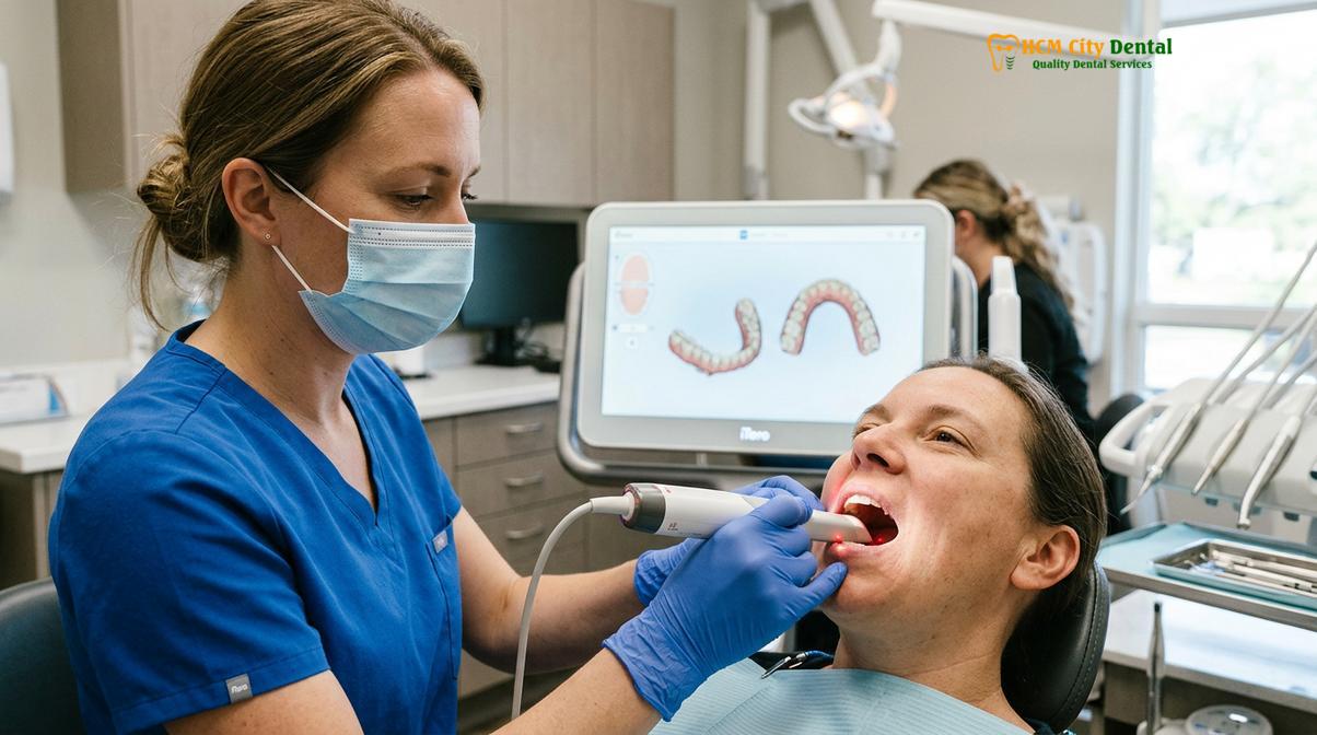
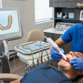
The fabrication of these conservative restorations relies heavily on digital dentistry. Rather than using messy, uncomfortable silicone impression materials that can distort, clinicians utilize advanced intraoral scanners. These devices capture highly precise 3D topographical data of the prepared tooth in a matter of seconds. This digital impression is then transmitted to a direct lab partnership where CAD/CAM technology is used to mill the restoration.
| Clinical Parameter | Traditional Full Crown | Ceramic Overlay |
|---|---|---|
| Tooth Structure Removal | Aggressive (60-75% removal, 360-degree shaving) | Conservative (Only damaged areas and occlusal surface) |
| Retention Mechanism | Mechanical retention (friction from parallel walls) | Chemical/Micromechanical resin bonding |
| Margin Location | Often subgingival (below gumline), risk of irritation | Supragingival (above gumline), easy to clean |
| Enamel Preservation | Minimal to none remaining on axial walls | Maximum preservation of peripheral enamel |
The Role of Residual Tooth Structure (Wall Thickness)
The clinical viability of an overlay is strictly dictated by the thickness and quality of the remaining tooth walls, which must be robust enough to support the bonded ceramic.
While the benefits of an inlay or onlay post-endodontic restoration are clear, it is crucial to understand that not every root-canal-treated tooth is a candidate for an overlay. The decision to pursue a conservative overlay versus a full crown is dictated almost entirely by the quantity and quality of the residual tooth structure. Specifically, the clinician must evaluate the wall thickness of the remaining buccal and lingual enamel and dentin.
During the cavity outline form preparation, the dentist meticulously removes any unsupported enamel, old restorative material, and active tooth decay. Once the tooth is clean, the remaining vertical walls are measured. Biomechanical studies indicate that for a tooth wall to withstand functional loading without flexing and breaking the adhesive bond, it must have a minimum thickness of 1.5 to 2.0 millimeters[3].
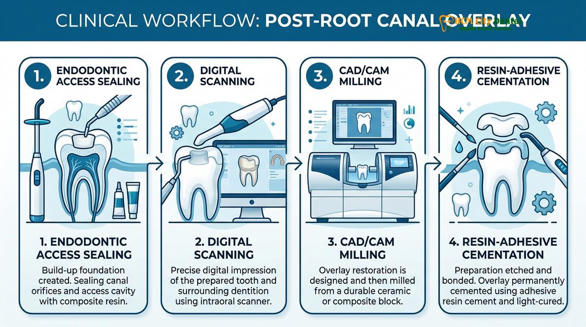
If the walls meet the thickness criteria, the clinician proceeds with immediate dentin sealing (IDS) and strict enamel bonding protocols. This involves isolating the tooth with a rubber dam to prevent moisture contamination from saliva or breath. The tooth surface is treated with a mild phosphoric acid etchant to create microscopic pores in the enamel, followed by the application of a primer and a silane coupling agent. A dual-cure resin-adhesive cement is then used to bond the CAD/CAM milled overlay directly to the tooth structure. This adhesive integration essentially fuses the ceramic and the tooth into a single, cohesive biomechanical unit, restoring much of the tooth’s original stiffness[5].
Preventing Vertical Root Fractures
Properly designed overlays distribute occlusal forces evenly across the tooth, significantly reducing the wedge effect that leads to devastating vertical root fractures.
One of the most devastating complications that can occur in an endodontically treated tooth is a vertical root fracture. This type of fracture originates in the root and propagates upward toward the chewing surface, or vice versa. Unlike a chipped cusp, a vertical root fracture is almost always a terminal event for the tooth, necessitating immediate extraction and replacement with a dental implant or bridge.
“The ultimate goal of post-endodontic restoration is not merely to fill a hole, but to re-establish the biomechanical harmony of the tooth. By capping the cusps with a bonded ceramic overlay, we convert destructive shear forces into manageable compressive forces, effectively neutralizing the wedge effect that leads to vertical root fractures.”
An overlay provides exceptional protection by acting as a rigid helmet over the vulnerable cusps. When the patient bites down, the force is absorbed by the high-strength ceramic and distributed evenly down the long axis of the root, exactly as nature intended. Furthermore, during the delivery of the restoration, the clinician will perform meticulous occlusal adjustments to ensure perfect bite alignment.
Dr. Cuong’s Decision Tree for Post-Root Canal Restoration
Treatment planning requires a personalized diagnostic approach, balancing the preservation of natural tooth structure with the mechanical demands of the patient’s bite.
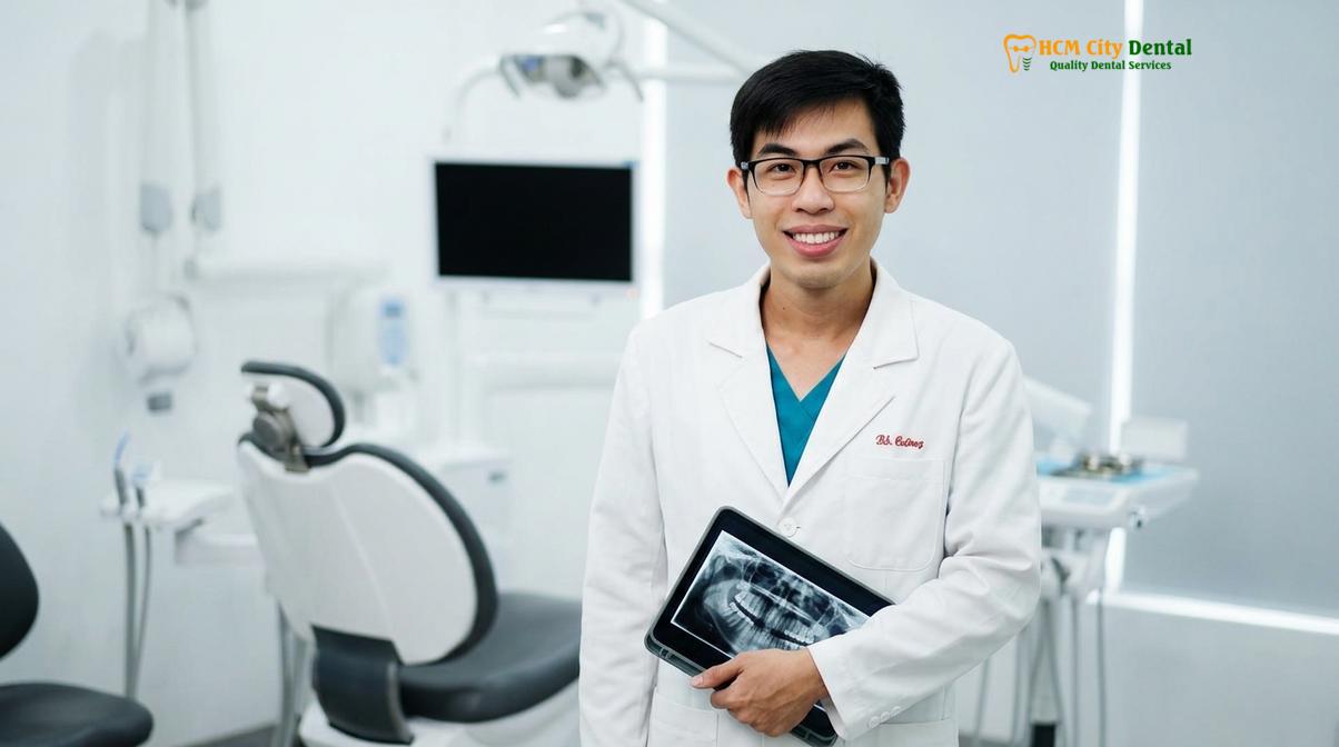
At HCMC Dental Clinic in Ho Chi Minh City, Dr. Nguyen Van Cuong emphasizes a biomimetic approach to post-endodontic care. Dr. Cuong meticulously evaluates each patient’s residual tooth structure to determine if a conservative overlay can safely replace the need for a full crown. By prioritizing the preservation of healthy enamel whenever clinically viable, he ensures that patients retain as much of their natural smile as possible while still receiving robust occlusal protection.
Clinical Case Study:
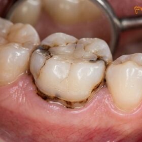
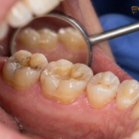
A recent patient visiting HCMC Dental Clinic in Ho Chi Minh City presented with a fractured upper premolar following endodontic therapy performed years prior. The tooth had been restored with a massive composite filling that eventually failed under chewing pressure. Upon clinical evaluation, it was determined that the buccal and lingual walls were still adequately thick, measuring approximately 2.0mm. Instead of grinding the tooth down for a traditional crown, a custom lithium disilicate overlay was digitally designed and milled. The restoration successfully capped the weakened cusps, seamlessly blending with the adjacent natural teeth and restoring full masticatory function without sacrificing the remaining healthy enamel.
When to Consult a Dentist: Important Clinical Notes
Recognizing the early warning signs of a failing post-endodontic restoration can mean the difference between saving a tooth and requiring an extraction.
Patients who have undergone root canal therapy must remain vigilant about the structural integrity of their treated teeth. According to guidelines from the Vietnam Odonto-Stomatology Association (VOSA) regarding conservative post-endodontic restorations, patients should seek immediate clinical evaluation if they experience specific warning signs[6].
Warning Signs of Structural Failure:
Contact your dental provider immediately if you experience a sharp pain when biting down and releasing, a feeling of looseness in the existing filling, or visible cracks propagating down the side of the tooth. These symptoms often indicate that the tooth is flexing dangerously under pressure.
“Delaying the placement of a definitive occlusal restoration after a root canal exponentially increases the risk of catastrophic tooth fracture. A temporary filling is not designed to withstand long-term masticatory forces and offers zero protection against the wedge effect.”
It is generally recommended to proceed with the final overlay or crown within a few weeks of completing the root canal procedure. Leaving a tooth unprotected for extended periods invites bacterial microleakage, which can re-infect the root canal system and necessitate a complex retreatment.
Conclusion & Next Steps
Choosing an overlay after a root canal is a scientifically backed, conservative approach that maximizes the retention of your natural tooth structure while providing essential protection against fractures. By leveraging digital scanning, CAD/CAM technology, and advanced resin bonding, modern dentistry offers a superior alternative to the aggressive enamel removal required for traditional full crowns.
If you have recently undergone endodontic therapy and want to explore conservative restoration options, contact HCMC Dental Clinic in Ho Chi Minh City today. Learn more about our advanced Inlays & Onlays tại HCMC Dental Clinic to see how we can protect your smile with precision biomimetic dentistry.
Frequently Asked Questions
Can you put an overlay on a root canal tooth?
Yes, an overlay is an excellent restoration for a root canal-treated tooth, provided there is sufficient healthy wall thickness remaining. It covers the chewing surface to prevent fractures while preserving the natural peripheral enamel that a traditional crown would otherwise grind away.
Why do root canal teeth crack?
Root canal teeth crack primarily because the internal dentin and pulp chamber roof have been removed, compromising the tooth’s structural architecture. Additionally, the loss of vital blood supply leads to dentin dehydration, increasing overall tooth brittleness under heavy biting forces.
Is a crown safer than an overlay after a root canal?
Not necessarily. While crowns provide excellent full-coverage protection, they require aggressive removal of healthy tooth structure. An overlay is often safer for the tooth’s long-term lifespan because it preserves the natural enamel, relies on advanced resin bonding, and leaves more tooth structure intact for future treatments.
How thick must the tooth walls be for an overlay?
For a successful overlay, the remaining vertical tooth walls should generally be at least 1.5 to 2.0 millimeters thick. If the walls are thinner than this threshold, they may flex under occlusal pressure, necessitating a full-coverage crown to provide a rigid ferrule effect.
How long does a dental overlay last after a root canal?
A well-maintained ceramic overlay can last a decade or longer. Longevity depends heavily on the precision of the CAD/CAM milling, the quality of the resin-adhesive cementation, the patient’s oral hygiene, and whether they wear a nightguard to protect against bruxism.
References
- Journal of Endodontics. Biomechanical properties of endodontically treated teeth. (2021).
- International Journal of Prosthodontics. Survival rates of ceramic overlays vs full crowns. (2020).
- Journal of Prosthetic Dentistry. The effect of wall thickness on fracture resistance. (2022).
- American Dental Association. Clinical guidelines for conservative restorations. (2019).
- Journal of Esthetic and Restorative Dentistry. Immediate dentin sealing and resin bonding protocols. (2023).
- Vietnam Odonto-Stomatology Association (VOSA). Guidelines on conservative post-endodontic restorations. (2022).