Recognizing failed dental inlay symptoms early—such as sharp pain when chewing, sudden temperature sensitivity, or visible margin discoloration—is critical to saving your tooth. A compromised seal allows bacteria to penetrate, leading to secondary decay beneath the restoration that requires immediate clinical intervention.
Clinical Summary:
Dental inlays and onlays are highly durable, conservative restorations, but they can fail due to microleakage, bonding degradation, or occlusal trauma. Symptoms of a failed restoration include localized bite sensitivity, foul odor, and margin discoloration. Advanced digital diagnostics, including transillumination and 3D intraoral scanning, are essential for detecting decay under porcelain inlay symptoms. Treatment involves careful removal of the defective material, cavity outline form refinement, and fabricating a precise CAD/CAM replacement to restore structural integrity and prevent further enamel loss.
Key Takeaways:
- Sharp pain upon biting or releasing pressure often indicates a cracked restoration or a compromised bonding layer.
- Microleakage at the tooth-restoration margin is the primary cause of secondary decay beneath inlays and onlays.
- Modern digital diagnostics, including transillumination and 3D scanning, can detect hidden failures before severe pain occurs.
- Replacing a failed inlay promptly prevents the need for more invasive procedures like root canal therapy or full crowns.
- High-strength materials like lithium disilicate and zirconia offer superior marginal sealing and longevity for replacement restorations.
How and Why Inlays and Onlays Fail
Dental inlays and onlays typically fail due to mechanical stress, degradation of the resin-adhesive cement, or recurrent bacterial infiltration at the margins. Understanding these failure mechanisms is crucial for preventing future complications.
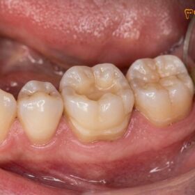
The foundation of modern restorative dentistry is built upon a strict preservation philosophy. Inlays & Onlays, as well as porcelain overlays, are conservative restorations designed to preserve maximum natural tooth structure. Unlike traditional full crowns that require aggressive 360-degree shaving of the enamel, partial coverage restorations only replace the damaged or decayed portions of the tooth. However, despite their high success rates, these restorations operate in a harsh oral environment subjected to immense biting forces, thermal fluctuations, and constant bacterial challenges. Over time, even the most meticulously placed ceramic piece can become a failed restoration if specific clinical or biomechanical factors are compromised [1].
One of the primary reasons for early failure is the degradation of the enamel bonding protocols. The success of an indirect restoration relies heavily on the micromechanical and chemical bond between the ceramic material, the resin-adhesive cement, and the natural tooth structure. If the tooth is not perfectly isolated from saliva and moisture during the cementation process, the bond strength is significantly reduced. Over time, the constant stress of mastication (chewing) can cause this weakened adhesive layer to break down, leading to microscopic gaps at the margins.
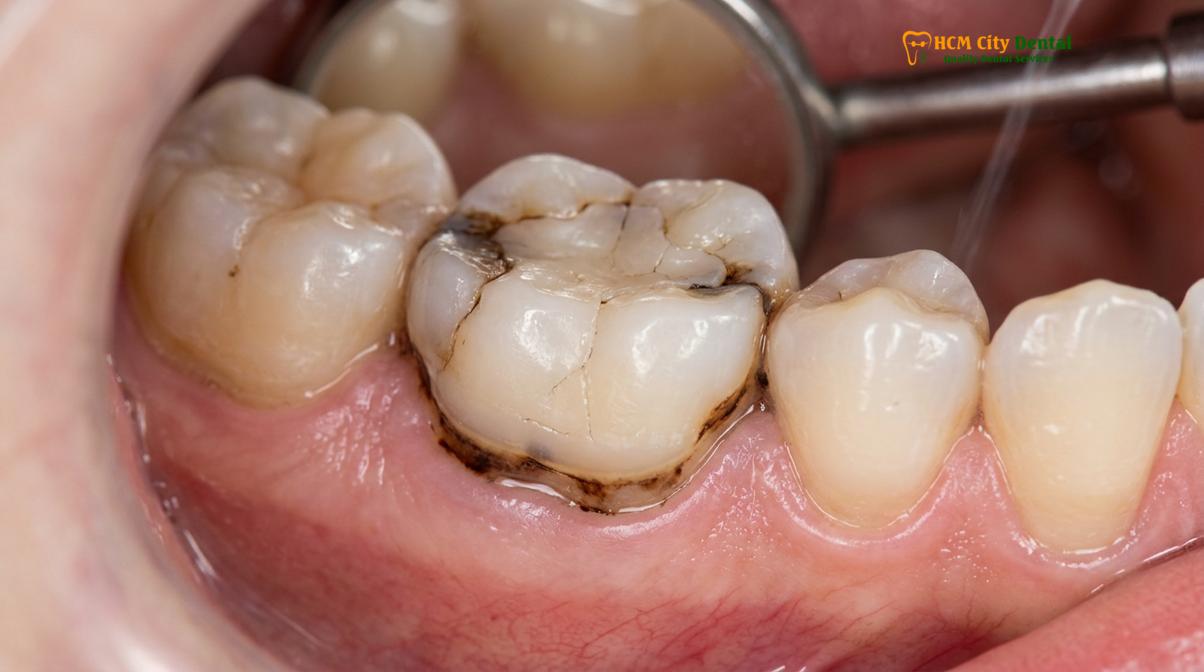
Another significant factor is occlusal trauma, particularly in patients who suffer from bruxism (chronic teeth grinding and clenching). The excessive forces generated during nocturnal grinding can exceed the flexural strength of the restorative material or the underlying tooth structure. This can lead to cohesive fractures within the ceramic itself or adhesive failures where the restoration debonds from the cavity preparation. Dr. Nguyen Van Cuong emphasizes a clinical perspective on conservative restoration material selection, noting that matching the restorative material’s compressive strength to the patient’s specific bite dynamics is essential for long-term success. For patients with heavy bites, restoring the vertical dimension of occlusion (VDO) and providing a protective nightguard are critical preventive measures [2].
Furthermore, errors in the initial cavity outline form can contribute to premature failure. If the preparation lacks adequate resistance and retention form, or if sharp internal line angles are left unrounded, stress concentrations can develop within the ceramic, leading to crack propagation. Additionally, if an inlay is used to replace a massive amalgam filling without accounting for the thin, unsupported enamel walls left behind, the remaining tooth structure may fracture under load. In such cases, extending the preparation to cover the weakened cusps—transitioning from an inlay to an onlay—is necessary to distribute occlusal forces evenly and protect the tooth.
Symptom Checklist: Pain, Odor, Sensitivity, and Mobility
Key indicators of a failing inlay include sharp bite sensitivity, persistent bad breath from trapped bacteria, thermal sensitivity, and any noticeable movement of the ceramic piece.
Identifying the early warning signs of a compromised restoration can mean the difference between a simple replacement and the need for extensive endodontic therapy. Patients should be highly vigilant regarding any changes in how their restored tooth feels or functions. One of the most common and immediate red flags is bite sensitivity. If you experience a sharp, localized pain when biting down on food, or specifically when releasing biting pressure, it strongly suggests that the restoration has cracked or that the adhesive bond has failed, allowing the ceramic piece to flex microscopically against the sensitive dentin.
“Patients often ignore mild thermal sensitivity, but when combined with localized pain upon chewing, it is a definitive clinical sign that the marginal seal of the restoration has been compromised and requires immediate evaluation.”
Thermal sensitivity is another classic symptom. While mild sensitivity to hot or cold is normal for a few days following the initial placement of a restoration, new or worsening thermal sensitivity months or years later is a clear indication of trouble. When the marginal seal breaks down, the insulating barrier is lost, allowing temperature changes to directly stimulate the pulpal nerves. If the pain lingers long after the hot or cold stimulus is removed, it may indicate that the dental pulp has become inflamed (irreversible pulpitis) due to bacterial invasion.
Foul odor or a persistent bad taste in the mouth, particularly localized to one specific tooth, is a strong indicator of microleakage. When the cement washes out or the margin degrades, a microscopic crevice is formed. This gap becomes a trap for food particles and plaque. Because this area cannot be cleaned with a toothbrush or floss, the trapped bacteria ferment the debris, producing sulfur compounds that cause halitosis and an unpleasant taste. If you are wondering how to detect failed onlay issues early, paying attention to localized odors during flossing is a highly effective method.
Finally, any perceptible mobility of the restoration is an absolute sign of failure. A properly bonded inlay or onlay should feel indistinguishable from the natural tooth. If you can feel the restoration shifting, clicking, or moving slightly when you apply pressure with your tongue or a finger, the adhesive bond has completely sheared. Even if the tooth is not currently painful, a loose restoration must be addressed immediately, as the underlying dentin is completely exposed to the oral environment and highly susceptible to rapid, aggressive decay.
Secondary Cavities: Decay Developing Under the Restoration
When the adhesive bond degrades, microleakage allows cariogenic bacteria to infiltrate the tooth, causing secondary decay that silently destroys the underlying dentin.
The most insidious consequence of a failed marginal seal is the development of secondary decay. Unlike primary cavities that form on the visible surfaces of the enamel, secondary caries develop underneath existing dental work. This occurs through a process known as microleakage. When the interface between the ceramic restoration and the natural tooth structure breaks down—whether due to polymerization shrinkage of the cement, mechanical wear, or chemical degradation—microscopic pathways open up. Saliva, carrying millions of cariogenic bacteria and fermentable carbohydrates, seeps into these pathways [3].
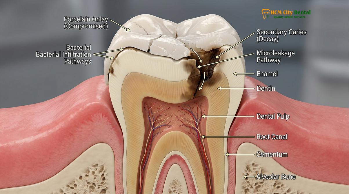
Because the environment beneath a leaking restoration is dark, moist, and completely inaccessible to oral hygiene implements, bacteria thrive. They produce acidic byproducts that rapidly demineralize the dentin. Dentin is significantly softer and more porous than enamel, meaning that once decay breaches the enamel margin, it spreads laterally and deeply at an accelerated rate. This is why decay under porcelain inlay symptoms are often not noticed until the cavity has grown large enough to approach the dental pulp, causing severe pain.
To understand the clinical implications, it is helpful to categorize the types of failure and their corresponding symptoms and causes:
| Type of Failure | Clinical Cause | Primary Symptoms | Recommended Action |
|---|---|---|---|
| Adhesive Debonding | Moisture contamination during cementation or heavy occlusal trauma. | Restoration feels loose, clicking sound when chewing, sudden sensitivity. | Immediate recementation or replacement if the cavity is contaminated. |
| Marginal Microleakage | Degradation of resin cement, poor initial marginal adaptation. | Localized bad breath, brown/black staining at the tooth junction, mild thermal sensitivity. | Thorough clinical exam, removal of restoration, and caries excavation. |
| Cohesive Fracture | Material fatigue, bruxism, inadequate ceramic thickness. | Sharp pain on release of bite, visible crack or missing piece of ceramic. | Complete restoration replacement using high-strength materials. |
| Secondary Decay | Prolonged microleakage allowing bacterial infiltration into dentin. | Deep throbbing pain, lingering sensitivity to sweets and temperature. | Removal of restoration, deep caries removal, potential pulp therapy. |
Preventing secondary decay relies entirely on achieving and maintaining a hermetic margin seal. This requires meticulous clinical technique during the preparation and bonding phases. Dentists must ensure that the cavity margins are placed on healthy, sound enamel whenever possible, as bonding to enamel is significantly stronger and more predictable than bonding to dentin. If you suspect that an old filling or inlay is leaking, seeking a professional evaluation to Replace Failing Fillings with Onlay is a proactive step to halt the progression of hidden decay and save the tooth from requiring a root canal.
Diagnosing Failures with X-Rays and Transillumination
Accurate detection of a failing restoration relies on advanced digital diagnostics, including high-resolution bitewing radiographs and fiber-optic transillumination, to reveal hidden marginal defects.
Because secondary decay and micro-fractures often occur beneath the surface or between the teeth, visual inspection alone is insufficient for a definitive diagnosis. Modern dental clinics employ a suite of digital diagnostics to accurately assess the integrity of existing restorations. The cornerstone of this diagnostic process is the high-resolution digital bitewing radiograph (X-ray). Bitewings provide a clear view of the interproximal areas (where teeth touch) and the internal adaptation of the restoration to the cavity floor. On an X-ray, secondary decay appears as a dark, radiolucent shadow beneath the bright, radiopaque restorative material [4].
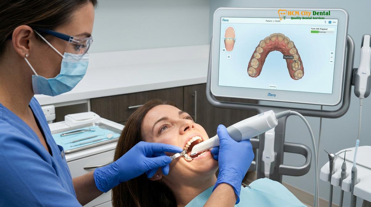
However, X-rays have limitations. Early-stage microleakage or very fine cracks in the ceramic may not be visible on a standard radiograph. To overcome this, clinicians utilize fiber-optic transillumination. This technique involves shining a high-intensity light through the tooth structure. Healthy enamel and dentin transmit light predictably, but cracks, fractures, and areas of demineralized decay disrupt the light beam, creating distinct shadows or visible fracture lines. This non-invasive diagnostic tool is incredibly effective for identifying compromised margins before they progress to massive structural failures.
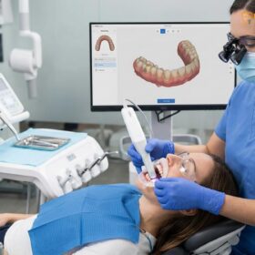
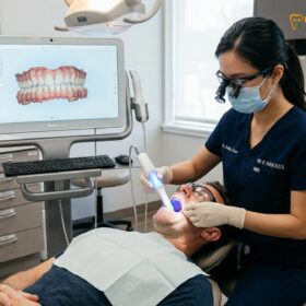
Furthermore, the integration of 3D intraoral scanners, such as the iTero scanner and Medit i700, has revolutionized diagnostic capabilities. These devices capture highly magnified, full-color 3D models of the dentition. The clinician can manipulate the digital model on a screen to inspect the margins of an inlay or onlay at a microscopic level. The software can even detect subtle changes in tooth wear or shifting over time, providing valuable data on occlusal forces. If a restoration is deemed to be failing, these same digital scans are used to plan the replacement, ensuring a seamless transition to a new, perfectly adapted ceramic piece.
The Replacement Process: Step-by-Step Removal and Preparation
Replacing a defective inlay involves carefully removing the old material, refining the cavity outline form, and utilizing CAD/CAM technology to mill a precise, biocompatible ceramic replacement.
When a restoration is diagnosed as failing, prompt restoration replacement is necessary to halt bacterial progression and restore the tooth’s structural integrity. The clinical workflow for replacing an inlay or onlay is highly precise, leveraging advanced technology to ensure a superior outcome compared to the original failing restoration.
The first step is profound local anesthesia and proper isolation, typically using a rubber dam. Isolation is critical to prevent saliva and bacteria from contaminating the surgical field once the tooth is opened. The clinician then uses specialized diamond burs to carefully section and remove the defective ceramic or composite material. Great care is taken to preserve as much of the remaining healthy tooth structure as possible. Once the old material is removed, the underlying cavity is thoroughly inspected. Any secondary decay or softened dentin is meticulously excavated until only hard, healthy tooth structure remains.
Clinical Case Review: A patient visited HCMC Dental Clinic in Ho Chi Minh City complaining of severe bite sensitivity in a lower molar with an old composite inlay. Upon examination, Dr. Cuong identified significant microleakage and secondary decay beneath the restoration. The treatment involved careful excavation of the decay and upgrading the restoration to a custom CAD/CAM milled e.max overlay. This approach successfully restored the vertical dimension of occlusion (VDO) and eliminated the patient’s pain, demonstrating the efficacy of precise digital workflows in managing complex restorative failures.
After the decay is removed, the cavity outline form is refined. The clinician ensures that all margins are smooth, typically creating a chamfer margin that provides a distinct finish line for the new restoration to seat against. Any sharp internal angles are rounded to prevent stress concentrations. If the decay was deep, a biocompatible base or liner may be placed to protect the pulp and block out undercuts.
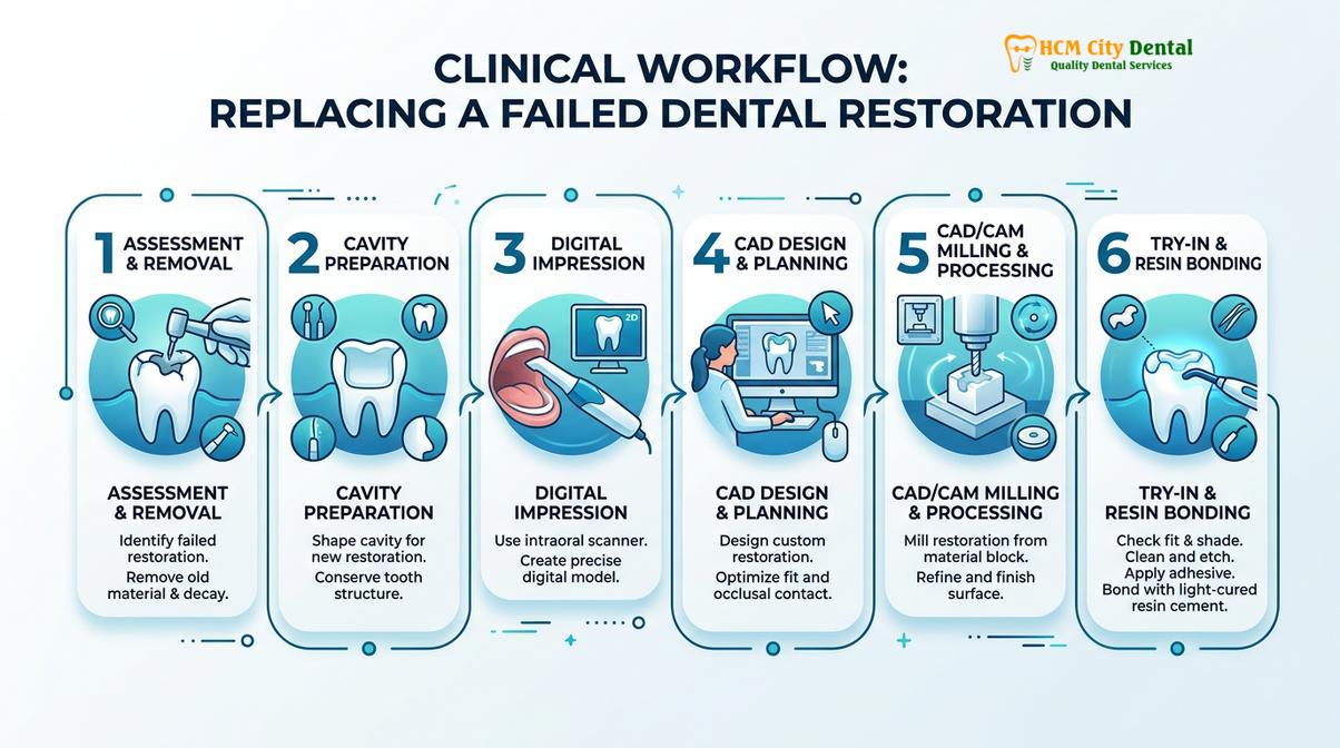
Next, the digital workflow takes over. Using the iTero or Medit i700 intraoral scanners, a highly accurate 3D digital impression is captured. This digital scanning eliminates the need for uncomfortable, gag-inducing silicone impressions. The digital data is instantly transmitted to the laboratory. HCMC Dental Clinic maintains a direct lab partnership, utilizing advanced CAD/CAM milling technology to fabricate the new restoration. This ensures an exact marginal fit, which is the most critical factor in preventing future secondary decay.
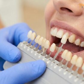
Material selection for the replacement is paramount. For the anterior aesthetic zone and premolars, high-end materials from Ivoclar, specifically IPS e.max lithium disilicate, are often chosen for their exceptional translucency and strong bonding capabilities. For posterior molars subjected to heavy biting forces, Dentsply Sirona Cercon HT Zirconia is frequently utilized due to its immense flexural strength. Both options provide 100% metal-free, biocompatible ceramic solutions that prevent gum line irritation and allergic reactions, aligning with a holistic dentistry approach.
Understanding the pricing structure is also important for patients planning their treatment. According to the latest clinic fee schedule, the estimated cost for a lab-made ceramic Inlay or Onlay for a walk-in patient is approximately $385 (10.0M VND). However, patients utilizing the WhatsApp pre-booking discount can secure this treatment from $231 (6.0M VND), representing a 40% savings. For more extensive damage requiring an Overlay or Partial Crown, the walk-in rate is ~$446 (11.6M VND), while the WhatsApp pre-booking discount brings the cost down to from $269 (7.0M VND). This transparent pricing, combined with a global warranty and remote WhatsApp checkup support, offers complete peace of mind for international and local patients alike.
Upgrading to a Mão (Full Crown) If Damage is Severe
When secondary decay or fractures compromise more than two-thirds of the tooth structure, upgrading from an inlay or onlay to a full-coverage crown becomes clinically necessary.
While the preservation philosophy dictates saving as much natural enamel as possible, there are clinical scenarios where a partial coverage restoration is no longer viable. If a failing inlay has allowed secondary decay to hollow out the tooth extensively, or if a significant portion of the buccal or lingual cusps has fractured off, the remaining tooth structure may be too weak to support a new onlay. In these cases, a crown upgrade is the most predictable and durable treatment option [5].
The decision between a Partial Crown vs Full Crown depends on the amount of solid, healthy dentin remaining after all decay and old restorative material have been removed. A full crown (mão răng) provides 360-degree coverage, acting like a protective helmet that binds the remaining tooth structure together and absorbs the full force of mastication. This is particularly important for teeth that have undergone root canal therapy, as they become more brittle over time.
Clinical Warning: Delaying the replacement of a loose, cracked, or leaking inlay can lead to irreversible pulp damage. Once bacteria penetrate the inner pulp chamber, a simple restoration is no longer sufficient, and the tooth will require endodontic (root canal) therapy or, in cases of catastrophic vertical root fracture, complete extraction.
When replacing cracked dental overlay restorations, the clinician must carefully evaluate the extent of the fracture line. If the crack extends below the gum line or into the root, a full crown may be required to stabilize the tooth. Modern full crowns utilize the same high-strength, biocompatible materials—such as monolithic zirconia or layered lithium disilicate—ensuring that the final result is both functionally robust and highly aesthetic. Patients seeking to understand the specific biomechanical differences between these options can review a detailed clinical comparison of Onlay vs Crown to make an informed decision regarding their long-term dental health.
When to See a Doctor
Dental restorations are not permanent, and ignoring the signs of failure can lead to severe dental emergencies. You should schedule an immediate clinical examination if you experience any of the following red flags:
- Severe, throbbing pain: Pain that wakes you up at night or radiates to your jaw and ear indicates that the dental pulp is actively infected.
- Visible swelling or pus: Swelling in the gums around the restored tooth, or a pimple-like bump (fistula) on the gums, is a sign of an active dental abscess requiring urgent antibiotic therapy and intervention.
- Complete dislodgement: If your inlay or onlay falls out completely, keep the ceramic piece safe and see a dentist immediately. Do not attempt to glue it back in yourself with household adhesives, as this is toxic and will ruin the cavity preparation.
“Immediate clinical evaluation is required if a dental restoration becomes completely dislodged, as the exposed dentin is highly susceptible to rapid bacterial colonization and severe hypersensitivity.”
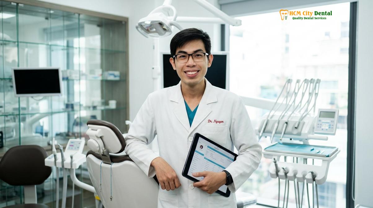
If you suspect your restoration is failing, or if you are experiencing unexplained bite sensitivity, do not wait for the pain to become unbearable. Early intervention is the key to preserving your natural tooth structure. Contact the expert team at HCMC Dental Clinic in Ho Chi Minh City for a comprehensive digital diagnostic evaluation and personalized treatment plan.
Frequently Asked Questions
What are the symptoms of a failed inlay?
The primary symptoms of a failed inlay include sharp pain when chewing, localized temperature sensitivity, visible discoloration around the tooth margin, and a foul odor or taste. These signs indicate that the marginal seal has degraded, allowing bacteria to penetrate the tooth structure. If you experience bite sensitivity or notice the restoration feels loose, immediate clinical evaluation is necessary to prevent further decay and protect the underlying dental pulp.
Can decay get under a porcelain onlay?
Yes, secondary decay can develop under a porcelain onlay if the adhesive bonding layer degrades or if microleakage occurs at the restoration margins. When the seal is compromised, cariogenic bacteria infiltrate the microscopic gaps between the ceramic and the natural tooth. Because this decay is hidden beneath the restoration, it often progresses silently until it reaches the sensitive dentin or pulp, making routine digital diagnostics and radiographic monitoring essential.
Does a cracked inlay need to be replaced immediately?
A cracked inlay requires prompt replacement to restore structural integrity and prevent catastrophic tooth fracture or severe bacterial infection. Even a microscopic hairline fracture compromises the hermetic seal of the restoration, allowing saliva and bacteria to seep into the underlying cavity preparation. Delaying treatment significantly increases the risk of irreversible pulpitis, which would necessitate root canal therapy rather than a simple restoration replacement.
How do you remove and replace a failed restoration?
Replacing a failed restoration involves carefully sectioning and removing the defective material using specialized dental burs, followed by thorough excavation of any secondary decay. The dentist then refines the cavity outline form and captures a precise 3D digital impression using an intraoral scanner. A new, custom-milled ceramic inlay or onlay is fabricated via CAD/CAM technology and permanently bonded to the tooth using advanced resin-adhesive cementation protocols.
How long should a dental inlay or onlay last before failing?
A well-placed dental inlay or onlay fabricated from high-quality ceramic can last between 10 to 15 years, and often much longer with excellent oral hygiene and regular professional maintenance. Longevity depends heavily on the precision of the marginal fit, the strength of the enamel bonding protocol, and the patient’s occlusal forces. Patients who suffer from bruxism should wear a custom nightguard to protect their restorations from excessive wear and premature failure.
References
- Journal of Prosthetic Dentistry. Clinical evaluation of ceramic inlays and onlays: A systematic review. (2021).
- International Journal of Prosthodontics. Microleakage and marginal adaptation of CAD/CAM restorations. (2020).
- Journal of Esthetic and Restorative Dentistry. Longevity and failure rates of lithium disilicate partial crowns. (2019).
- Operative Dentistry. Secondary caries detection under indirect restorations using digital transillumination. (2022).
- American Dental Association. Clinical guidelines for the diagnosis and management of defective dental restorations. (2018).