Color matching dental inlays requires a precise blend of clinical photography, digital shade analysis, and advanced ceramic materials. By evaluating the hue, chroma, and value of adjacent enamel, dentists can fabricate custom-milled restorations that seamlessly integrate with your natural tooth structure, aiming for optimal aesthetic and functional outcomes.
Clinical Summary:
Achieving an invisible margin with a ceramic inlay relies heavily on the “chameleon effect” of modern glass-ceramics and meticulous laboratory communication. Unlike traditional silver fillings, aesthetic dentistry utilizes high-translucency materials like lithium disilicate to mimic natural optical refraction. The clinical workflow involves digital scanning, shade mapping using standardized guides, and custom staining by master ceramists. When bonded correctly using resin-adhesive cementation, these conservative restorations preserve healthy tooth structure while providing long-lasting, biocompatible, and visually discreet repairs.
Key Takeaways:
- Dental inlays preserve significantly more natural tooth structure compared to full-coverage crowns.
- Advanced intraoral scanners eliminate the need for uncomfortable traditional silicone impressions.
- Lithium disilicate ceramics offer superior optical refraction, blending smoothly with adjacent enamel.
- Custom staining in a dental laboratory ensures the restoration matches the exact hue, chroma, and value of your teeth.
- Resin-adhesive cementation creates a watertight margin seal, helping to prevent secondary decay and microleakage.
The Challenge of Mimicking Natural Teeth
Replicating the complex optical properties of natural dentition requires understanding how light penetrates translucent enamel and reflects off the underlying opaque dentin.
The human tooth is not a monochromatic block of white; it is a highly complex, multi-layered anatomical structure with varying degrees of translucency, opalescence, and fluorescence. The outer layer, the enamel, is composed of densely packed hydroxyapatite crystals that allow light to pass through. Beneath the enamel lies the dentin, which is more opaque and carries the primary color or “hue” of the tooth. When a tooth suffers from extensive tooth decay or requires the replacement of a large silver filling, restoring it to its original natural appearance presents a significant clinical challenge. The restorative material must not only withstand heavy occlusal (biting) forces but also mimic this intricate interplay of light and structure[1].
This is where the preservation philosophy of modern aesthetic dentistry becomes paramount. Inlays, onlays, and porcelain overlays are conservative restorations designed to preserve maximum natural tooth structure. Unlike traditional full-coverage crowns, which require a 360-degree shaving of the tooth, an inlay only replaces the damaged portion within the cusps. By preserving the healthy perimeter of the natural tooth, the dentist maintains the original enamel frame, which is crucial for achieving a natural color match. The remaining natural enamel acts as a visual anchor, allowing the new restoration to blend in rather than stand out.
Furthermore, choosing a lab-milled inlay over a massive direct composite filling addresses a critical biomechanical issue: preventing polymerization shrinkage. When large amounts of composite resin are cured with a dental light, the material shrinks slightly, creating stress on the cavity walls and potentially leading to micro-gaps or cuspal deflection. A custom-milled ceramic inlay is fabricated outside the mouth and bonded passively, eliminating this shrinkage stress while providing a highly polished, non-porous surface that maintains its color match over many years of use.
Using the VITA Shade Guide and Digital Photography
Dentists utilize standardized shade guides and high-resolution digital imaging to communicate precise color data to the dental laboratory.
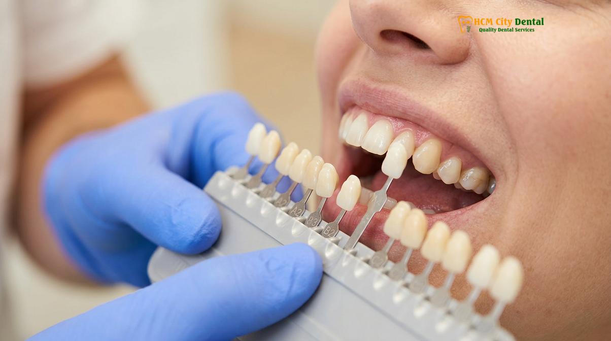
The foundation of an invisible restoration begins with accurate shade selection. Historically, dentists relied solely on visual comparison using a standard shade tab. Today, the process of evaluating shade for dental restorations is a highly systematic protocol. The most advanced system used in clinical practice is the VITA 3D-Master guide, which categorizes tooth color based on three distinct dimensions: value (lightness or darkness), chroma (the intensity or saturation of the color), and hue (the actual color family, such as reddish-yellow or brownish-yellow)[2].
Because teeth dehydrate rapidly when a patient’s mouth is open during a dental procedure, their value temporarily increases, making them appear whiter and more opaque. Therefore, a skilled clinician will always perform the color match process at the very beginning of the appointment, before any drilling or isolation takes place. The dentist will hold the shade guide directly next to the target tooth and adjacent teeth, utilizing natural, color-corrected lighting to avoid metamerism—a phenomenon where colors appear to match under one light source but look completely different under another.
“The integration of digital shade analysis has revolutionized restorative dentistry. By combining high-definition intraoral scans with polarized photography, we eliminate the subjective variables of human vision, providing the laboratory with an exact topographic and chromatic map of the patient’s dentition.”
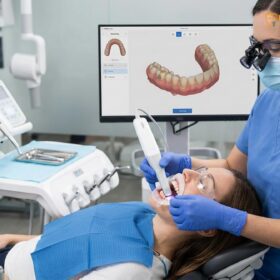
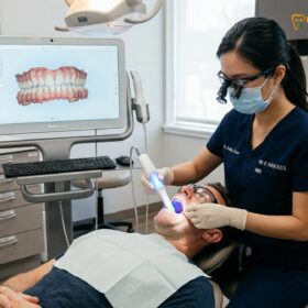
Beyond visual guides, modern clinics employ advanced digital technology. Digital scanning eliminates uncomfortable silicone impressions, providing precise 3D data for the custom lab-milled restorations. These advanced scanners do more than just capture the physical geometry of the cavity preparation; they also record high-definition color data. When combined with polarized clinical photography—which removes surface glare to reveal the internal dentin structure—the dental laboratory receives a comprehensive digital dossier, ensuring the final ceramic piece is a perfect match[5].
Chameleon Effect: How Light Interacts with Glass Ceramics
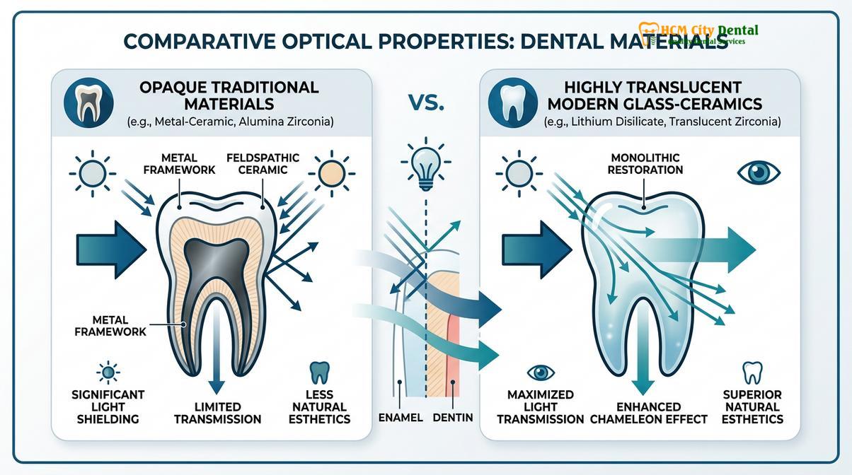
The chameleon effect allows highly translucent ceramics to draw in and reflect the color of surrounding natural tooth structure, creating an invisible margin.
One of the most fascinating phenomena in modern restorative dentistry is the chameleon effect. This optical illusion occurs when a restorative material is capable of absorbing, scattering, and reflecting the light from the adjacent natural tooth structure. Instead of blocking light like older metal-based or highly opaque materials, a translucent ceramic allows light to penetrate its surface, bounce off the surrounding natural cavity walls, and reflect back to the observer’s eye. This causes the restoration to visually adopt the color characteristics of the host tooth, effectively blurring the transition line between the artificial material and the natural enamel[3].
To achieve this seamless integration, material selection is critical. For the anterior aesthetic zone and premolars, dentists frequently utilize high-end materials like lithium disilicate. Lithium disilicate is a glass-ceramic characterized by its needle-like crystal structure embedded in a glassy matrix. This unique composition provides exceptional flexural strength while mimicking the optical refraction of natural enamel. When light hits the inlay, it scatters through the crystals in a manner almost identical to how light scatters through natural enamel rods.
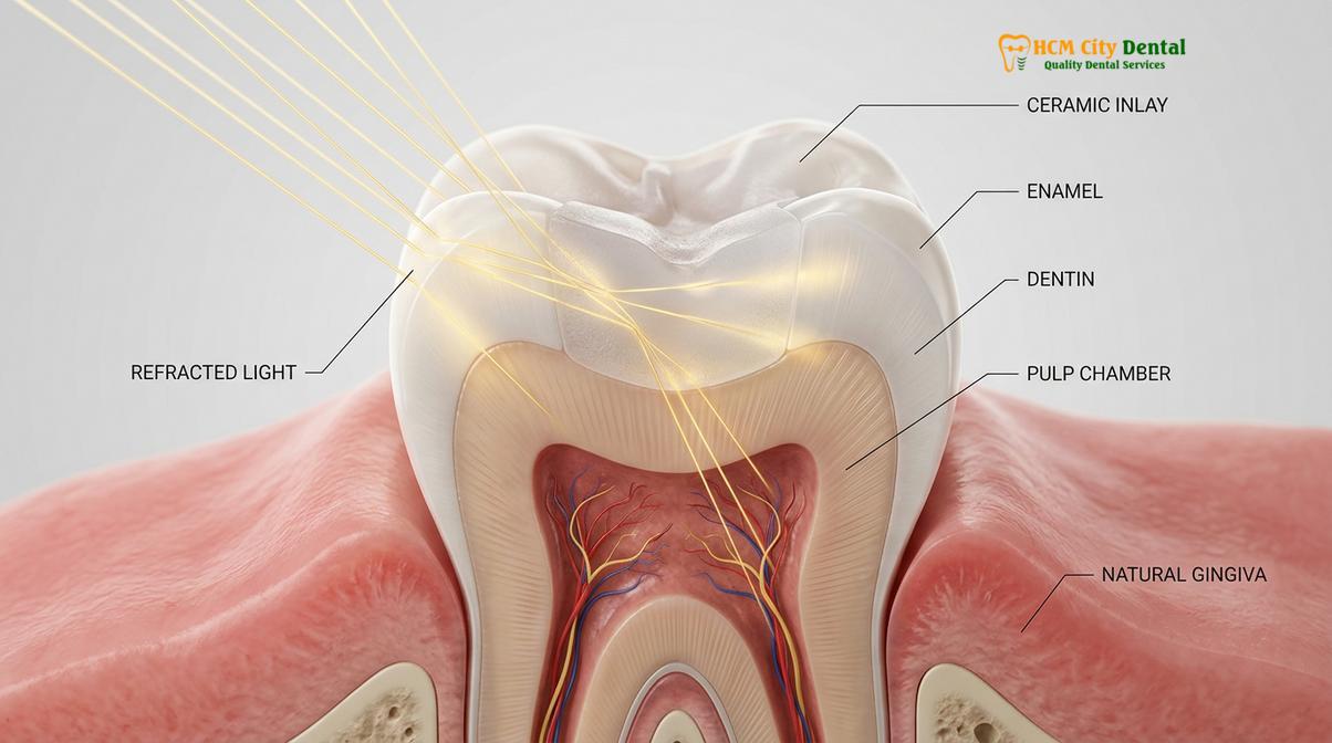
The degree of translucency must be carefully prescribed by the dentist based on the depth of the cavity and the color of the underlying dentin. If the underlying tooth structure is heavily discolored (perhaps from a previous root canal or amalgam staining), a highly translucent ceramic would allow that darkness to show through, resulting in a grayish appearance. In such cases, the dentist will select a lower translucency or medium opacity block to mask the discoloration while still maintaining surface vitality. Understanding this delicate balance of optical refraction is what separates a standard filling from a truly biomimetic restoration.
Custom Staining in the Dental Laboratory
Master ceramists apply microscopic layers of specialized tints to the milled ceramic to replicate natural anatomical characterizations like fluorosis spots or developmental grooves.
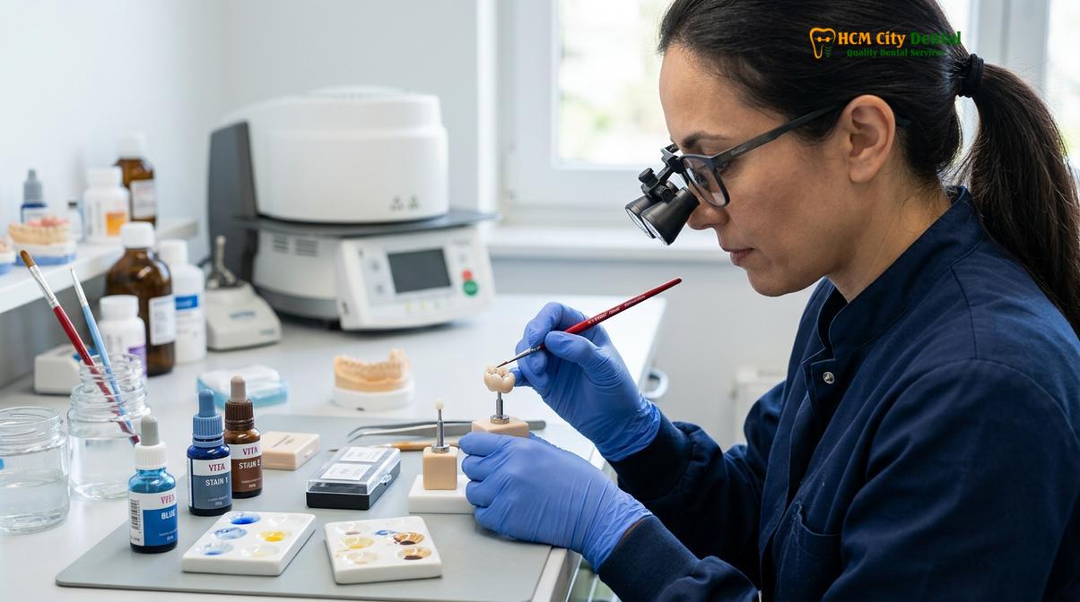
While CAD/CAM technology can mill a ceramic block to microscopic precision, a freshly milled inlay is essentially monochromatic. To transform this uniform piece of ceramic into a lifelike tooth replica, it must undergo custom staining in the dental laboratory. This process requires the artistic eye of a master ceramist who interprets the digital photographs and shade maps provided by the dentist.
Through a direct lab partnership and CAD/CAM workflow, clinics ensure an exact marginal fit to prevent secondary decay, while the ceramist focuses on aesthetics. Using fine brushes, the technician applies liquid metallic oxide stains to the surface of the milled restoration. They may add a touch of ochre to the central fissure to mimic natural occlusal staining, a hint of blue to the marginal ridges to simulate enamel opalescence, or subtle white striations to replicate hypocalcification spots. Once the custom staining is complete, the restoration is coated with a clear glaze and placed into a high-temperature ceramic furnace.
| Material Type | Primary Indication | Translucency Level | Flexural Strength | Color Matching Capability |
|---|---|---|---|---|
| Lithium Disilicate | Premolars, Anterior Inlays, High Aesthetic Zones | High to Very High | ~400 – 500 MPa | Excellent (Strong Chameleon Effect) |
| High-Translucency Zirconia | Molars, Heavy Bruxism, Large Onlays | Medium to High | ~900 – 1200 MPa | Very Good (Requires expert layering) |
| Composite Resin (Direct) | Small Cavities, Temporary Repairs | Variable | ~100 – 150 MPa | Good (Prone to staining over time) |
During the firing process, the stains fuse permanently into the glass matrix, and the ceramic crystallizes, reaching its final strength. For patients with heavy posterior biting forces, dentists may opt for high-translucency zirconia. While older generations of zirconia were notoriously opaque and chalky, modern high-translucency zirconia allows for excellent color matching while providing unparalleled fracture resistance. The laboratory technician will carefully infiltrate the pre-sintered zirconia with coloring liquids before firing, ensuring the shade is built into the core of the material rather than just painted on the surface.
Preventing the ‘Fake Tooth’ Look
Achieving a natural appearance depends heavily on precise cavity preparation, margin design, and the correct application of translucent bonding cements.
Even with a perfectly fabricated aesthetic ceramic inlay blend, the final result can be compromised if the clinical execution during the delivery appointment is flawed. Preventing the “fake tooth” look requires meticulous attention to the margin sealing and the bonding protocol. The preparation of the tooth must feature smooth, flowing cavity outline forms and precise chamfer margins. Sharp internal angles can create stress concentrations in the ceramic, while poorly defined margins can lead to a visible transition line between the tooth and the restoration[4].
The cementation process is arguably the most critical step in ensuring the color match porcelain inlay remains invisible. Unlike traditional crowns that can be cemented with opaque luting agents, translucent ceramic inlays require resin-adhesive cementation. The dentist must select a resin cement shade that complements the ceramic. If the cement is too white or opaque, it will create a visible “halo” or white line at the margin. Conversely, if the cement is too dark, it will lower the overall value of the restoration, making it look gray.
“The ultimate success of an aesthetic restoration lies not just in the material, but in the meticulous isolation and bonding protocol that prevents microleakage and ensures seamless optical integration.”
The enamel bonding protocols involve isolating the tooth (often with a rubber dam) to prevent moisture contamination. The internal surface of the ceramic is etched with hydrofluoric acid and treated with a silane coupling agent to create a chemical bond. Simultaneously, the natural tooth structure is etched and primed. When the resin cement is applied and light-cured, it creates a monolithic, watertight seal. This biocompatibility is a major advantage; the metal-free ceramic prevents gum line irritation and respects the body’s natural tissues, aligning with the latest standards from the Vietnam Odonto-Stomatology Association (VOSA)[6].
Clinical Cases of Seamless Transitions
Real-world clinical applications demonstrate how conservative ceramic restorations can completely rejuvenate a damaged tooth while remaining visually undetectable.
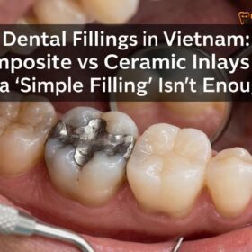
The true test of color matching dental inlays is seen in daily clinical practice, particularly when replacing old, failing restorations. Consider a patient presenting with a large, fractured amalgam (silver) filling that has caused the surrounding enamel to turn gray. The clinical objective is not only to remove the decay and the metal but to restore the tooth’s original vitality and strength without resorting to an aggressive full crown.
Clinical Case Review: Amalgam Replacement
A patient visiting HCMC Dental Clinic in Ho Chi Minh City presented with a failing, leaking silver filling on a lower first molar, causing thermal sensitivity. The clinical team utilized an advanced intraoral scanner to capture the preparation digitally. A custom-milled ceramic inlay was fabricated with a medium translucency block and custom surface staining to match the adjacent premolars. Following strict resin-adhesive cementation, the final restoration was completely indistinguishable from the natural tooth structure, resolving the sensitivity and restoring full occlusal function.
Dr. Nguyen Van Cuong is a leading restorative specialist who oversees the digital workflow and aesthetic protocols at the clinic. With extensive experience in biomimetic dentistry, Dr. Cuong ensures that every ceramic restoration is meticulously color-matched to the patient’s natural dentition. His approach combines advanced intraoral scanning with artistic laboratory communication to deliver highly functional and visually undetectable results.
Beyond aesthetics, these restorations play a crucial role in restoring the vertical dimension of occlusion for bruxism patients who have worn down their natural teeth. By carefully designing the occlusal anatomy of the onlays, dentists can rebuild the patient’s bite alignment. Patients also enjoy complete peace of mind with comprehensive post-treatment support, ensuring that their investment in custom-milled inlays and onlays is protected long after they leave the clinic.
When to See a Doctor for Restorative Evaluation
Prompt clinical evaluation is necessary if you experience thermal sensitivity, notice visible cracks, or suspect a failing existing restoration.
While high-quality ceramic restorations are designed for longevity, natural teeth are still susceptible to new decay, and older traditional fillings inevitably degrade over time. It is highly recommended to schedule a professional dental evaluation if you notice any sharp pain when biting down, lingering sensitivity to hot or cold beverages, or visible dark margins around an old filling. These symptoms often indicate microleakage, where bacteria have penetrated beneath the existing restoration, risking further damage to the underlying dentin and dental pulp.
Additionally, if you are unhappy with the aesthetic appearance of large, discolored composite resins or dark silver amalgams, a consultation can determine if you are a candidate for a conservative ceramic inlay or onlay. Early intervention allows the dentist to preserve the maximum amount of healthy enamel, ensuring a stronger, more aesthetic final result. Regular biannual checkups and professional cleanings are also essential to monitor the integrity of all dental restorations and maintain optimal oral health.

Frequently Asked Questions
How do dentists color-match inlays?
Dentists color-match inlays by using standardized shade guides, digital intraoral scanners, and high-resolution clinical photography. These tools capture the exact hue, chroma, and value of the adjacent natural teeth, allowing the dental laboratory to formulate a precise ceramic blend that seamlessly integrates with the patient’s unique smile.
Will a ceramic inlay stain over time?
High-quality ceramic inlays are highly resistant to staining because their non-porous, glazed surface helps prevent pigment absorption. Unlike composite resin fillings that may discolor from coffee, tea, or tobacco, lab-milled porcelain and lithium disilicate restorations generally maintain their original custom-matched shade for the lifespan of the restoration.
What is the chameleon effect in dentistry?
The chameleon effect in dentistry refers to the optical ability of translucent restorative materials to draw in and reflect the color of surrounding natural tooth structure. This light-scattering property helps the margins of a ceramic inlay blend smoothly into the adjacent enamel, masking the transition line.
Can you whiten an inlay after placement?
No, you cannot whiten a ceramic inlay after it has been permanently placed and bonded. Dental bleaching agents only affect natural tooth enamel; therefore, dentists highly recommend completing any desired teeth whitening treatments before the final shade selection and fabrication of your custom inlay.
How long does it take to custom stain a dental inlay?
Custom staining a dental inlay typically takes a master ceramist several hours of meticulous layering and furnace firing. While the CAD/CAM milling process is rapid, the artistic application of microscopic tints and the final glazing cycle require precise temperature control to achieve lifelike optical properties.
References
- Journal of Esthetic and Restorative Dentistry. Optical properties and translucency of lithium disilicate ceramics.
- International Journal of Prosthodontics. Clinical evaluation of color match in CAD/CAM ceramic restorations.
- Journal of Dentistry. The chameleon effect of highly translucent restorative materials.
- American Dental Association. Clinical guidelines for conservative indirect restorations.
- Journal of Prosthetic Dentistry. Accuracy of digital intraoral scanners in shade determination.
- Vietnam Odonto-Stomatology Association (VOSA). Guidelines on aesthetic restorative materials and clinical protocols.
Achieving a flawless, natural-looking smile requires precision, advanced materials, and an expert clinical eye. If you are looking for expertly crafted restorations, contact HCMC Dental Clinic in Ho Chi Minh City today. Our dedicated team utilizes state-of-the-art digital shade matching and custom laboratory staining to ensure your Inlays & Onlays blend seamlessly with your natural teeth.