For many international patients suffering from terminal dentition, the journey to restoring a functional, confident smile is fraught with clinical complexities and prohibitive costs. This comprehensive full mouth rehabilitation case study vietnam details the transformative journey of a 62-year-old American veteran who traveled to HCMC Dental Clinic in Ho Chi Minh City to reclaim his oral health. Facing severe bone loss, failing conventional bridges, and the inability to chew solid foods, the patient required a sophisticated, predictable surgical intervention.
Through advanced digital dentistry, 3D virtual surgical planning, and the proven All-on-4 treatment protocol, the clinical team successfully reconstructed both his upper and lower arches. This clinical review explores the biomechanical principles, surgical execution, and restorative phases involved in delivering a world-class, fixed implant-supported prosthesis, demonstrating why Vietnam has become a premier destination for complex dental reconstructions.
Clinical Summary:
This case study examines the dual-arch All-on-4 rehabilitation of a 62-year-old male patient presenting with generalized severe chronic periodontitis, terminal dentition, and significant posterior bone atrophy. Utilizing a fully digital workflow, the surgical team achieved high primary implant stability, allowing for the immediate loading of fixed provisional bridges. Following a dedicated osseointegration period, the patient was successfully restored with definitive CAD/CAM titanium-zirconia hybrid prostheses, resulting in the complete restoration of masticatory function, occlusal vertical dimension (OVD), and facial aesthetics.
Key Takeaways:
- Patient Profile: 62-year-old American veteran with terminal dentition and severe posterior bone resorption.
- Surgical Protocol: Dual-arch All-on-4 utilizing tilted posterior implants to avoid anatomical structures and maximize anterior bone utilization.
- Immediate Loading: Fixed acrylic provisional bridges delivered rapidly, supported by high primary stability.
- Final Prosthesis: Precision-milled monolithic zirconia over a rigid titanium framework for maximum durability and aesthetic vitality.
- Clinical Outcome: Restoration of natural bite force, improved nutritional intake, and significant cost savings compared to US dental estimates.
Clinical Assessment: Terminal Dentition, Bone Resorption & Chewing Inability
The foundation of any successful full-arch reconstruction begins with a meticulous clinical and radiographic assessment. When dealing with an american patient dental implant story of this magnitude, understanding the historical progression of the patient’s oral decline is critical for formulating a predictable treatment plan. Modern digital dentistry has significantly improved the survival rates and predictability of these complex interventions, allowing for highly accurate preoperative evaluations [1].
Initial Presentation and Chief Complaints
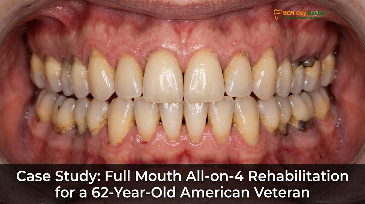
The patient presented to HCMC Dental Clinic in Ho Chi Minh City with a chief complaint of chronic dental pain, mobile teeth, and an inability to masticate properly. His dental history revealed decades of piecemeal dentistry, including multiple failing root canals, recurrent decay under old porcelain-fused-to-metal (PFM) bridges, and advanced, untreated periodontal disease.
Clinically, the patient exhibited severe mobility in his remaining anterior teeth. His posterior segments were entirely edentulous, having lost his molars over a decade prior. This prolonged absence of posterior teeth led to severe posterior bite collapse, a loss of occlusal vertical dimension (OVD), and a resulting “sunken” facial appearance. The psychological toll was equally profound; he reported severe social anxiety and a diet restricted entirely to soft, processed foods, which was negatively impacting his systemic health and gastrointestinal function.
Radiographic Findings and Bone Density Analysis
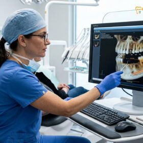
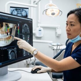
A comprehensive diagnostic workup was initiated, beginning with a high-resolution 3D Cone Beam Computed Tomography (CBCT) scan. The radiographic analysis revealed a challenging anatomical landscape:
* Maxilla (Upper Jaw): Severe pneumatization (expansion) of the maxillary sinuses bilaterally, leaving minimal residual alveolar bone height in the posterior regions. The anterior maxilla showed moderate horizontal bone loss but retained adequate density for implant anchorage.
* Mandible (Lower Jaw): Significant vertical resorption in the posterior mandible, with the inferior alveolar nerve canal positioned dangerously close to the alveolar crest. The anterior symphysis region, however, presented dense, high-quality cortical bone.
In cases of such severe atrophy, traditional implant approaches would require extensive, multi-stage bone grafting procedures. For the maxilla, this would involve bilateral open sinus floor elevations packed with particulate bone grafts, followed by a prolonged healing period before implants could even be placed. However, the All-on-4 concept was specifically developed to bypass these extensive grafting procedures by tilting the posterior implants, anchoring them into the dense anterior bone while avoiding the sinus cavities and mandibular nerves [2].
To understand the financial implications of such complex procedures, many international patients consult our comprehensive guide on understanding dental implant costs to ensure transparency before traveling.
3D CBCT Virtual Surgical Planning & Flapless Implant Placement
The success of a fixed bridge on implants case study relies heavily on the precision of the surgical execution. At HCMC Dental Clinic, the transition from terminal dentition to a fixed implant bridge is orchestrated entirely through a digital workflow, ensuring maximum safety and predictability.
Digital Workflow and Surgical Guide Fabrication
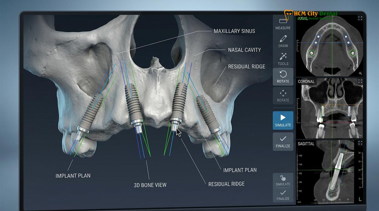
The surgical team utilized a dual-scan protocol. The patient’s existing dentition and soft tissues were captured using an intraoral scanner, creating a highly accurate digital file. This surface data was then superimposed over the DICOM data generated by the CBCT scan.
Using specialized implant planning software, the clinical team virtually performed the extractions, leveled the bone (alveoloplasty) to create a flat restorative platform, and positioned the four implants in each arch.
* Anterior Implants: Placed axially (straight) in the anterior region.
* Posterior Implants: Tilted distally to increase the antero-posterior (A-P) spread, which is crucial for minimizing cantilever length on the final prosthesis and distributing occlusal forces evenly across the arch.
Once the virtual plan was finalized, custom surgical guides were 3D printed. These guides allow for a minimally invasive surgical approach, significantly reducing post-operative swelling, pain, and healing time.
The Surgical Phase: Achieving High Primary Stability
On the day of surgery, the patient was placed under conscious sedation for maximum comfort. The remaining failing teeth were atraumatically extracted. Thorough surgical debridement was performed to remove all granulation tissue and bacterial load from the periodontal infections.
The surgical guides were pinned into place, and the osteotomies (bone preparations) were performed with precise depth and angulation control. Premium implants were utilized due to their aggressive thread design, which is engineered to compress bone during insertion, thereby maximizing primary stability.
“The paradigm of implant dentistry has shifted dramatically. With 3D virtual planning and tilted implant protocols, we can now offer patients who were previously told they didn’t have enough bone a fixed, functional smile in a single surgical visit, bypassing months of invasive bone grafting.”
For an immediate loading protocol to be viable, the implants must achieve a high degree of mechanical fixation in the bone. This is measured using resonance frequency analysis. High insertion torque and stability quotients are generally required to safely attach a provisional bridge on the same day. In this case, all eight implants achieved excellent primary stability.
Immediate Fixed Provisional Teeth Delivered Within 24 Hours
One of the most profound psychological benefits of the All-on-4 protocol is the immediate transformation. Unlike traditional two-stage implant surgery, which leaves the patient wearing a removable, often uncomfortable healing denture for months, the immediate loading protocol provides fixed, non-removable teeth almost instantly.
The Science of Immediate Loading
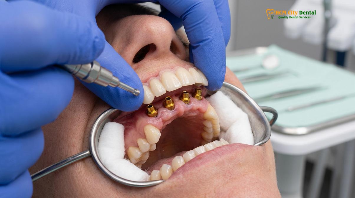
Following implant placement, straight and angled Multi-Unit Abutments (MUAs) were attached to the implants. These abutments serve to correct the angulation of the tilted posterior implants, bringing the restorative platforms parallel to one another.
A pre-milled, high-density acrylic provisional bridge was then retrofitted to the abutments. Temporary titanium cylinders were picked up inside the acrylic bridge using a strong dual-cure resin. This process transforms a standard denture into a fixed, screw-retained provisional bridge.
The biological success of immediate loading relies on the principle of “cross-arch stabilization.” By rigidly splinting the four implants together with the provisional bridge, any micromotion is kept below the critical threshold. This rigid splinting prevents fibrous encapsulation and promotes true osseointegration. While some patients with healthy natural teeth might only need cosmetic enhancements, which are highly durable [3], terminal dentition requires this robust structural replacement. Furthermore, strict adherence to international safety protocols ensures that materials used in these provisional and final stages meet the highest global standards [4].
Restoring the Occlusal Vertical Dimension (OVD)
Beyond mere function, the provisional phase is critical for aesthetic and functional prototyping. The patient’s severe posterior bite collapse had resulted in a loss of OVD. The provisional bridge was designed to open his bite back to its natural, youthful dimension. This not only improved his facial profile—smoothing out deep nasolabial folds and supporting his lips—but also relieved stress on his temporomandibular joints (TMJ).
The provisional phase serves as a “test drive.” Over the subsequent healing months, the patient evaluates the aesthetics, speech phonetics, and chewing function of the temporary bridge. Any necessary adjustments are noted and seamlessly incorporated into the design of the final zirconia prosthesis.
For patients considering different full-arch configurations, exploring an All-on-6 cost guide can provide insight into when six implants might be recommended over four, depending on bone density and bite force requirements.
Healing Phase & Final Zirconia-Titanium Hybrid Bridge Delivery
After a dedicated healing period, the patient returned to HCMC Dental Clinic for the final restorative phase. Radiographic evaluation confirmed that all implants had successfully osseointegrated, achieving biological stability. The soft tissues (gums) had also matured and stabilized around the abutments. Throughout this process, the clinic maintained rigorous infection control standards in alignment with national health guidelines set forth by the Vietnam Ministry of Health [5] and advanced aerosol management protocols [6].
Osseointegration and Soft Tissue Maturation
The transition from the provisional to the final prosthesis is a meticulous process. New, highly accurate impressions were taken to capture the exact 3D position of the healed implants.
Engineering the Final Prosthesis
The final prosthesis chosen for this severe bone loss implant success saigon case was a premium Zirconia-Titanium hybrid bridge. This represents the pinnacle of modern dental engineering:
1. Titanium Framework: A custom CAD/CAM milled titanium bar forms the hidden skeleton of the bridge. This provides absolute rigidity, preventing the zirconia from flexing and fracturing under heavy occlusal loads.
2. Monolithic Zirconia: The teeth and gingival base are milled from a solid block of high-translucency monolithic zirconia. Zirconia is exceptionally strong, highly resistant to chipping, and completely impervious to staining or plaque accumulation.
3. Pink Porcelain Layering: To achieve a lifelike appearance, master ceramists hand-layer pink porcelain over the gingival areas of the zirconia, perfectly mimicking the natural stippling, color variations, and vascularity of healthy gum tissue.
“The integration of a milled titanium framework with monolithic zirconia offers an unparalleled combination of structural integrity and aesthetic vitality, ensuring the patient’s new smile is built to withstand the test of time.”
Patients often wonder about the timeline for such comprehensive care. Reviewing how long the dental implant procedure takes can help set realistic expectations for the healing and restorative phases.
Clinical Outcome: Masticatory Efficiency, Nutrition & Facial Support
The culmination of this full mouth rehabilitation yielded life-changing results for the patient. By replacing failing dentition with a rigid, implant-supported framework, the clinical team successfully restored the patient’s ability to chew a normal, healthy diet.
Patient Outcome Metrics
Pre-Treatment: Restricted to a soft food diet, severe pain upon chewing, collapsed facial profile, and high social anxiety due to aesthetic concerns.
Post-Treatment: Full restoration of masticatory function allowing for a diverse diet including fibrous vegetables and proteins. The restored occlusal vertical dimension provided immediate facial support, reducing the appearance of premature aging. The patient reported a complete elimination of dental pain and a profound improvement in self-confidence.
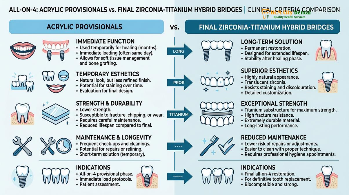
To better understand the transition from the healing phase to the final outcome, the following table outlines the key differences between the provisional and definitive prostheses used in this case:
| Feature | Provisional Bridge (Healing Phase) | Final Hybrid Bridge (Definitive Phase) |
|---|---|---|
| Material | High-density PMMA Acrylic | Monolithic Zirconia over Titanium Bar |
| Durability | Moderate (designed for short-term use) | Extremely High (highly resistant to fracture) |
| Aesthetics | Good, but prone to staining over time | Excellent, lifelike translucency, stain-resistant |
| Function | Soft-solid diet only | Full masticatory function restored |
For those navigating the financial aspects of such care, understanding independent dental insurance options can be a valuable step in the planning process. Additionally, reviewing the pros and cons of dental implants can help patients make informed decisions about their long-term oral health investments.
When to Seek Immediate Dental Evaluation
Recognizing the signs of terminal dentition early can provide patients with more treatment options and potentially preserve valuable jawbone. It is highly recommended to seek a comprehensive dental evaluation if you experience any of the following symptoms:
* Severe Tooth Mobility: Teeth that feel loose or shift during chewing are often a sign of advanced periodontal disease and loss of supporting bone.
* Chronic Dental Pain or Infections: Recurrent abscesses, persistent bad breath, or constant pain indicate deep-seated infections that can affect systemic health.
* Inability to Chew Solid Foods: If your diet has become restricted to soft foods due to missing teeth or pain, your nutritional intake and gastrointestinal health may be compromised.
* Failing Dental Work: Old bridges that are loose, or multiple teeth that have broken off at the gum line, require professional assessment to prevent further complications.
* Changes in Facial Structure: A sunken appearance around the mouth or deepening wrinkles can indicate severe bone loss following the extraction of multiple teeth.
Important Clinical Note: Delaying treatment for severe periodontal disease or failing dentition can lead to progressive bone loss. Once the alveolar bone resorbs beyond a certain point, even advanced protocols like All-on-4 may require supplementary bone grafting, increasing both the complexity and the healing time of the procedure.
Frequently Asked Questions
How severe was the patient’s bone loss prior to All-on-4 surgery?
The patient presented with severe posterior maxillary and mandibular bone resorption, leaving less than 4mm of vertical bone height in the molar regions. This necessitated the All-on-4 protocol, which utilizes tilted posterior implants to bypass the sinus cavities and mandibular nerves, maximizing the use of available dense anterior bone without requiring extensive grafting.
Was the patient able to eat solid foods with immediate provisional teeth?
Yes, but with strict modifications. The immediate provisional bridge is fixed and functional, allowing the patient to eat a soft-solid diet immediately. However, hard, crunchy, or chewy foods must be strictly avoided during the initial healing phase to prevent micromotion and support successful osseointegration of the implants.
What implant brand and bridge materials were used for this case?
This case utilized premium implants recognized for their high primary stability and success in immediate loading protocols. The final prosthesis was a state-of-the-art hybrid bridge featuring a precision-milled CAD/CAM titanium framework for structural integrity, overlaid with monolithic zirconia teeth and custom-layered pink porcelain for optimal gingival aesthetics.
How much did the American veteran save compared to US dental estimates?
The patient saved a significant percentage on the total cost of treatment. While a dual-arch All-on-4 rehabilitation with premium zirconia in the United States typically carries a high premium, the comprehensive treatment in Vietnam—including all diagnostics, surgery, provisional bridges, and final zirconia prostheses—was completed at a highly competitive rate without compromising on material quality.
What maintenance protocol keeps full mouth implants healthy long-term?
Long-term success requires a meticulous daily hygiene routine, including the use of a water flosser, super floss, and non-abrasive gel toothpaste to clean beneath the bridge. Additionally, patients are often advised to wear a custom occlusal night guard to protect the zirconia from bruxism, and attend professional maintenance visits regularly for radiographic evaluation.
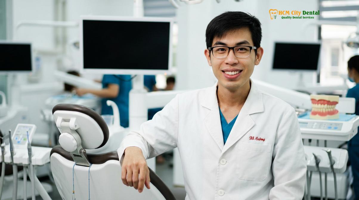
Dr. Nguyen Van Cuong is a leading specialist in complex oral rehabilitations at HCMC Dental Clinic. With extensive training in digital implantology, Dr. Cuong focuses on restoring severe atrophy cases using advanced surgical protocols. His commitment to precision ensures that international patients receive world-class care tailored to their unique anatomical needs. Patients seeking the highest standard of care often consult our guide on the best implant dentist in Ho Chi Minh City to understand the rigorous credentials required for such transformative surgeries.
If you are considering a similar transformation, contact HCMC Dental Clinic in Ho Chi Minh City to explore your options. Learn more about our comprehensive care at our Comprehensive Dental Care at HCMC Dental Clinic hub.
References
- Journal of the American Dental Association. Implant survival rates in digital dentistry. (2022).
- International Journal of Oral and Maxillofacial Implants. All-on-4 full arch rehabilitation. (2021).
- Journal of Esthetic and Restorative Dentistry. Minimal prep porcelain veneer longevity. (2020).
- British Dental Journal. Dental tourism safety protocols and material traceability. (2023).
- Vietnam Ministry of Health (MOH). National guidelines on infection control in dental facilities. (2021).
- Journal of Clinical Periodontology. Infection control and aerosol management. (2021).