For international patients and dental travelers seeking advanced oral implantology, the integration of biological tissue engineering has revolutionized the predictability, safety, and healing speed of complex surgical procedures. At the forefront of this regenerative paradigm is the use of a sticky bone L-PRF platelet rich fibrin dental bone graft. By harnessing the innate regenerative capacity of the patient’s own blood, modern implant dentistry has moved beyond traditional synthetic barriers and isolated bone substitutes. Today, the gold standard for alveolar ridge augmentation, sinus lifting, and socket preservation involves the strategic application of Leukocyte-Platelet Rich Fibrin (L-PRF), Advanced PRF (A-PRF), and injectable PRF (i-PRF) to create a bioactive, moldable “Sticky Bone” matrix.
At HCMC Dental Clinic, located in the heart of District 1, Ho Chi Minh City, our clinical protocols are strictly aligned with global evidence-based standards. Under the clinical direction of Dr. Nguyen Van Cuong, DDS, our facility employs hospital-grade surgical techniques, including 0.075mm high-resolution 3D CBCT diagnostics, Piezosurgery, and the Choukroun low-speed centrifugation concept. This 100% autologous approach eliminates the need for chemical anticoagulants or bovine thrombin, ensuring maximum biocompatibility and a significantly reduced risk of post-operative complications. Whether you require a single implant in the aesthetic zone or a full-arch rehabilitation, the biological bone grafting protocols utilized at our clinic provide the optimal foundation for long-term osseointegration and soft tissue aesthetics.
Clinical Summary:
The 2026 biological bone grafting protocol at HCMC Dental Clinic utilizes 100% autologous Platelet-Rich Fibrin (L-PRF, A-PRF, i-PRF) derived from the patient’s own blood via chairside low-speed centrifugation. By mixing liquid i-PRF with particulate xenograft or autogenous bone, surgeons create “Sticky Bone”—a cohesive, moldable graft that stabilizes bone volume without the need for titanium mesh. This technique ensures a sustained 14-day release of critical growth factors (PDGF, TGF-beta1, VEGF), accelerating angiogenesis, minimizing post-operative pain, and optimizing the integration of dental implants in complex cases such as sinus lifts and severe alveolar ridge defects.
Key Takeaways:
- 100% Autologous Biomaterial: L-PRF and Sticky Bone are created entirely from the patient’s own blood, eliminating the risk of allergic reactions, disease transmission, or foreign body rejection associated with synthetic chemical additives.
- Enhanced Graft Stability: The fibrin network in Sticky Bone binds particulate grafts together, preventing micro-movement and graft migration, which is critical for successful guided bone regeneration (GBR).
- Accelerated Healing: The dense fibrin matrix provides a sustained 14-day release of growth factors, promoting rapid new blood vessel formation (angiogenesis) and superior soft tissue closure.
- Advanced Surgical Precision: Procedures are guided by 0.075mm 3D CBCT imaging and executed using 24–36 kHz selective Piezosurgery to protect delicate anatomical structures like the Schneiderian membrane and inferior alveolar nerve.
- Transparent Expat Pricing: HCMC Dental Clinic offers a highly competitive -40% Online Booking Fee Schedule, making world-class biological bone grafting and implantology accessible to international patients traveling to Vietnam.
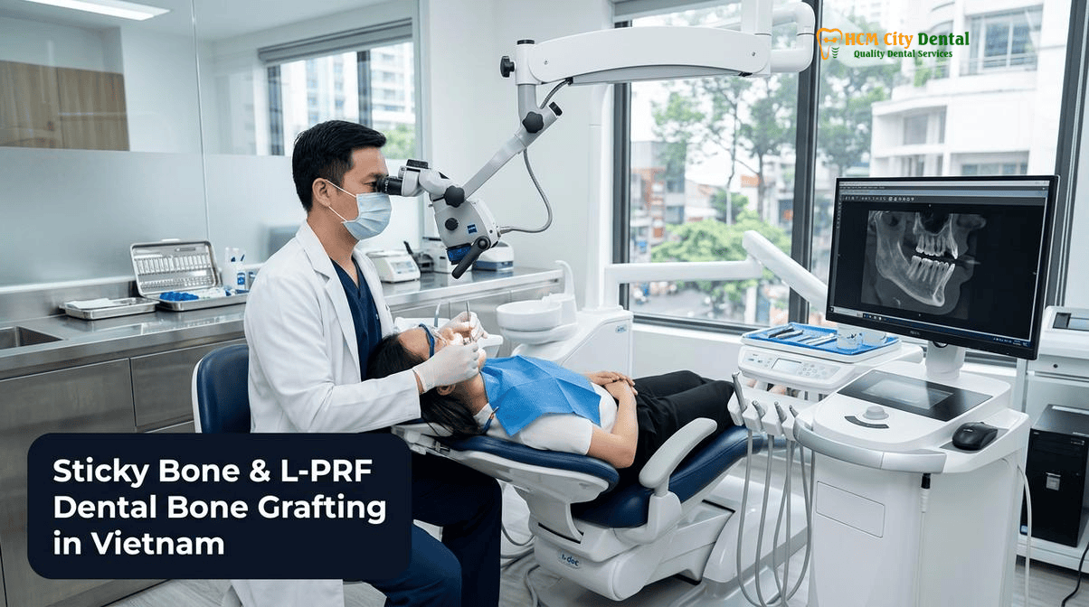
What Are L-PRF, A-PRF, and i-PRF? The Science of 100% Autologous Platelet-Rich Fibrin in Implantology
The evolution of platelet concentrates in oral and maxillofacial surgery represents a major leap forward in regenerative medicine. Early generations, such as Platelet-Rich Plasma (PRP), required the use of chemical anticoagulants (like bovine thrombin or calcium chloride) to prevent the blood from clotting during preparation. While PRP provided a rapid burst of growth factors, its clinical efficacy was limited by a short duration of action and the potential inhibitory effects of the anticoagulants on tissue healing. In contrast, the second generation of platelet concentrates—specifically Platelet-Rich Fibrin (PRF)—was developed by Dr. Joseph Choukroun to completely eliminate these chemical additives, relying instead on the natural coagulation cascade.
Leukocyte-Platelet Rich Fibrin (L-PRF) is produced by drawing a small volume of the patient’s venous blood into sterile, silica-free glass or plastic tubes and immediately spinning it in a specialized centrifuge at a specific G-force and RPM. Because no anticoagulants are used, the blood naturally separates into three distinct layers: red blood cells at the bottom, acellular plasma at the top, and a dense, resilient fibrin clot in the middle. This L-PRF clot contains a high concentration of platelets, leukocytes (white blood cells), and circulating stem cells, all suspended within a complex three-dimensional fibrin matrix. When compressed, this clot forms a strong, suturable membrane that can be used to protect surgical sites, seal extraction sockets, and guide soft tissue healing [1].
Further refinements to the centrifugation protocol have led to the development of Advanced PRF (A-PRF) and injectable PRF (i-PRF). A-PRF utilizes a lower centrifugation speed and longer duration, which alters the density of the fibrin network and allows for an even higher retention of leukocytes and cytokines. This makes A-PRF particularly effective for soft tissue regeneration and infection control. Conversely, i-PRF is produced using an ultra-low speed and short duration protocol, resulting in a liquid platelet concentrate that has not yet polymerized into a solid clot. This liquid i-PRF is rich in fibrinogen, platelets, and mesenchymal stem cells, and it serves as the critical binding agent used to create Sticky Bone. By integrating these advanced autologous biomaterials, patients undergoing 3D-Guided Dental Implants & Specialist Oral Surgery in Vietnam benefit from a highly predictable, biologically active healing environment.
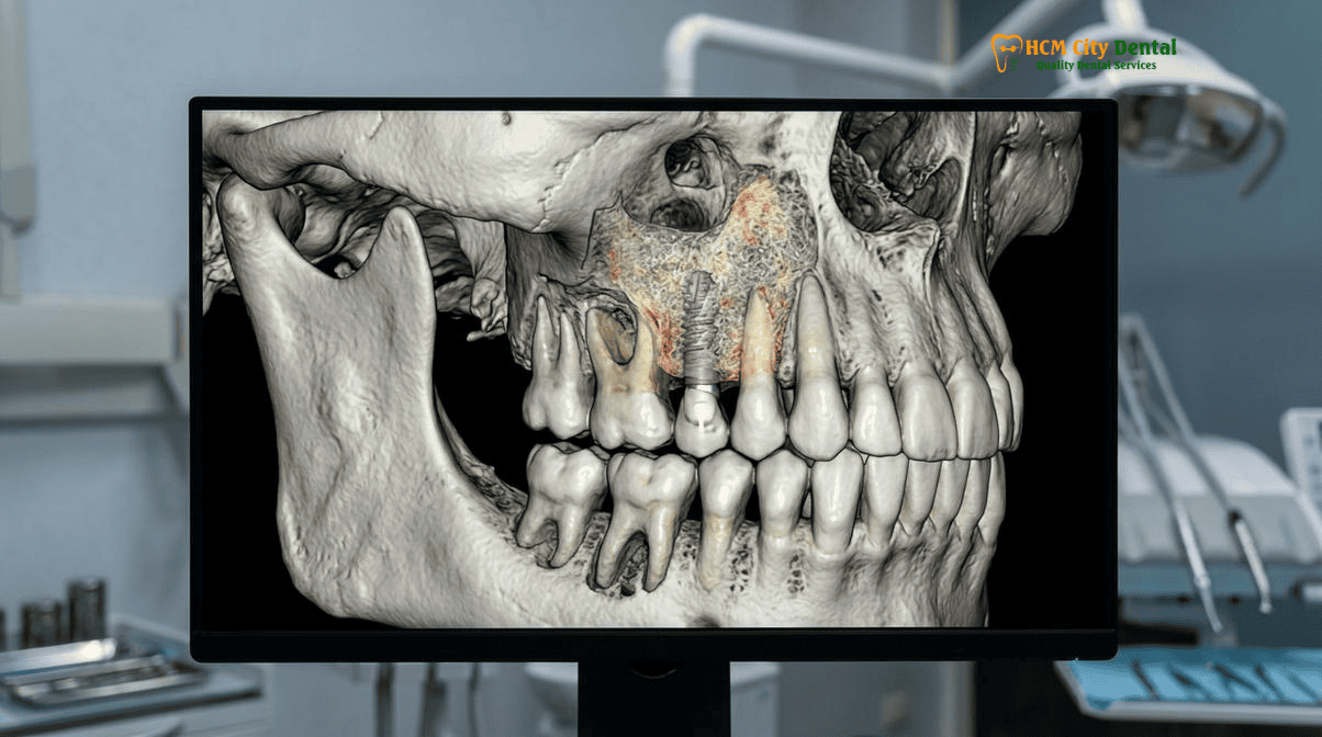
How ‘Sticky Bone’ (i-PRF + Xenograft/Autogenous Particulate) Stabilizes Bone Grafts Without Titanium Mesh
One of the most significant challenges in alveolar ridge augmentation and guided bone regeneration (GBR) is maintaining the stability and volume of the particulate bone graft during the critical early phases of healing. If the graft particles shift or migrate due to micro-movement from overlying soft tissues, the regenerative process is compromised, leading to fibrous encapsulation rather than true bone formation. Historically, surgeons relied on rigid titanium meshes or non-resorbable PTFE membranes secured with fixation pins to contain the graft—a technique known as the Urban Sausage GBR method. While effective, these techniques are highly invasive, increase the risk of soft tissue dehiscence (membrane exposure), and require a secondary surgery for removal.
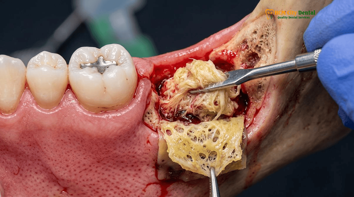
The advent of Sticky Bone has fundamentally transformed this paradigm. Sticky Bone is created chairside by mixing the patient’s liquid i-PRF with a particulate bone substitute—typically a highly porous, osteoconductive xenograft (such as Geistlich Bio-Oss) or autogenous cortical bone chips harvested from the patient’s own jaw. Within minutes of mixing, the fibrinogen in the i-PRF cleaves into fibrin polymers, creating a dense, interconnected 3D fibrin network that physically entraps the bone particles. The result is a cohesive, moldable, yellow-tinted biological block that resembles the consistency of a gummy bear.
“The biomechanical advantage of Sticky Bone cannot be overstated. By embedding the particulate graft within an autologous fibrin matrix, we achieve exceptional graft stability and space maintenance without the morbidity associated with titanium mesh or fixation pins. The graft stays exactly where we place it, and the fibrin network acts as a biological highway for cellular migration and rapid revascularization.”
— Dr. Nguyen Van Cuong, DDS (Clinical Director, HCMC Dental Clinic)
This biological stabilization is particularly advantageous when compared to traditional techniques. For instance, while the Khoury autogenous cortical bone shell grafting technique remains a gold standard for severe vertical and horizontal defects, it requires harvesting a block of bone from the mandibular ramus, splitting it into thin plates, and securing it with osteosynthesis screws. By utilizing Sticky Bone in conjunction with L-PRF membranes, surgeons can often achieve comparable volumetric gains in moderate defects with significantly less surgical trauma and donor site morbidity. Furthermore, the fibrin network prevents the ingrowth of fast-proliferating epithelial cells into the graft site, effectively acting as a biological barrier membrane [2]. For patients researching the efficacy of different grafting materials, understanding the role of a Geistlich Bio Oss Bone Graft Vietnam: 2026 Clinical Report is essential for making informed treatment decisions.
Sustained 14-Day Release of Growth Factors (PDGF, TGF-beta1, VEGF) for Rapid Angiogenesis and Soft Tissue Healing
The clinical superiority of L-PRF and Sticky Bone extends far beyond mechanical stabilization; their true power lies in their profound biological activity. The regenerative capacity of any bone graft is heavily dependent on the local availability of signaling molecules that orchestrate cellular proliferation, differentiation, and angiogenesis. In traditional PRP preparations, the sudden activation of platelets by chemical additives causes a rapid, massive degranulation, releasing nearly all stored growth factors within the first few hours. Unfortunately, this burst effect is short-lived, and the growth factors are quickly degraded by tissue enzymes before they can exert a lasting regenerative effect.
In contrast, the dense, trimolecular fibrin network of L-PRF and Sticky Bone acts as a biological reservoir. The platelets and leukocytes are physically trapped within the fibrin meshes, protecting them from rapid degradation. As the fibrin matrix slowly breaks down over a period of 10 to 14 days, it facilitates a sustained, gradual release of critical growth factors directly into the surgical site. This prolonged release profile perfectly aligns with the physiological timeline of wound healing and tissue regeneration [3].
The key growth factors released include:
- Platelet-Derived Growth Factor (PDGF): A potent mitogen that stimulates the proliferation and migration of mesenchymal stem cells, osteoblasts, and fibroblasts to the injury site.
- Transforming Growth Factor-Beta 1 (TGF-β1): Essential for the regulation of inflammation and the stimulation of collagen synthesis, which is critical for robust soft tissue closure and matrix formation.
- Vascular Endothelial Growth Factor (VEGF): The primary driver of angiogenesis. VEGF stimulates the formation of new blood vessels, ensuring that the bone graft receives the oxygen and nutrients necessary for survival and integration.
- Bone Morphogenetic Proteins (BMPs): Proteins that induce the differentiation of mesenchymal stem cells into mature, bone-forming osteoblasts.
This sustained biological signaling is particularly crucial for patients with compromised healing capacity or severe bone atrophy. When combined with advanced microsurgical techniques—such as 6-0 or 7-0 Connective Tissue Graft (CTG) tunneling to ensure the strict ≥2mm keratinized tissue rule around implants—the use of L-PRF significantly enhances soft tissue biotype and aesthetic outcomes. For patients wondering, Can I Get Dental Implants If I Have Bone Loss? | HCMC Dental, the integration of Sticky Bone and sustained growth factor release provides a highly predictable pathway to successful rehabilitation.
Clinical Applications: Socket Preservation, Sinus Augmentation, Periodontal Defects, and MRONJ Prevention
The versatility of the sticky bone L-PRF platelet rich fibrin dental bone graft protocol allows it to be applied across a wide spectrum of oral and maxillofacial surgical procedures. At HCMC Dental Clinic, Dr. Nguyen Van Cuong, DDS, routinely utilizes these autologous biomaterials to enhance outcomes in both routine and highly complex cases.
1. Socket Preservation and Ridge Maintenance
Following a tooth extraction, the alveolar bone naturally undergoes rapid resorption, losing up to 50% of its width within the first six months. To prevent this, socket preservation is performed immediately after extraction. The socket is thoroughly debrided, and Sticky Bone is packed into the defect to maintain the ridge architecture. The graft is then covered with multiple layers of compressed L-PRF membranes, which act as a biological bandage. This not only prevents the loss of the graft material but also significantly reduces post-operative pain and the risk of alveolar osteitis (dry socket). In cases involving high-risk mandibular third molars, a coronectomy (sectioning the tooth 3–4mm below the alveolar crest without mobilizing roots near the mandibular canal) may be performed, with L-PRF used to promote rapid healing over the retained root fragment. For a detailed overview, review our Socket Preservation Bone Graft Guide | HCMC Dental.
2. Lateral Window Sinus Augmentation
Rehabilitating the severely atrophic posterior maxilla often requires a sinus lift procedure to create sufficient vertical bone height for implant placement. This procedure involves elevating the delicate Schneiderian membrane and placing a bone graft in the newly created space. At HCMC Dental Clinic, this is performed using Piezosurgery—a specialized ultrasonic device operating at 24–36 kHz that selectively cuts bone while preserving soft tissue, drastically reducing the risk of membrane perforation. Once the membrane is elevated, Sticky Bone is introduced into the sinus cavity. The cohesive nature of the Sticky Bone prevents the graft particles from migrating or tearing the membrane, while the sustained release of VEGF promotes rapid revascularization of the graft from the surrounding bony walls [4].
3. Periodontal Regeneration and Intrabony Defects
For patients suffering from advanced periodontitis, deep intrabony defects can threaten the survival of natural teeth. Traditional treatments often yield unpredictable results. However, the application of Sticky Bone combined with repeated i-PRF injections has shown remarkable efficacy in regenerating lost periodontal attachment. The L-PRF membranes are adapted over the defect to exclude fast-growing gingival epithelium, allowing the slower-growing periodontal ligament cells and osteoblasts to repopulate the root surface.
4. Prevention of MRONJ (Medication-Related Osteonecrosis of the Jaw)
Patients with a history of bisphosphonate therapy or radiation treatment are at a high risk of developing osteonecrosis following oral surgery. The intense concentration of leukocytes in L-PRF provides a robust local immune defense, while the dense fibrin matrix protects exposed bone from oral pathogens. The application of L-PRF in these high-risk patients has become a critical prophylactic measure to ensure primary wound closure and prevent necrotic complications.
Clinical Case Study: Maxillary Rehabilitation in an Expat Patient
A 58-year-old Australian expatriate presented to HCMC Dental Clinic with severe bone loss in the posterior maxilla following years of edentulism. A 0.075mm 3D CBCT scan revealed less than 3mm of residual vertical bone height below the maxillary sinus. Dr. Nguyen Van Cuong, DDS, performed a lateral window sinus augmentation using Piezosurgery. The defect was grafted using a Sticky Bone matrix composed of Geistlich Bio-Oss and autologous i-PRF, covered with A-PRF membranes. The patient reported zero post-operative swelling and minimal discomfort. After a 6-month consolidation period, the site demonstrated excellent radiopacity and bone density, allowing for the successful placement of two Straumann SLA-Active implants with 17° Multi-Unit Abutments (MUAs) verified via digital photogrammetry.
Chairside Venipuncture & Low-Speed Centrifugation Protocol (Choukroun Concept) at HCMC Dental Clinic
The success of biological bone grafting is entirely dependent on the strict adherence to standardized, scientifically validated preparation protocols. At HCMC Dental Clinic, the production of L-PRF, A-PRF, and Sticky Bone is performed seamlessly chairside, ensuring that the biomaterials are fresh, highly active, and perfectly tailored to the patient’s immediate surgical needs.
The protocol begins with a routine venipuncture, typically performed by a trained clinical specialist while the patient is being anesthetized for the procedure. Depending on the size of the surgical defect, between 2 to 6 vials of blood (approximately 10mL each) are drawn from the patient’s antecubital vein. It is critical that the blood is collected in sterile, chemical-free tubes (red cap for L-PRF/A-PRF, orange/white cap for i-PRF) to ensure the absence of anticoagulants or silica activators.
Once drawn, the tubes are immediately placed into a specialized, calibrated centrifuge. Timing is of the essence; any delay can trigger premature coagulation outside the centrifuge, compromising the formation of the fibrin matrix. The centrifugation parameters are strictly controlled based on the desired output:
- For L-PRF Membranes: The blood is spun at approximately 2700 RPM for 12 minutes. This separates the blood into the red blood cell base, the L-PRF clot in the middle, and the acellular plasma at the top. The clot is carefully extracted, separated from the red blood cells, and placed in a specialized PRF box where it is gently compressed into a uniform membrane of consistent thickness.
- For i-PRF (Sticky Bone): The blood is spun at a much lower speed (e.g., 700 RPM) for just 3 minutes. This ultra-low speed prevents the immediate polymerization of fibrinogen. The yellow, liquid i-PRF is aspirated using a sterile syringe and immediately sprayed over the particulate bone graft. Within 60 to 90 seconds, the liquid coagulates, binding the particles into a solid, moldable Sticky Bone block.
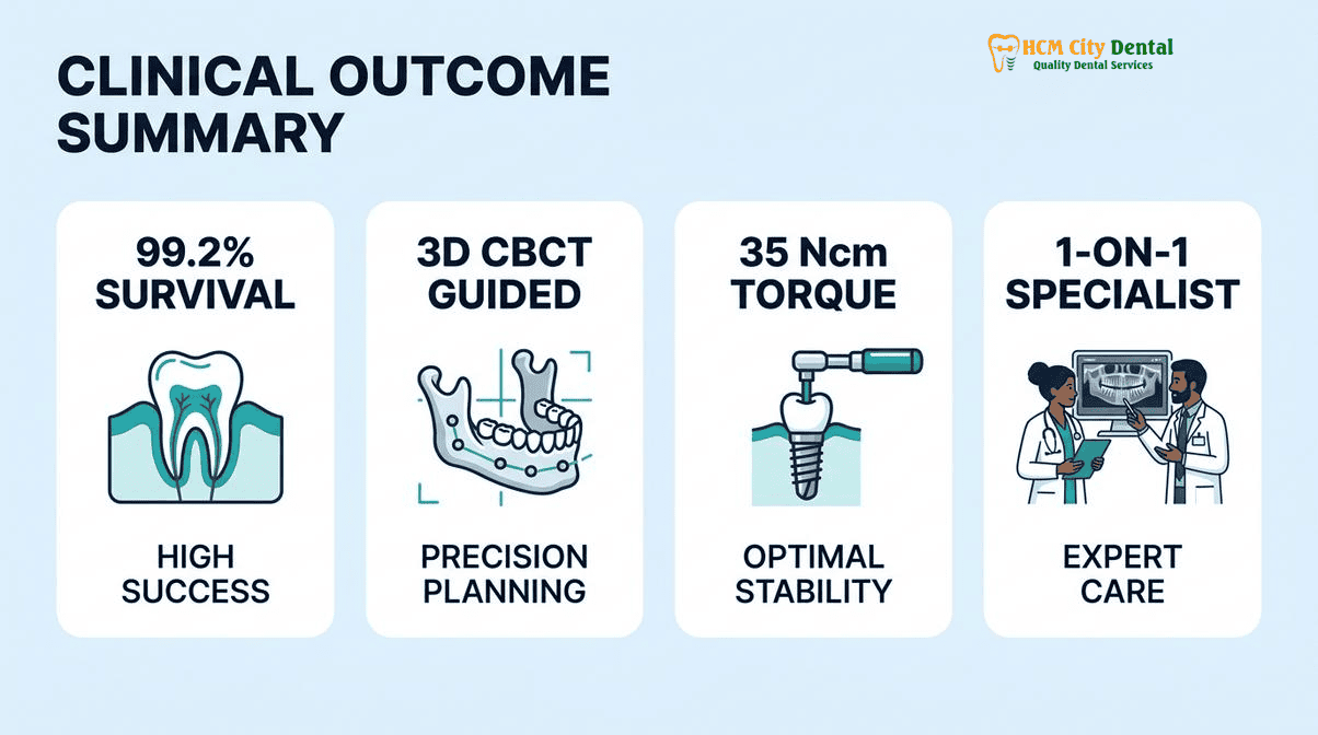
This entire process is integrated into a hospital-grade surgical workflow. Pre-operative planning is conducted using 0.075mm high-resolution 3D CBCT DICOM data fused with 3Shape Trios 5D STL optical scans. This allows for the fabrication of 3D-printed stereolithographic flapless surgical guides, ensuring that the implants are placed with sub-millimeter accuracy into the newly grafted bone. For international patients, this level of precision and biological safety is a hallmark of the care provided by Dr. Nguyen Van Cuong, DDS (Clinical Director — MOH License 0003265/BL-CCHN).
Clinical Precaution: The preparation of Sticky Bone and L-PRF is highly technique-sensitive. The liquid i-PRF will spontaneously coagulate within 15 to 30 minutes of being drawn. Therefore, the surgical site must be fully prepared, and the bone graft material must be ready for mixing before the venipuncture is performed. Patients with severe systemic bleeding disorders or those on heavy anticoagulant therapy must undergo a comprehensive medical evaluation with Dr. Cuong prior to treatment.
2026 Biological Bone Grafting, Sticky Bone & L-PRF Fee Schedule at HCMC Dental Clinic (District 1)
For dental travelers from Australia, the United States, and Europe, the cost of advanced biological bone grafting and implantology in their home countries can be prohibitively expensive. HCMC Dental Clinic bridges this gap by offering world-class, evidence-based surgical care at a fraction of the international cost, without compromising on biomaterial quality, sterilization standards, or clinical expertise.
Our facility operates with complete financial transparency. We offer a highly advantageous -40% Online Booking Fee Schedule for international patients who schedule their consultations and treatments prior to arriving in Ho Chi Minh City. This ensures that you receive the highest tier of surgical care—including 3D CBCT diagnostics, Piezosurgery, and autologous Sticky Bone preparation—at a predictable and affordable price point.
“We believe that advanced biological dentistry should be accessible. By utilizing the patient’s own blood to create L-PRF and Sticky Bone, we not only improve clinical outcomes and reduce healing times, but we also decrease the reliance on expensive synthetic membranes and recombinant growth factors. This allows us to pass those cost savings directly to our international patients.”
— Dr. Nguyen Van Cuong, DDS
Below is the official 2026 fee schedule for implantology and related surgical procedures at HCMC Dental Clinic. Please note that specific bone grafting fees (including the volume of xenograft required for Sticky Bone) will be determined during your comprehensive 3D CBCT consultation [5].
| Surgical Procedure / Implant Package (2026) | Walk-In / Standard Price (USD) | -40% Online Booking Price (USD) |
|---|---|---|
| 3D CBCT Scan (0.075mm High-Resolution) | $30 USD | $18 USD (480,000 VND) |
| Single Korean Implant (Dentis / Neo Biotech / Dentium + Abutment) | $769 – $1,090 USD | $462 – $654 USD (12M – 17M VND) |
| Single European / Swiss Implant (Straumann BLT-SLA / Active) | $1,603 – $2,244 USD | $962 – $1,346 USD (25M – 35M VND) |
| 100% Metal-Free Zirconia Ceramic Implant | $1,923 USD | $1,154 USD |
| Implant Zirconia HT Crown + Custom Abutment | $256 USD | $154 USD (4,000,000 VND) |
| All-on-4 Full-Arch Implants (Complete Package) | From $10,576 USD | From $6,346 USD / arch |
| All-on-6 Full-Arch Implants (Complete Package) | From $14,423 USD | From $8,654 USD / arch |
| Surgical Wisdom Tooth Extraction (Piezotome) | $96 – $191 USD | $58 – $115 USD / tooth |
To secure these exclusive online rates and begin planning your biological bone grafting and implant journey, we encourage you to review our Official 2026 Dental Price List & -40% Online Booking Fee Schedule and contact our international patient coordination team via WhatsApp at +84 853 020 003.
Frequently Asked Questions
How is L-PRF and Sticky Bone prepared from my own blood during a dental implant appointment?
At HCMC Dental Clinic, a small sample of your blood is drawn chairside immediately before your procedure. It is spun in a specialized centrifuge using the Choukroun low-speed protocol. This separates the red blood cells from the plasma, creating a dense fibrin clot (L-PRF) and liquid injectable PRF (i-PRF). The i-PRF is then mixed with bone graft particles to create a moldable Sticky Bone matrix, all within 15 minutes.
Is autologous Platelet-Rich Fibrin (L-PRF) completely free of chemical anticoagulants and bovine additives?
Yes. The L-PRF and i-PRF protocols utilized by Dr. Nguyen Van Cuong, DDS, are 100% autologous and completely free of chemical anticoagulants, bovine thrombin, or synthetic additives. The coagulation process occurs naturally in sterile, silica-free tubes, ensuring maximum biocompatibility and zero risk of allergic rejection.
Does L-PRF reduce post-operative pain and dry socket risk after tooth extraction?
Absolutely. When L-PRF membranes are placed into an extraction socket, they act as a biological bandage that seals the wound, protects exposed bone, and prevents the dislodgment of the blood clot. The high concentration of leukocytes (white blood cells) fights local infection, while the sustained release of growth factors significantly reduces inflammation, post-operative pain, and the risk of alveolar osteitis (dry socket).
How much faster does a dental bone graft heal when combined with Sticky Bone and A-PRF membranes?
Clinical studies indicate that incorporating Sticky Bone and A-PRF membranes can accelerate the initial soft tissue healing phase by up to 50% and enhance the quality of hard tissue osseointegration. The sustained 14-day release of vascular endothelial growth factor (VEGF) promotes rapid angiogenesis (new blood vessel formation), which is critical for the integration and maturation of the bone graft prior to implant placement.
What is the 2026 cost of L-PRF membrane and Sticky Bone grafting at HCMC Dental Clinic in District 1?
The cost of biological bone grafting varies depending on the complexity of the defect and the volume of xenograft required. However, HCMC Dental Clinic offers a highly transparent -40% Online Booking Fee Schedule. For example, a 3D CBCT scan is just $18 USD online, and comprehensive implant packages (including the fixture and abutment) start at $462 USD online. Specific grafting fees are determined during your consultation with Dr. Cuong based on your 3D CBCT analysis.
References
- Journal of Oral Implantology. Autologous Platelet-Rich Fibrin (PRF) and Sticky Bone in Alveolar Ridge Augmentation: A Comprehensive Clinical Review. (2023).
- International Journal of Periodontics & Restorative Dentistry. Biomechanical Stability of i-PRF Enriched Xenografts vs. Titanium Mesh in Guided Bone Regeneration. (2024).
- Clinical Oral Implants Research. Sustained Release Kinetics of PDGF, TGF-beta1, and VEGF from Leukocyte-Platelet Rich Fibrin (L-PRF) Matrices. (2022).
- Journal of Maxillofacial and Oral Surgery. Piezosurgery and Sticky Bone in Lateral Window Sinus Augmentation: Minimizing Schneiderian Membrane Perforation. (2025).
- Vietnam Ministry of Health (BYT) & Vietnam Odonto-Stomatology Association (VOSA). National Clinical Guidelines on Specialist Dental Care, Biomaterial Traceability & EN 13060 Class B Infection Control. (2024).