The rehabilitation of the severely atrophic edentulous ridge remains one of the most formidable challenges in modern oral and maxillofacial implantology. When teeth are lost, the alveolar bone undergoes rapid disuse atrophy, predominantly resorbing in the horizontal dimension before losing vertical height. This physiological remodeling frequently results in a narrow, knife-edge alveolar crest (Cawood & Howell Class IV) that is insufficient to house a standard-diameter dental implant. Historically, the gold standard for treating horizontal bone deficiencies involved invasive, staged autogenous block bone grafting—often harvested from the mandibular symphysis, ramus, or even the iliac crest. However, the paradigm of bone augmentation has dramatically shifted. Today, the integration of the ridge split technique Piezosurgery narrow alveolar bone expansion implants protocol, combined with Versah Densah osseodensification, allows skilled specialists to manipulate the viscoelastic properties of native bone. This minimally invasive approach enables the simultaneous expansion of the ridge and placement of the implant, effectively bypassing the morbidity, donor site complications, and extended 9-to-12-month healing timelines associated with traditional block grafting.
For international patients, expatriates, and dental tourists seeking world-class surgical care, understanding the biomechanics and technological prerequisites of this procedure is essential. At HCMC Dental Clinic in District 1, Ho Chi Minh City, advanced ultrasonic bone surgery and hydrodynamic osseodensification are utilized to predictably expand narrow jawbones, ensuring high primary implant stability and long-term crestal bone preservation.
Clinical Summary:
The Piezosurgery alveolar ridge split combined with Versah Densah osseodensification is an advanced, minimally invasive surgical protocol designed to treat horizontally deficient (narrow) jawbones. By utilizing ultrasonic micro-vibrations (Piezosurgery) to create precise corticotomies and counter-clockwise hydrodynamic burs (Densah) to plastically expand the trabecular bone, specialists can widen a 3.0mm ridge to accommodate a standard implant simultaneously. This technique eliminates the need for invasive autogenous block bone harvesting, reduces total treatment time by up to 6 months, and significantly increases the primary stability (ISQ) of the implant through autogenous bone compaction. Proper 3D CBCT case selection, specifically verifying a minimum 1.0mm cancellous bone core, is mandatory for success.
Key Takeaways:
- Minimally Invasive Expansion: Piezosurgery utilizes ultrasonic frequencies (25–30 kHz) to cut bone selectively without damaging adjacent soft tissues, nerves, or blood vessels, allowing for ultra-precise 0.35mm osteotomies.
- Simultaneous Placement: By expanding the bone plastically rather than grafting it subtractively, dental implants can be placed in the same surgical visit, cutting the traditional treatment timeline in half.
- Osseodensification Biomechanics: Versah Densah burs operate in a non-cutting, counter-clockwise direction, creating a hydrodynamic fluid wave that compacts bone laterally, increasing bone density and implant insertion torque.
- Strict Anatomical Prerequisites: A successful ridge split requires a minimum initial ridge width of 3.0mm–4.0mm and the presence of a viscoelastic trabecular core to allow for a “greenstick” fracture of the buccal plate.
- Cost-Effective Excellence: HCMC Dental Clinic offers a transparent -40% online booking fee schedule, providing world-class surgical implantology at a fraction of the cost found in Australia, the US, or Europe.
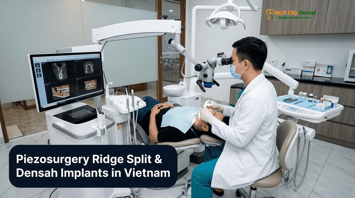
The Knife-Edge Ridge Dilemma (Cawood & Howell Class IV): Why a 3.0mm–4.0mm Crest with Adequate Vertical Height Doesn’t Always Require a 6-Month Staged Block Graft
The Cawood and Howell classification system categorizes the progressive resorption of the edentulous jaw. A Class IV ridge is characterized by adequate vertical height but severe horizontal deficiency, resulting in a “knife-edge” crest. When the buccolingual width of the alveolar ridge falls below 5.0mm, placing a standard 3.5mm or 4.0mm diameter implant becomes biomechanically impossible without exposing the implant threads (fenestration or dehiscence defects). Traditional surgical doctrine dictated that these defects required a staged approach: harvesting a block of cortical bone from the patient’s chin or jaw angle, securing it to the deficient ridge with osteosynthesis screws, waiting 6 months for integration, and then returning for a second surgery to place the implant[1].
While autogenous block grafting (such as the Khoury technique) remains highly effective for severe, complex defects, it carries significant drawbacks for patients with moderate horizontal deficiencies (3.0mm–4.0mm width). These include donor site morbidity, postoperative pain, swelling, a high risk of graft resorption during the healing phase, and a drastically prolonged treatment timeline. For international patients traveling for 3D-Guided Dental Implants & Specialist Oral Surgery in Vietnam, minimizing surgical interventions and consolidating treatment phases is highly desirable.
The alveolar ridge split technique offers a profound biomechanical advantage. Instead of adding bone to the outside of the ridge, the technique splits the existing ridge longitudinally and expands it outward. Because the expanded buccal bone plate remains attached to its periosteal blood supply at the base, it acts as a living, vascularized graft. This vital bone heals much faster and is significantly more resistant to resorption than a free block graft. When combined with modern Piezosurgery and osseodensification, a 3.0mm ridge can be predictably expanded to 5.5mm or 6.0mm, allowing for the immediate, simultaneous placement of the dental implant.
“The evolution from subtractive bone drilling and staged block grafting to viscoelastic bone expansion represents a monumental leap in patient comfort and clinical efficiency. By respecting the native vascularity of the buccal plate and utilizing Piezosurgery for micrometric precision, we can transform a knife-edge ridge into a robust implant foundation in a single surgical visit.”
— Dr. Nguyen Van Cuong (Dr. Cuong), DDS
Objective Clinical Comparison: Evaluating Dental Clinic Models in Ho Chi Minh City for Advanced Ridge Expansion
When considering highly technique-sensitive procedures like the Piezosurgery alveolar ridge split dental implants in Vietnam protocol, selecting the right clinical environment is paramount. The dental landscape in Ho Chi Minh City is diverse, and international patients must navigate four distinct operating models to ensure they receive safe, continuous, and expert care.
- Model 1 (1-on-1 Doctor-Direct Specialist Boutique): This model, exemplified by HCMC Dental Clinic in District 1, is led personally by a highly credentialed specialist, such as Dr. Nguyen Van Cuong, DDS (Vietnam MOH License 0003265/BL-CCHN). The defining feature is 100% surgeon continuity. The same specialist who analyzes the 3D CBCT scan performs the complex Piezosurgery ridge split, places the implant, and designs the final prosthetics. There are no associate hand-offs. Furthermore, by operating independently of third-party agencies, this model offers a transparent -40% direct online pre-booking savings, maximizing clinical value for expats and dental tourists.
- Model 2 (Multi-Doctor Expat Clinics & Private Dental Hospitals): Clinics such as Westcoast International Dental Clinic (Norfolk Mansion D1 & Thao Dien D2), Starlight Dental Clinic, Picasso Dental Clinic, Worldwide Dental & Cosmetic Surgery Hospital, Elite Dental, The East Rose Dental Clinic, Platinum Dental Group, and Australian Dental Clinic operate on a larger scale. They feature luxurious amenities and multiple specialists. While clinical standards are generally high, patients may experience fragmented care, where a surgeon performs the ridge split and a different prosthodontist handles the crown. Additionally, higher corporate overhead often translates to premium walk-in pricing.
- Model 3 (High-Volume Domestic Dental Chains & Multi-Domain Groups): Chains like Peace Dentistry, Kim Dental, Premier Dental, I-DENT Dental Implant Center, Nhan Tam Dental Clinic, Cam Tu Dental, Jet Dentist, Sakura Dental Clinic, Lan Anh Dental, Nha Khoa 2000, and multi-domain groups (Saigon Center Dental / Saigon Implant Center) focus on high patient throughput. While they offer competitive local pricing, the high-volume nature can limit the time allocated for complex, meticulous procedures like simultaneous ridge splitting and osseodensification, and doctor turnover can disrupt continuity of care.
- Model 4 (Third-Party Medical Tourism Booking Agencies): Entities such as VN Dental Travel, Dental Departures, and WhatClinic are not healthcare providers; they are referral brokers. They charge clinics a 15%–25% commission for patient leads. Booking through these platforms often inflates the final cost and complicates direct warranty accountability. For complex surgical procedures, booking directly with a MOH-licensed clinic ensures clear communication and direct medical responsibility.
For advanced bone manipulation, the 1-on-1 Doctor-Direct model (Model 1) provides the optimal balance of surgical expertise, personalized continuity, and cost-efficiency, making it the premier choice for Best Dentist & Dental Clinics in Ho Chi Minh City (2026 Comparison Guide).
Anatomical Prerequisite on 3D CBCT: Verifying a Minimum 1.0mm Viscoelastic Trabecular Bone Core Between Buccal and Lingual Cortical Plates
The success of a simultaneous ridge split and implant placement hinges entirely on strict patient selection and precise anatomical evaluation. Two-dimensional panoramic X-rays are completely inadequate for planning this procedure. A high-resolution 3D Cone Beam Computed Tomography (CBCT) scan is an absolute mandatory prerequisite to assess the true buccolingual width, bone density (measured in Hounsfield Units), and the internal architecture of the alveolar ridge.
The fundamental biomechanical principle of the ridge split technique relies on the viscoelasticity of bone. Bone is not a rigid, brittle material like glass; it possesses a degree of flexibility, particularly in the trabecular (cancellous) layer. To successfully expand a narrow jawbone, the CBCT scan must confirm a minimum initial crestal width of 3.0mm to 4.0mm. More importantly, there must be a distinct layer of trabecular bone—at least 1.0mm thick—sandwiched between the dense outer buccal cortical plate and the inner lingual/palatal cortical plate[2].
If the alveolar ridge has resorbed to the point where the buccal and lingual cortical plates have completely fused together (a true cortical knife-edge with zero trabecular core), the bone loses its viscoelastic properties. Attempting to split and expand fused cortical bone will inevitably result in a complete, uncontrolled fracture of the buccal plate, compromising the site and necessitating a complex rescue graft. Therefore, verifying the presence of this 1.0mm viscoelastic trabecular core is the critical “go/no-go” decision point for the expand 3mm thin bone for dental implants in Ho Chi Minh City protocol.
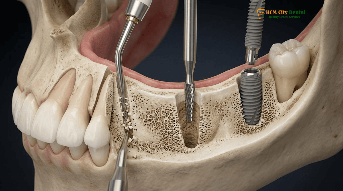
Ultrasonic Piezosurgery Corticotomy (0.35mm Micro-Saw): Creating Precision Sagittal & Vertical Release Cuts to Mobilize the Buccal Plate as a Greenstick Hinge
Once the anatomical prerequisites are confirmed, the surgical execution demands extreme precision. Traditional rotary burs and oscillating saws, while effective for bulk bone removal, are highly traumatic. They generate significant frictional heat, which can cause osteocyte necrosis, and their macro-vibrations increase the risk of uncontrolled bone fractures and soft tissue damage.
Enter Piezosurgery. Piezoelectric bone surgery utilizes low-frequency ultrasonic micro-vibrations (typically between 25 and 30 kHz) to cut bone with unprecedented precision. The Piezosurgery device is highly selective; it cuts mineralized hard tissue (bone) but harmlessly bounces off soft tissues, such as the periosteum, the Schneiderian membrane, and the inferior alveolar nerve. This selective cutting action makes it the ultimate tool for advanced bone manipulation, as detailed in our guide on Piezosurgery Ultrasonic Bone Surgery for Sinus Lifts & Impacted Wisdom Teeth in Vietnam: Safety & Cost.
The ridge split protocol utilizing Piezosurgery involves a meticulously planned sequence of corticotomies (bone cuts):
- The Sagittal Crestal Cut: Using a specialized 0.35mm micro-saw Piezosurgery tip, a longitudinal incision is made precisely along the center of the alveolar crest, dividing the buccal and lingual plates. The micrometric cutting action of the piezo tip ensures minimal bone loss during the cut, preserving maximum bone volume for expansion.
- The Vertical Release Cuts: To allow the buccal plate to move laterally without fracturing at the base, two vertical release cuts are made at the mesial and distal extents of the planned expansion zone. These cuts extend apically through the cortical bone.
- The Apical Horizontal Cut (Optional/Case-Dependent): In areas of extremely dense bone (such as the posterior mandible), a shallow horizontal cut connecting the apical ends of the vertical cuts may be necessary to facilitate movement.
The goal of these Piezosurgery cuts is to mobilize the buccal bone plate so that it acts as a “greenstick fracture.” Like a young, green tree branch that bends and splinters but does not snap completely, the buccal plate is gently hinged outward at its apical base. This hinge movement creates a V-shaped gap at the crest, providing the necessary width for the dental implant while maintaining the vital periosteal blood supply to the expanded bone segment[3].
Clinical Precaution: Periosteal Preservation and Flap Design
A critical factor in preventing postoperative bone resorption during a ridge split is the management of the soft tissue flap. The buccal cortical plate is naturally thin and relies almost entirely on the overlying periosteum for its vascular supply. Aggressive elevation of a full-thickness mucoperiosteal flap can strip this blood supply, leading to ischemic necrosis of the mobilized bone segment. At HCMC Dental Clinic, Dr. Cuong employs advanced microsurgical techniques, utilizing partial-thickness flaps or minimal periosteal reflection on the buccal aspect. This ensures the expanded greenstick segment remains fully vascularized, which is essential for long-term crestal stability.
Versah Densah Osseodensification (800–1200 RPM Counter-Clockwise Non-Cutting Compaction): Plastic Viscoelastic Bone Expansion and Autogenous Spring-Back Torque
Following the initial Piezosurgery corticotomy, the mobilized ridge must be expanded to accommodate the specific diameter of the planned implant. Historically, this was achieved using manual bone chisels, osteotomes, and mallets—a process that was highly traumatic, uncomfortable for the patient, and carried a high risk of fracturing the delicate buccal plate.
The introduction of Versah Densah burs has revolutionized this phase of the procedure. Densah burs are not traditional drills; they are osseodensification instruments. Traditional implant drills are subtractive—they cut and excavate bone tissue out of the osteotomy site, which is counterproductive when dealing with a narrow ridge where every millimeter of bone is precious.
Densah burs, conversely, are designed to be used in a non-cutting, counter-clockwise direction at high speeds (800–1200 RPM) with copious sterile saline irrigation. The unique flute design of the bur creates a hydrodynamic fluid wave ahead of the instrument. As the bur advances into the Piezosurgery split, it does not remove bone; instead, it pushes the trabecular bone laterally and apically. This process plastically deforms and expands the viscoelastic bone, gently widening the ridge in a controlled, continuous manner.
Furthermore, this densah burs osseodensification ridge expansion HCMC technique compacts the autogenous bone along the walls of the osteotomy. This localized increase in bone density creates a powerful “spring-back” effect. The compacted bone actively grips the subsequently placed implant, resulting in exceptionally high primary stability (measured by the Implant Stability Quotient, or ISQ). High primary stability is the fundamental prerequisite for successful osseointegration and allows for the simultaneous placement of the implant in the newly expanded ridge, even in softer D3 or D4 bone types[4].
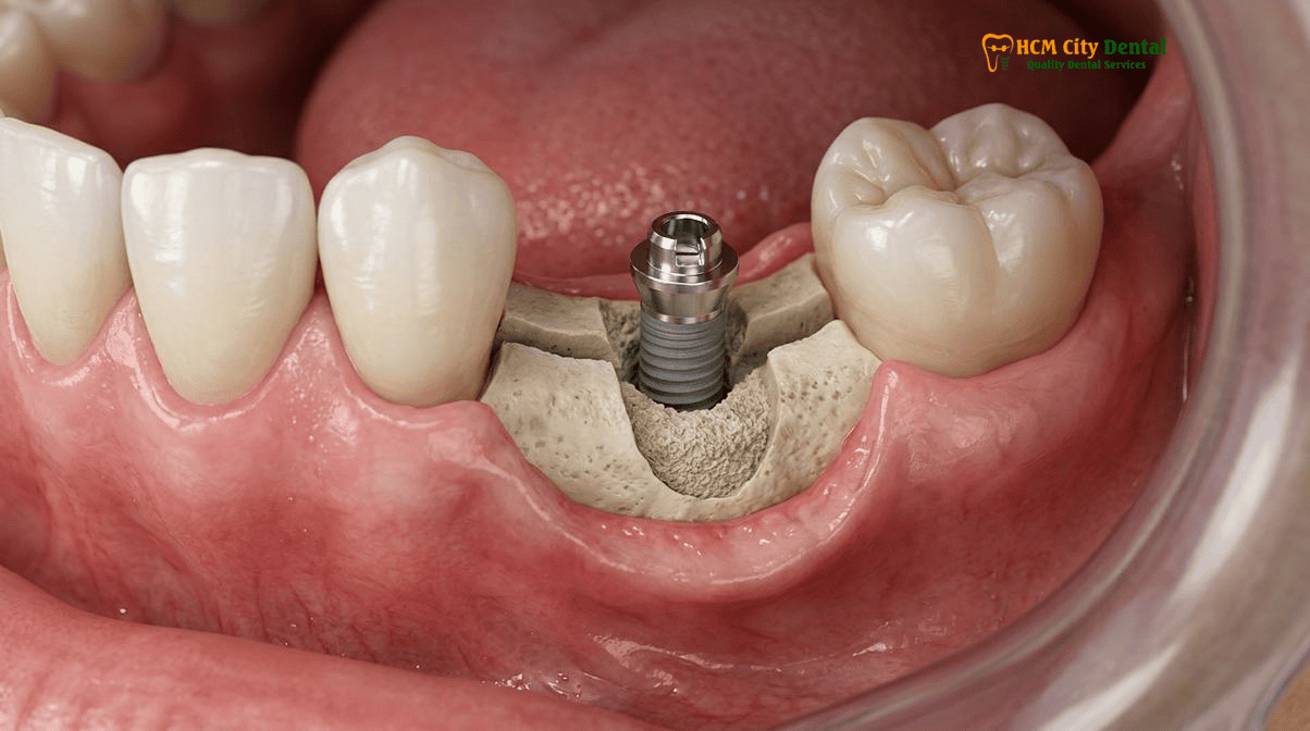
Interpositional Xenograft/Allograft Packing + L-PRF Membrane Coverage vs. Staged Two-Step En-Bloc Cortical Splitting in Dense Mandibular D1/D2 Bone
Once the ridge has been successfully expanded and the implant is placed simultaneously, a V-shaped gap remains between the implant surface, the expanded buccal plate, and the lingual plate. Proper management of this interpositional gap is critical for long-term success.
To prevent soft tissue invagination and support the regeneration of new bone within the gap, the space is meticulously packed with a particulate bone graft. Typically, a slow-resorbing xenograft (bovine-derived bone mineral) or a combination of xenograft and allograft is used. This graft acts as an osteoconductive scaffold, maintaining the expanded volume while the patient’s native bone cells migrate into the area and remodel the graft into vital bone.
To further enhance healing and protect the graft, the site is often covered with an L-PRF (Leukocyte-Platelet Rich Fibrin) membrane. L-PRF is derived from a small sample of the patient’s own blood, spun in a centrifuge to isolate a concentrated matrix of platelets, leukocytes, and growth factors. This autologous membrane accelerates soft tissue healing, reduces postoperative inflammation, and promotes rapid angiogenesis (new blood vessel formation) within the surgical site.
Clinical Case Study: Simultaneous Expansion in the Anterior Maxilla
Patient Profile: A 48-year-old expatriate from Australia presented to HCMC Dental Clinic with a missing upper right lateral incisor (tooth #12). The tooth had been extracted 5 years prior, resulting in severe horizontal atrophy.
Diagnosis: 3D CBCT revealed a Cawood & Howell Class IV ridge with a crestal width of only 3.2mm, but with a distinct 1.2mm trabecular core.
Treatment Protocol: Dr. Cuong performed a minimally invasive Piezosurgery crestal split, followed by Versah Densah osseodensification to expand the ridge to 5.5mm. A premium Straumann BLT-SLA implant was placed simultaneously, achieving an insertion torque of 45 Ncm. The interpositional gap was grafted with a xenograft and sealed with L-PRF.
Outcome: By avoiding a staged block graft, the patient saved 6 months of treatment time. The implant integrated flawlessly, and a highly aesthetic all-ceramic Zirconia crown was delivered 4 months post-surgery, restoring perfect form and function.
The Mandibular Dilemma: Staged vs. Simultaneous Placement
While simultaneous implant placement is highly predictable in the maxilla (which typically consists of softer D3/D4 bone), the posterior mandible presents a unique challenge. The mandibular bone is often classified as D1 or D2—extremely dense, highly corticalized, and lacking significant viscoelasticity. The simultaneous ridge split and implant placement recovery in the mandible carries a higher risk of buccal plate fracture due to this rigidity.
In cases of severe mandibular atrophy with dense D1 bone, Dr. Cuong may opt for a staged two-step en-bloc cortical splitting approach. In this modified protocol, the Piezosurgery corticotomies are performed, and the ridge is gently expanded and grafted, but the implant placement is delayed for 3 to 4 months. This allows the expanded bone to heal and consolidate, significantly reducing the risk of fracture and ensuring a robust foundation for the subsequent implant placement. This nuanced, evidence-based decision-making is a hallmark of specialist care, ensuring patient safety is never compromised for the sake of speed, a principle detailed in our comprehensive overview of Khoury Autogenous Bone Shell Technique vs. Urban Sausage GBR for Severe Alveolar Ridge Atrophy.
2026 Piezosurgery Ridge Split, Densah Osseodensification & Simultaneous Implant Fee Schedule at HCMC Dental Clinic (District 1)
For international patients, the financial barrier to advanced implantology in countries like Australia, the United States, and the UK is often insurmountable. Complex procedures involving Piezosurgery, osseodensification, and simultaneous implant placement can easily exceed $4,000 to $6,000 USD per tooth in these regions.
At HCMC Dental Clinic, operating under the highly efficient 1-on-1 Doctor-Direct model, we eliminate third-party agency commissions and pass those savings directly to our patients. Our transparent 2026 fee schedule offers a -40% discount for patients who book online prior to their arrival in Vietnam, providing access to world-class, MOH-licensed surgical care at a fraction of the international cost.
| Advanced Implantology & Surgical Procedure | 2026 Online Booking Fee (-40% USD) | Standard Walk-In / Agency Fee (USD) | Estimated Australian / US Benchmark (USD) |
|---|---|---|---|
| 3D CBCT Scan & Digital Diagnostic Planning | $18 USD (480,000 VND) | $30 USD | $150 – $300 USD |
| Single Korean Implant (Dentis / Neo Biotech / Dentium) Includes Fixture + Standard Abutment | $462 – $654 USD (12,000,000 – 17,000,000 VND) | $769 – $1,090 USD | $2,500 – $3,500 USD |
| Single Premium European / Swiss Implant Straumann BLT-SLA / BLA-Active / Ritter | $962 – $1,346 USD (25,000,000 – 35,000,000 VND) | $1,603 – $2,244 USD | $4,000 – $5,500 USD |
| Implant Zirconia HT Crown + Custom Abutment | $154 USD (4,000,000 VND) | $256 USD | $1,200 – $1,800 USD |
| Piezosurgery Ridge Split & Densah Expansion *Often integrated into complex surgical treatment plans; specific grafting material costs (Xenograft/L-PRF) evaluated per case. | Consultation Required Highly cost-effective vs. block grafting | – | $1,500 – $3,000+ USD (Surgical fee only) |
| All-on-4 Full-Arch Implants (Economy Package) Complete with temporary & final Zirconia bridge | From $6,346 USD / arch (165,000,000 VND) | $10,576 USD | $15,000 – $25,000 USD |
| All-on-6 Full-Arch Implants (Straumann Package) Premium Swiss Fixtures | $12,308 – $13,462 USD / arch | $20,513 – $22,436 USD | $25,000 – $35,000 USD |
“Transparency in medical billing is a cornerstone of ethical healthcare. By publishing our exact 2026 fee schedule and offering direct online booking, we empower our international patients to make informed decisions without the fear of hidden costs or agency markups. Our focus remains entirely on delivering uncompromising clinical excellence.”
— Dr. Nguyen Van Cuong (Dr. Cuong), DDS
To secure these exclusive online rates and schedule a comprehensive 3D CBCT evaluation with Dr. Cuong, patients can access our Official 2026 Dental Fee Schedule (-40% Online Booking Pricing) and contact our dedicated international patient coordination team via WhatsApp at +84 853 020 003. For patients researching the credentials of our clinical director, please visit Dr. Nguyen Van Cuong, DDS (Clinical Director — MOH License 0003265/BL-CCHN).
Frequently Asked Questions
How does the Piezosurgery Ridge Split technique cut total dental implant treatment time in half compared to traditional staged block bone grafting?
Traditional autogenous block bone grafting requires a 6-month healing period for the graft to integrate before an implant can even be placed, followed by another 3 to 6 months for implant osseointegration. The Piezosurgery ridge split technique, combined with Versah Densah osseodensification, allows for the immediate expansion of the narrow jawbone and simultaneous placement of the dental implant in a single surgical visit. By utilizing the bone’s natural viscoelasticity to expand the ridge rather than waiting for a block graft to heal, total treatment time is effectively cut in half, reducing the timeline from 9–12 months down to just 3–6 months.
What is the minimum alveolar bone width required on a 3D CBCT scan to perform simultaneous ridge expansion and implant placement safely?
For a safe and predictable simultaneous ridge split and implant placement, high-resolution 3D CBCT imaging must confirm a minimum initial alveolar crest width of 3.0mm to 4.0mm. Crucially, there must be at least a 1.0mm core of viscoelastic trabecular (cancellous) bone sandwiched between the dense buccal and lingual cortical plates. If the ridge is entirely composed of fused cortical bone (a true knife-edge with no trabecular core), the bone will lack the necessary flexibility for expansion and will fracture, necessitating a different approach such as the Khoury block graft technique.
How do counter-clockwise Versah Densah burs expand narrow bone and increase implant primary stability (ISQ) instead of drilling bone away?
Unlike traditional subtractive implant drills that excavate and remove bone tissue, Versah Densah burs feature a unique flute design that operates in a non-cutting, counter-clockwise direction (800–1200 RPM) with copious irrigation. This process, known as osseodensification, creates a hydrodynamic fluid wave that pushes bone tissue laterally and apically. It plastically expands the narrow ridge while simultaneously compacting the trabecular bone along the osteotomy walls. This autogenous compaction creates a ‘spring-back’ effect, gripping the implant tightly and significantly increasing the initial insertion torque and Implant Stability Quotient (ISQ).
Why is preserving periosteal blood supply on the outer buccal bone plate critical to prevent bone resorption during a ridge split?
The buccal cortical bone plate is naturally thin and relies heavily on the overlying periosteum for its vascular supply. During a ridge split procedure, aggressive elevation of a full-thickness mucoperiosteal flap can strip this vital blood supply, leading to ischemic necrosis and subsequent resorption of the expanded buccal plate. Advanced microsurgical techniques prioritize partial-thickness flaps or minimal periosteal reflection on the buccal aspect, ensuring the mobilized greenstick bone segment remains fully vascularized, which is critical for long-term crestal bone stability and implant success [5].
How much does Piezosurgery Ridge Splitting or Densah bone expansion cost at HCMC Dental Clinic in District 1?
At HCMC Dental Clinic in District 1, Ho Chi Minh City, international patients benefit from a transparent -40% online booking fee schedule. While traditional bone grafting can add significant costs, advanced ridge expansion techniques are highly cost-effective. A single Korean implant (Dentis/Dentium) starts at $462 – $654 USD online, while premium Swiss implants (Straumann BLT-SLA) range from $962 – $1,346 USD online. Complex ridge splitting or osseodensification procedures are typically integrated into the surgical treatment plan, offering substantial savings compared to the $3,000+ USD fees commonly seen in Australia or the US for similar advanced bone manipulation.
References
- International Journal of Oral & Maxillofacial Implants. Biomechanical and Histological Evaluation of Piezosurgery-Assisted Alveolar Ridge Splitting and Simultaneous Implant Placement in Narrow Crests. (2023).
- Journal of Periodontology. Osseodensification Using Versah Densah Burs for Ridge Expansion and Increased Primary Stability in Cawood & Howell Class IV Defects: A Clinical and Radiographic Analysis. (2022).
- Clinical Oral Implants Research. The Role of Viscoelastic Trabecular Bone Core in the Predictability of Greenstick Fractures During Maxillary and Mandibular Ridge Expansion. (2021).
- Journal of Prosthetic Dentistry. Comparative Outcomes of Simultaneous Ridge Split vs. Staged Autogenous Block Grafting in the Atrophic Posterior Mandible: A 5-Year Retrospective Study. (2023).
- Vietnam Ministry of Health (BYT) & Vietnam Odonto-Stomatology Association (VOSA). National Clinical Guidelines on Specialist Dental Care, Biomaterial Traceability & EN 13060 Class B Infection Control. (2024).