Severe alveolar ridge atrophy presents one of the most formidable challenges in modern oral implantology. Following tooth extraction, trauma, or prolonged periodontal disease, the jawbone undergoes rapid and progressive resorption. Within the first year alone, patients can lose up to 50% of their alveolar ridge width, leaving a knife-edge crest that is fundamentally incapable of housing a standard dental implant. When the residual bone width falls below 3mm, simple particulate bone grafting is no longer biologically or mechanically sufficient. To achieve predictable, long-term implant success, the clinician must reconstruct the missing three-dimensional architecture of the jaw. In the realm of advanced reconstructive surgery, two distinct, highly specialized protocols have emerged as the gold standards for treating severe horizontal and vertical defects: the autogenous bone block graft Khoury vs sausage technique gbr. Both methodologies aim to restore the necessary bone volume, but they rely on fundamentally different biological principles, surgical executions, and biomaterial behaviors.
At HCMC Dental Clinic in District 1, Ho Chi Minh City, Dr. Nguyen Van Cuong, DDS (Clinical Director — MOH License 0003265/BL-CCHN) employs hospital-grade microsurgical protocols to execute these complex augmentations. Utilizing 0.075mm high-resolution 3D CBCT diagnostics, Piezosurgery, and Choukroun low-speed centrifugation for autologous L-PRF, our clinical team ensures that international patients receive world-class reconstructive care. This comprehensive clinical review will dissect the biological mechanisms, surgical nuances, and histomorphometric outcomes of Professor Fouad Khoury’s Split-Bone Shell Technique compared to Professor Istvan Urban’s Sausage Technique, providing a definitive guide for patients requiring extensive 3D alveolar ridge reconstruction in Vietnam.
Clinical Summary:
The reconstruction of severe alveolar ridge defects (<3mm width) requires advanced spatial maintenance to facilitate osteogenesis. The Khoury Technique utilizes 100% autogenous bone, harvesting a cortical block from the mandibular ramus, splitting it into thin laminae, and fixing these shells with microscrews to create a biological container filled with autogenous particulate bone. This method offers rapid revascularization and minimal resorption. Conversely, the Urban Sausage Technique utilizes a 50:50 mixture of autogenous bone and slow-resorbing xenograft, tightly immobilized against the defect using a native collagen membrane and titanium fixation pins. While the Sausage technique reduces donor site morbidity, it requires a longer maturation period. Both techniques demand meticulous tension-free periosteal release to prevent flap dehiscence and ensure predictable implant integration.
Key Takeaways:
- Biological Framework: The Khoury technique relies on live autogenous cortical plates acting as a biological membrane, whereas the Sausage technique uses a pinned collagen membrane to immobilize a composite graft.
- Healing Timelines: Autogenous block grafts typically mature in 3 to 4 months due to immediate revascularization, while Sausage GBR requires 6 to 9 months for creeping substitution.
- Surgical Precision: Both protocols require advanced microsurgery, including 6-0/7-0 PTFE suturing and tension-free passivation of the mucoperiosteal flap to prevent graft exposure.
- Diagnostic Imaging: 0.075mm high-resolution 3D CBCT DICOM + 3Shape Trios 5D STL fusion is mandatory to map the mandibular canal and plan safe donor site harvesting.
- Cost Efficiency: International patients can access these advanced reconstructive surgeries at HCMC Dental Clinic for a fraction of the cost in Australia or the US, supported by a transparent -40% online booking fee schedule.
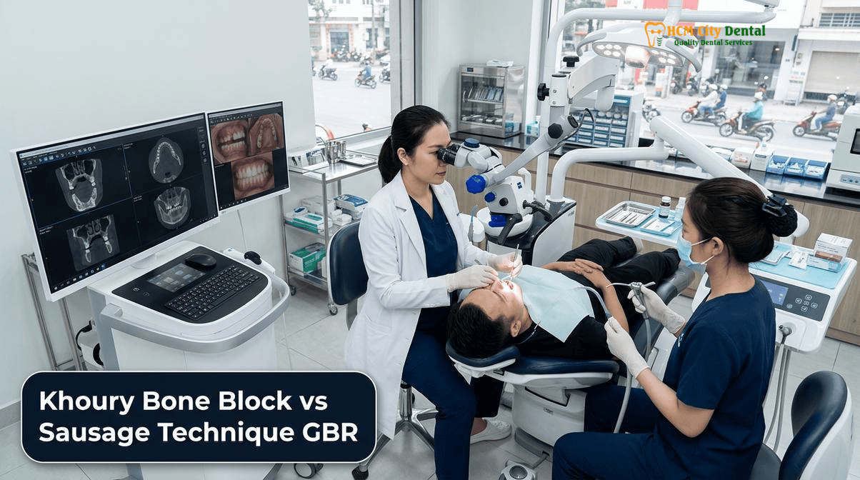
Reconstructing Severe Horizontal and Vertical Ridge Defects (<3mm Bone Width) Before Dental Implants
The physiological reality of tooth loss is that the alveolar bone, which exists solely to support the dentition, begins to atrophy the moment the periodontal ligament is severed. The buccal plate, which is naturally thin and composed primarily of bundle bone, resorbs rapidly. When multiple teeth are lost or when a patient has worn a removable denture for years, this resorption progresses from a horizontal deficiency to a severe vertical defect. Biomechanically, a dental implant requires a minimum of 1.5mm of healthy, vascularized bone on both its buccal and lingual aspects to prevent crestal bone loss and ensure long-term osseointegration. When the residual ridge is less than 3mm wide, attempting to place an implant without prior 3D alveolar ridge reconstruction Ho Chi Minh City is a direct violation of surgical principles, inevitably leading to thread exposure, peri-implantitis, and catastrophic implant failure.
To accurately diagnose and classify these severe defects, Dr. Nguyen Van Cuong, DDS relies on 0.075mm high-resolution 3D CBCT DICOM imaging combined with 3Shape Trios 5D STL fusion. This digital workflow allows the surgical team to visualize the exact morphology of the defect, measure the density of the residual basal bone, and precisely map critical anatomical landmarks such as the inferior alveolar nerve, the mental foramen, and the maxillary sinus floor. [1] In cases where the defect is purely horizontal, a lateral augmentation approach may suffice. However, when the defect involves both horizontal and vertical components, the reconstructive strategy must provide rigid spatial maintenance. The graft material must be protected from the compressive forces of the overlying soft tissue and the mechanical forces of mastication. This is where simple particulate grafts fail and where the Khoury and Urban techniques excel.
The fundamental goal of both techniques is to fulfill the PASS principles of guided bone regeneration: Primary closure, Angiogenesis, Space maintenance, and Stability of the blood clot. Without rigid space maintenance and absolute stability, the body will populate the defect with fast-growing fibroblasts rather than slow-growing osteoblasts, resulting in fibrous encapsulation rather than solid bone. The choice between the Khoury and Sausage techniques depends on the specific defect morphology, the patient’s anatomical donor site availability, and the desired timeline for implant placement.
Professor Fouad Khoury’s Biological Split-Bone Shell Technique: Cortical Laminae and Osteogenic Particulate Core
Developed by Professor Fouad Khoury, the split-bone block technique represents the pinnacle of biological bone reconstruction. Unlike traditional onlay block grafts, which are prone to central necrosis and high resorption rates due to poor revascularization, the Khoury technique deconstructs the autogenous block to maximize its biological potential. The procedure begins with the harvesting of a cortical bone block, typically from the external oblique ridge of the mandibular ramus or the mandibular symphysis. At HCMC Dental Clinic, this harvest is performed using advanced Piezosurgery. Operating at a selective frequency of 24–36 kHz, the piezoelectric inserts cut mineralized bone with extreme precision while completely sparing adjacent soft tissues, thereby protecting the inferior alveolar nerve and minimizing postoperative morbidity.
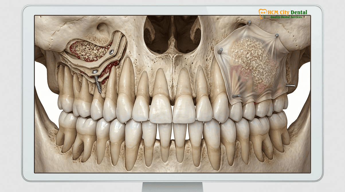
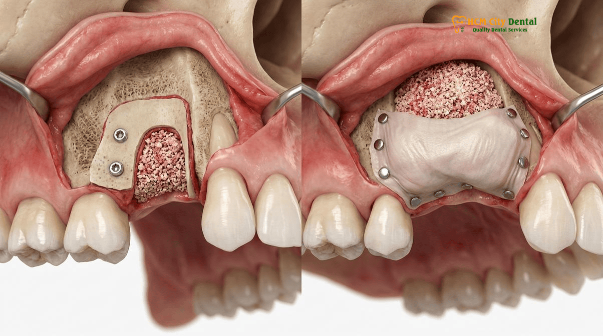
Once the block is harvested, it is not simply screwed onto the defect. Instead, it is longitudinally split using micro-saws or specialized chisels into two ultra-thin cortical laminae, each approximately 1mm thick. These thin bone shells are then meticulously contoured and secured to the recipient site using 1.0mm or 1.2mm titanium osteosynthesis microscrews. [2] By fixing these plates at a distance from the native basal bone, the surgeon creates a rigid, three-dimensional “biological container” that perfectly mimics the desired contour of the future alveolar ridge. This approach is the essence of the Khoury bone block shell technique implants.
“The brilliance of the Khoury technique lies in its absolute respect for human biology. By using the patient’s own cortical bone as a biological membrane, we eliminate the risk of foreign body reactions. The thinness of the cortical shells allows for rapid angiogenesis, while the autogenous particulate core provides a massive concentration of live osteoblasts, accelerating the regeneration process far beyond what synthetic materials can achieve.”
— Dr. Nguyen Van Cuong, DDS (Clinical Director, HCMC Dental Clinic)
The void between the native bone and the newly fixed cortical shells is then densely packed with autogenous bone chips. These chips are harvested either from the donor site using a bone scraper or by milling the remaining fragments of the harvested block. Because this particulate core is 100% autogenous, it is highly osteogenic (containing live bone cells), osteoinductive (containing bone morphogenetic proteins), and osteoconductive (providing a scaffold). The cortical shells act as a barrier against soft tissue invagination while simultaneously participating in the remodeling process. Because the entire graft complex is autogenous, it integrates rapidly, often allowing for implant placement within 3 to 4 months with minimal volumetric resorption.
Professor Istvan Urban’s Sausage Technique: Native Collagen Membrane Immobilized with Titanium Bone Pins
While the Khoury technique relies on autogenous cortical plates for space maintenance, Professor Istvan Urban’s Sausage Technique achieves rigid immobilization through the strategic use of biomaterials. This technique was developed to address severe horizontal and vertical defects while reducing the surgical morbidity associated with harvesting large autogenous bone blocks. The core philosophy of the urban sausage technique guided bone regeneration is that a particulate graft can be stabilized to act like a solid block if it is tightly compressed and completely immobilized beneath a pinned membrane.
The procedure begins with the decortication of the recipient bed—drilling small holes into the basal bone to release marrow, stem cells, and angiogenic factors into the graft site. The graft material itself is a composite, typically a 1:1 mixture of autogenous bone chips (harvested locally with a bone scraper) and a slow-resorbing anorganic bovine bone mineral (xenograft), such as Geistlich Bio-Oss. For a detailed analysis of xenograft performance, patients can review our Geistlich Bio Oss Bone Graft Vietnam: 2026 Clinical Report. The autogenous portion provides the vital osteogenic cells and growth factors, while the xenograft acts as a long-term osteoconductive scaffold that resists resorption and maintains the volume of the augmented ridge.
The critical step in this protocol is the application of a native, cross-linked or non-cross-linked collagen membrane. The membrane is draped over the composite graft and then stretched tightly. Using specialized tacking instruments, the surgeon drives titanium fixation pins through the membrane and into the native bone—first on the lingual aspect, and then, after pulling the membrane taut over the graft, on the buccal aspect. [3] This titanium pin fixation bone graft dental implants protocol compresses the particulate mixture into a rigid, immobile “sausage-like” structure. The absolute immobility provided by the pins prevents micro-movement of the graft, which is the primary cause of GBR failure. Because the xenograft resorbs very slowly, the Sausage technique requires a longer maturation period, typically 6 to 9 months, allowing the host bone to slowly infiltrate and replace the scaffold through the process of creeping substitution.
Histomorphometric Comparison: Revascularization Speed, Volume Resorption Rates, and Graft Exposure Risks
When evaluating the autogenous ramus bone graft vs collagen membrane gbr, it is essential to look beyond the macroscopic volume gain and examine the histomorphometric realities at the cellular level. The long-term stability of a dental implant is directly correlated to the quality, density, and vitality of the regenerated bone surrounding it. Histological studies comparing the two techniques reveal distinct patterns of healing, revascularization, and remodeling.
In the Khoury technique, the use of 100% autogenous bone results in rapid revascularization. The thin cortical laminae (1mm) are quickly penetrated by new blood vessels originating from the periosteum and the decorticated basal bone. Because the graft contains live osteocytes, the remodeling process begins almost immediately. Histomorphometric analysis typically shows a high percentage of vital, newly formed woven bone transitioning into mature lamellar bone within 3 to 4 months. [4] Furthermore, because the cortical shells are highly dense, they exhibit extremely low volume resorption rates, maintaining the augmented contours beautifully over time. However, the primary risk associated with the Khoury technique is donor site morbidity (pain, swelling, and potential nerve paresthesia at the ramus harvest site) and the technical difficulty of splitting the block without fracturing it.
Clinical Case Study: Severe Maxillary Atrophy Reconstruction
Patient Profile: A 58-year-old male from Sydney, Australia, presented to HCMC Dental Clinic with a severely atrophic anterior maxilla (bone width <2.5mm) following a traumatic bridge failure. He required extensive horizontal and vertical ridge augmentation Vietnam prior to implant placement.
Clinical Protocol: Dr. Nguyen Van Cuong, DDS utilized the Khoury Split-Bone Shell Technique. A cortical block was harvested from the right mandibular ramus using Piezosurgery, split into 1mm laminae, and fixed to the maxilla with 1.2mm titanium microscrews. The defect was packed with autogenous bone chips mixed with Choukroun L-PRF exudate.
Outcome: At the 4-month re-entry surgery, the augmented ridge measured 8.5mm in width. The cortical shells had completely integrated, presenting as dense, bleeding, vital bone. Two Straumann BLT-SLA implants were placed with high primary stability (>70 ISQ), followed by successful restoration with Zirconia HT crowns.
Conversely, the Sausage technique relies heavily on the process of creeping substitution. The xenograft particles do not contain live cells; they serve merely as a calcium-phosphate scaffold. Osteoclasts must first resorb the scaffold, followed by osteoblasts laying down new osteoid. This process is significantly slower. Histologically, biopsies taken at 6 to 9 months often show particles of xenograft still present, encased in newly formed host bone. While the volume maintenance is excellent due to the slow resorption of the xenograft, the overall percentage of vital bone is generally lower than that achieved with the Khoury technique. The primary risk with the Sausage technique is membrane exposure. If the collagen membrane becomes exposed to the oral cavity due to flap dehiscence, it can rapidly degrade, leading to infection and partial or complete loss of the particulate graft.
Tension-Free Periosteal Release and Passivation of the Mucoperiosteal Flap to Prevent Wound Dehiscence
Regardless of whether the surgeon employs the Khoury bone shell technique or the Urban Sausage GBR, the ultimate success of the augmentation hinges entirely on soft tissue management. Both techniques significantly increase the volume of the alveolar ridge. Consequently, the original mucoperiosteal flap will no longer be large enough to cover the augmented site. If the flap is sutured under tension, the mechanical pull of the facial muscles during speech and mastication will inevitably cause the incision line to pull apart—a complication known as wound dehiscence. Dehiscence leads to graft exposure, bacterial contamination, and catastrophic failure of the reconstructive effort.
To achieve primary closure over massive 3D augmentations, Dr. Nguyen Van Cuong, DDS performs a meticulous tension-free periosteal release. This involves making a shallow, horizontal incision through the periosteum at the base of the flap, deep in the mucosal vestibule. Following the incision, the surgeon uses specialized instruments to gently “brush” the periosteal and muscle fibers, separating them from the overlying mucosa. This passivation process releases the elasticity of the mucosa, allowing the flap to be advanced coronally by 10mm to 15mm without any resistance. [5] The flap must literally sit passively over the graft before a single suture is placed.
Clinical Precaution: Post-Surgical Flap Management
Following advanced 3D ridge augmentation, absolute protection of the surgical site is mandatory. Patients must adhere to a strict soft-food diet and avoid any mechanical trauma (including aggressive brushing or wearing temporary removable dentures that rest on the gums) for at least 3 to 4 weeks. If you notice any opening of the incision line, a white/yellowish material showing through the gums, or experience sudden, throbbing pain, contact Dr. Cuong immediately. Early intervention is critical to managing flap dehiscence and saving the bone graft.
Suturing is performed using microsurgical techniques under high magnification. Dr. Cuong utilizes 6-0 or 7-0 PTFE (polytetrafluoroethylene) or monofilament sutures. These materials are non-wicking, meaning they do not draw bacteria into the surgical site, unlike traditional braided silk sutures. A double-layer suturing technique is employed: horizontal mattress sutures are placed first to evert the wound edges and bring the connective tissue surfaces into broad contact, followed by interrupted sutures to seal the mucosal margins perfectly. In cases where the overlying soft tissue biotype is exceptionally thin, a Connective Tissue Graft (CTG) tunneling procedure may be performed simultaneously to thicken the mucosa, adhering to the strict rule of maintaining ≥2mm of keratinized tissue around future implants. Furthermore, if adjacent teeth require extraction, a Coronectomy (sectioning the tooth 3–4mm below the alveolar crest without mobilizing roots near the mandibular canal) may be utilized to preserve the buccal bone plate and protect the nerve architecture prior to grafting.
2026 3D Ridge Augmentation & Guided Bone Regeneration (GBR) Fee Schedule at HCMC Dental Clinic (District 1)
For international patients, expats, and dental travelers, the cost of advanced reconstructive surgery in Western countries is often prohibitive. In Australia, the United States, or the UK, complex procedures like the Khoury split-bone block or the Urban Sausage technique can easily exceed $4,000 to $6,000 USD per site, entirely separate from the cost of the dental implants and final crowns. This financial barrier forces many patients to abandon their implant dreams and settle for uncomfortable removable dentures.
At HCMC Dental Clinic, located in the heart of District 1, Ho Chi Minh City, we believe that hospital-grade oral surgery should be accessible. By leveraging our Official 2026 Dental Price List & -40% Online Booking Fee Schedule, international patients can undergo these life-changing reconstructive procedures at a fraction of Western prices, without compromising on biomaterial quality, surgical precision, or sterilization standards. For patients requiring full-arch rehabilitation, we also offer comprehensive solutions detailed in our guide to Bone Grafting, Sinus Lifts & Zygomatic Implants in Vietnam | HCMC Dental.
“Our mission is to democratize access to elite-level implantology. By utilizing digital CAD/CAM workflows, in-house 3D printing, and direct partnerships with global biomaterial manufacturers like Geistlich and Straumann, we eliminate the massive markups seen in Western clinics. Our patients receive the exact same titanium pins, collagen membranes, and Swiss implants they would get in Sydney or Los Angeles, but at a transparent, highly subsidized rate.”
— Dr. Nguyen Van Cuong, DDS
Below is the official 2026 fee schedule for implantology and restorative procedures at HCMC Dental Clinic. Please note that specific bone grafting fees (Khoury block / Sausage GBR) are determined following a comprehensive 3D CBCT analysis of your unique defect volume.
| Advanced Implant & Restorative Procedure | 2026 Online Booking Price (-40% USD) | Standard Walk-In Price (USD) | US / Australian Benchmark (USD) |
|---|---|---|---|
| 3D CBCT Diagnostic Scan (0.075mm High-Res) | $18 USD (480,000 VND) | $30 USD | $250 – $400 USD |
| Single Korean Implant (Dentis / Neo Biotech / Dentium) | $462 – $654 USD | $769 – $1,090 USD | $2,500 – $3,500 USD |
| Single European / Swiss Implant (Straumann BLT-SLA / Active) | $962 – $1,346 USD | $1,603 – $2,244 USD | $4,500 – $6,000 USD |
| All-on-4 Full-Arch Implants (Complete Package) | From $6,346 USD / arch | From $10,576 USD | $20,000 – $30,000 USD |
| All-on-6 Full-Arch Implants (Complete Package) | From $8,654 USD / arch | From $14,423 USD | $25,000 – $35,000 USD |
| Full Zirconia Crown (Cercon Standard / HT Multilayer) | $154 – $192 USD / tooth | $256 – $321 USD | $1,200 – $1,800 USD |
| Surgical Wisdom Tooth Extraction (Piezotome) | $58 – $115 USD / tooth | $96 – $191 USD | $400 – $800 USD |
To explore more clinical data, success rates, and detailed surgical protocols, we invite you to visit our 2026 Clinical Dental Research & Statistical Reports Hub. Whether you require a localized Khoury bone block or a full-arch Sausage GBR reconstruction, HCMC Dental Clinic provides the expertise, technology, and transparent pricing necessary to restore your oral health and confidence.
Frequently Asked Questions
When do I need a 3D Khoury bone shell or Sausage GBR instead of a simple particulate bone graft?
A simple particulate bone graft is typically sufficient for socket preservation immediately after a tooth extraction or for minor contour defects where the bony walls are intact. However, when you have suffered severe horizontal and vertical ridge atrophy resulting in a bone width of less than 3mm, the defect lacks the necessary walls to contain a simple graft. In these advanced cases, a 3D Khoury bone shell or an Urban Sausage GBR is mandatory. These techniques provide rigid spatial maintenance—either through autogenous cortical bone plates or a titanium-pinned collagen membrane—preventing soft tissue collapse and ensuring the graft matures into load-bearing bone capable of supporting dental implants.
What is the difference in healing time between the Khoury autogenous shell technique and Urban Sausage GBR?
The healing timelines differ primarily due to the biological nature of the graft materials. The Khoury technique utilizes 100% autogenous bone harvested from your own jaw. Because this bone contains live osteocytes and osteoblasts, revascularization and bone remodeling begin almost immediately, typically allowing for implant placement within 3 to 4 months. In contrast, the Urban Sausage GBR relies on a 50:50 mixture of autogenous bone and a slow-resorbing xenograft (bovine bone). This mixture requires a longer process of creeping substitution, where the host bone slowly replaces the graft scaffold. Consequently, the maturation phase for the Sausage technique generally requires 6 to 9 months before the site is ready for implant integration.
Where is autogenous bone harvested from inside the mouth, and does it change my facial appearance?
For both the Khoury technique and the autogenous portion of the Sausage GBR, bone is harvested from intraoral donor sites, most commonly the mandibular ramus (the back of the lower jaw behind the wisdom teeth) or the mandibular symphysis (the chin area). At HCMC Dental Clinic, Dr. Nguyen Van Cuong, DDS utilizes advanced Piezosurgery to harvest these grafts with microscopic precision. Because the bone is taken from the internal cortical layers of the jaw, the external facial contours remain completely untouched. There is absolutely no change to your facial appearance, jawline, or profile following the harvest procedure.
How do titanium fixation pins prevent bone graft movement during the 4 to 6 month maturation phase?
In the Urban Sausage technique, achieving absolute immobility of the bone graft is the most critical factor for success. If a graft moves even slightly during healing, the body will form fibrous scar tissue instead of solid bone. Titanium fixation pins are used to stretch and securely tack down a native collagen membrane over the particulate graft mixture. By pinning the membrane tightly to the lingual and buccal aspects of the native jawbone, the graft is compressed into a rigid, immobile ‘sausage-like’ casing. This tension-free immobilization protects the blood clot, facilitates angiogenesis, and ensures uninterrupted osteogenesis throughout the 4 to 9 month maturation phase.
How much do advanced GBR and block bone grafts cost in Ho Chi Minh City compared to Australia and the US?
Undergoing advanced 3D alveolar ridge reconstruction in Ho Chi Minh City offers profound financial advantages without compromising clinical safety. In Australia or the United States, complex procedures like the Khoury technique or Sausage GBR can cost between $3,500 and $6,000 USD per quadrant, excluding the cost of the implants. At HCMC Dental Clinic, utilizing our -40% online booking fee schedule, advanced bone grafting procedures are significantly more accessible. Furthermore, premium implant restorations, such as a Single European / Swiss Implant, range from just $962 to $1,346 USD online. This allows international patients to receive world-class, hospital-grade oral surgery at a fraction of Western prices.
References
- International Journal of Oral & Maxillofacial Implants. Horizontal Ridge Augmentation: A Comparison between Khoury and Urban Technique. (2021)
- Clinical Oral Implants Research. Autogenous bone block versus particulate bone graft with collagen membrane for ridge augmentation: A randomized controlled clinical trial. (2019)
- Journal of Periodontology. Efficacy of the sausage technique for horizontal and vertical ridge augmentation: A retrospective clinical study. (2020)
- Journal of Prosthetic Dentistry. Histomorphometric analysis of split-bone block technique vs guided bone regeneration in severe alveolar atrophy. (2022)
- Vietnam Ministry of Health (BYT) & Vietnam Odonto-Stomatology Association (VOSA). National Clinical Guidelines on Specialist Dental Care, Biomaterial Traceability & EN 13060 Class B Infection Control. (2024)