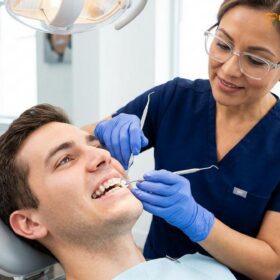
When patients mention, “i have my teeth checking by the dentist,” they are referring to a comprehensive clinical dental examination. This routine procedure involves diagnostic imaging, periodontal assessment, and professional prophylaxis to detect early signs of decay, prevent gum disease, and maintain optimal oral health.
Clinical Summary:
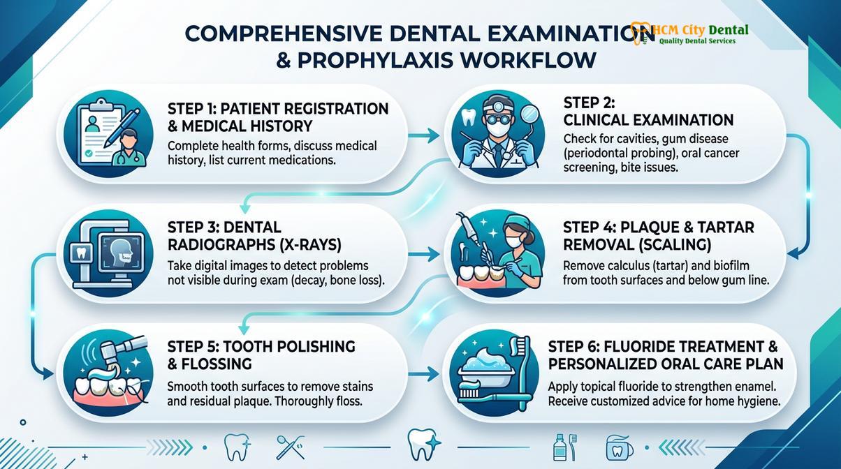
A comprehensive dental examination is a foundational diagnostic procedure in modern dentistry designed to evaluate the complete oral and maxillofacial complex. The clinical workflow encompasses a meticulous visual and tactile inspection, periodontal probing to assess gingival health, and radiographic analysis to detect interproximal caries and alveolar bone pathology. Following the diagnostic phase, a professional dental cleaning (prophylaxis) is performed using ultrasonic and hand instrumentation to remove biofilm and calculus. This preventative approach is critical for mitigating the progression of dental caries and periodontal disease, while also serving as a primary screening mechanism for oral cancer and systemic health indicators manifesting in the oral cavity.
Key Takeaways:
- A standard dental check-up includes visual inspection, periodontal charting, and radiographic imaging.
- Professional prophylaxis utilizes ultrasonic scaling to remove hardened calculus that cannot be eliminated by brushing alone.
- Routine examinations are crucial for the early detection of oral cancer and asymptomatic dental caries.
- Financial planning for dental care involves understanding insurance coverage and alternative payment structures.
- Systemic conditions, such as diabetes and cardiovascular disease, are closely linked to periodontal health.
The Clinical Workflow of a Comprehensive Dental Examination
A comprehensive dental examination follows a systematic workflow, including visual inspection, periodontal probing, and radiographic imaging to evaluate the overall health of teeth and surrounding tissues.
The foundation of preventative dentistry lies in the comprehensive oral evaluation. When a patient presents to the clinic for a routine check-up, the clinician initiates a highly structured diagnostic protocol. This process is not merely a cursory glance at the dentition; it is a rigorous medical assessment of the hard and soft tissues of the oral cavity, the temporomandibular joint (TMJ), and the associated maxillofacial structures. According to Dr. Nguyen Van Cuong at HCMC Dental Clinic, a thorough examination is the most critical step in formulating an accurate, individualized treatment plan that addresses both immediate concerns and long-term oral health stability[1].
Visual and Tactile Inspection
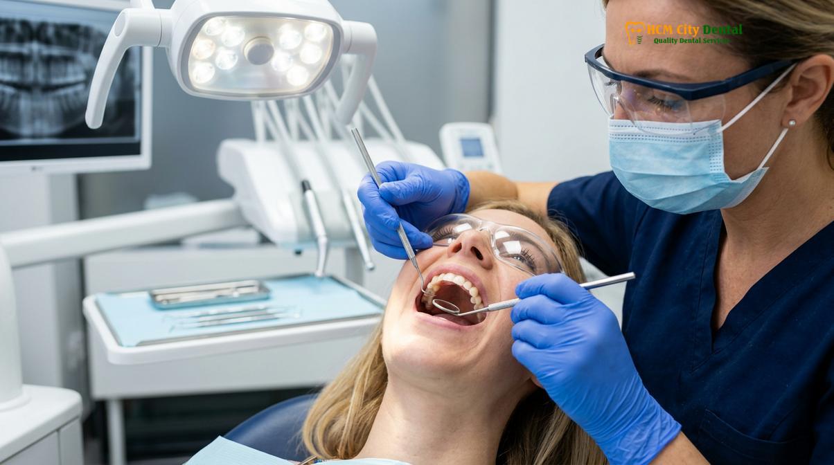
The initial phase of the examination involves a meticulous visual and tactile inspection. The clinician utilizes a dental mirror, an explorer, and high-intensity operatory lighting to evaluate each tooth surface. The primary objective is to identify clinical signs of dental caries, defective restorations, enamel attrition, abrasion, and erosion. The explorer, a specialized tactile instrument, is gently guided over the occlusal fissures and smooth surfaces of the teeth. A “catch” or softening of the enamel indicates demineralization and potential carious lesions. Furthermore, the clinician assesses the integrity of existing dental work, checking for marginal leakage around crowns, bridges, and composite fillings.
Beyond the hard tissues, the visual inspection encompasses a thorough evaluation of the oral mucosa, including the buccal mucosa, palate, floor of the mouth, and the lateral borders of the tongue. Any abnormalities, such as leukoplakia (white patches), erythroplakia (red patches), or ulcerations that have not healed within a standard timeframe, are documented and may warrant further investigation or biopsy. This soft tissue evaluation is a critical component of opportunistic oral cancer screening.
Periodontal Probing and Charting
Following the visual assessment, the clinician performs periodontal probing to evaluate the health of the periodontium—the specialized tissues that surround and support the teeth. Using a calibrated periodontal probe, the clinician measures the depth of the gingival sulcus (the space between the tooth and the gum tissue) at six specific sites around each tooth. Healthy sulcular depths typically range from 1 to 3 millimeters. Depths of 4 millimeters or greater often indicate the presence of periodontal pockets, which are clinical markers of gingival inflammation and potential alveolar bone loss[2].
During this process, the clinician also records instances of bleeding on probing (BOP), which is a primary indicator of active gingival inflammation. The accumulation of these metrics allows the dentist to diagnose the patient’s periodontal status, ranging from localized gingivitis to advanced periodontitis. Accurate periodontal charting is essential for determining the appropriate type of dental cleaning required, whether it be a standard prophylaxis or therapeutic scaling and root planing.
Radiographic Analysis (X-rays)
While visual and tactile inspections are indispensable, they are limited to the visible surfaces of the teeth and gums. Radiographic analysis is mandatory for a complete diagnostic picture. Dental X-rays allow the clinician to visualize the interproximal spaces (areas between the teeth), the subgingival root structures, and the supporting alveolar bone. Bitewing radiographs are typically taken to detect interproximal caries and evaluate the crestal bone levels. Periapical radiographs provide a comprehensive view of the entire tooth, from the crown to the apex of the root, enabling the detection of periapical abscesses, cysts, and endodontic pathology.
In modern clinical settings, digital radiography has largely replaced traditional film, offering significant advantages. Digital X-rays require substantially less radiation exposure, provide immediate high-resolution images, and allow for digital enhancement (such as adjusting contrast and magnification) to improve diagnostic accuracy[3]. For patients requiring a broader overview of the maxillofacial complex, a panoramic radiograph or a Cone Beam Computed Tomography (CBCT) scan may be prescribed to evaluate the TMJ, impacted wisdom teeth, and overall bone architecture.
Professional Dental Cleaning (Prophylaxis) Explained
Professional dental cleaning involves the meticulous removal of plaque and calculus using ultrasonic scalers and hand instruments, followed by polishing to prevent periodontal disease.
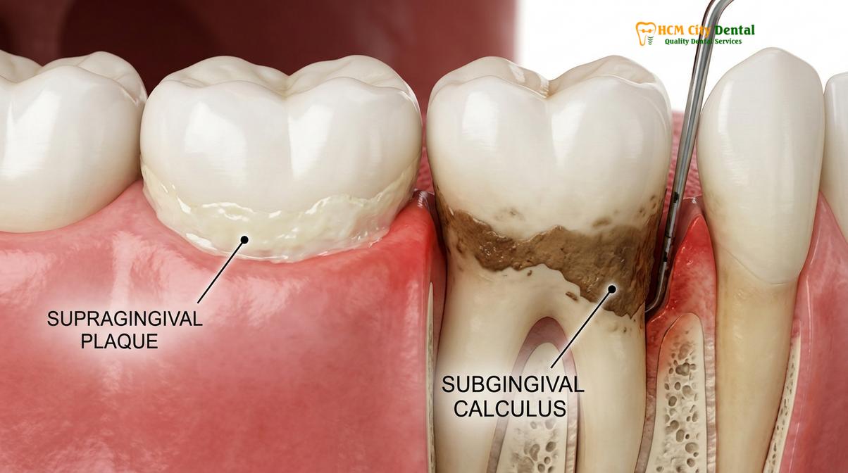
A cornerstone of the routine dental visit is the professional prophylaxis, commonly referred to as a Dental Cleaning. Even with rigorous home care, including twice-daily brushing and daily flossing, microscopic bacterial biofilm (plaque) inevitably accumulates in hard-to-reach areas. Over time, this plaque mineralizes through the precipitation of calcium and phosphate ions from saliva, forming calculus (tartar). Calculus is a porous, calcified substance that harbors pathogenic bacteria and cannot be removed by a standard toothbrush. Its presence acts as a chronic irritant to the gingival tissues, initiating an inflammatory cascade that leads to gingivitis and, if left untreated, periodontitis.
Ultrasonic Scaling and Hand Instrumentation
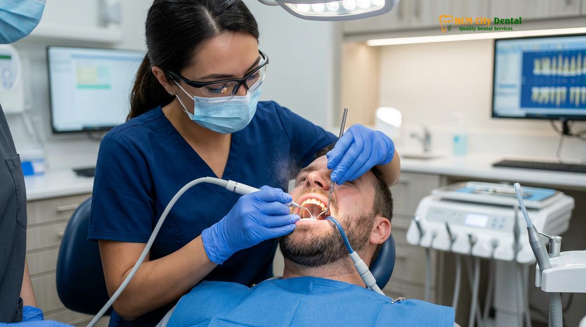
The removal of calculus is achieved through a process known as scaling. Modern dental prophylaxis heavily relies on ultrasonic scalers. These sophisticated devices utilize high-frequency vibrational energy (typically ranging from 25,000 to 30,000 cycles per second) to fracture and dislodge calculus deposits from the tooth surfaces. The ultrasonic tip is continuously cooled by a stream of water, which serves a dual purpose: it prevents thermal damage to the tooth and creates a phenomenon known as cavitation. The rapid formation and collapse of microscopic water bubbles generate shockwaves that disrupt bacterial cell walls, effectively flushing the periodontal pockets of pathogens and endotoxins.
Following ultrasonic debridement, the clinician utilizes specialized hand instruments, such as curettes and scalers, to perform fine scaling. Hand instrumentation allows for precise tactile feedback, enabling the clinician to detect and remove residual calculus deposits, particularly in subgingival areas and complex root anatomies. The meticulous combination of ultrasonic and hand scaling ensures that the tooth surfaces are completely free of calcified irritants, creating a biologically acceptable environment for the gingival tissues to heal and reattach.
“The mechanical disruption of the oral biofilm and the complete removal of calculus are the most critical interventions in arresting the progression of periodontal disease and maintaining the integrity of the dentogingival junction.” — Journal of Periodontology
Polishing and Fluoride Application
Once the tooth surfaces are thoroughly debrided, the next step is coronal polishing. This procedure utilizes a slow-speed handpiece equipped with a soft rubber cup and a specialized prophylaxis paste. The paste contains mild abrasives that gently remove extrinsic stains caused by coffee, tea, tobacco, and certain foods. Beyond aesthetic improvements, polishing creates a microscopically smooth enamel surface, which makes it more difficult for new bacterial biofilm to adhere to the teeth.
The final step in many preventative workflows is the application of professional-grade fluoride. Fluoride therapy is a highly effective pharmacological intervention for caries prevention. When applied topically via gels, foams, or varnishes, fluoride ions integrate into the hydroxyapatite structure of the enamel, converting it into fluorapatite. Fluorapatite is significantly more resistant to the acidic byproducts generated by cariogenic bacteria. Furthermore, fluoride promotes the remineralization of early, non-cavitated carious lesions, effectively reversing the initial stages of tooth decay.
Financial Considerations and Dental Coverage
Understanding the financial aspects of dental care involves evaluating insurance policies, average treatment costs, and alternative payment structures to ensure accessible oral health maintenance.
While the clinical benefits of routine dental examinations are undeniable, patients must also navigate the financial aspects of oral healthcare. A common barrier to consistent preventative care is a lack of understanding regarding treatment costs and insurance coverage. Patients frequently inquire about the average cost of dental services to budget appropriately for their bi-annual visits. Addressing these financial considerations transparently is essential for fostering a trusting patient-provider relationship.
Navigating Dental Insurance
For many patients, the first step in managing healthcare expenses is to find dental insurance that aligns with their clinical needs and financial capabilities. Dental insurance operates differently from standard medical insurance; it is primarily designed to emphasize preventative care rather than catastrophic coverage. Most comprehensive dental plans follow a “100-80-50” coverage structure. This typically means that preventative services, such as routine examinations, X-rays, and prophylaxis, are covered at 100%. Basic restorative procedures, like composite fillings, are often covered at 80%, while major procedures, such as crowns or a dental bridge, are covered at 50%.
When evaluating policies, patients must consider the dental insurance cost, which includes monthly premiums, annual deductibles, and yearly maximums. A common question during financial consultations is, “how much is dental insurance going to save me annually?” The answer depends heavily on the patient’s oral health status. For individuals requiring only preventative maintenance, a basic plan may suffice. However, patients with a history of extensive restorative needs may benefit from a premium plan with higher annual maximums, despite the higher monthly cost.
Out-of-Pocket Expenses and Alternatives
For patients without insurance coverage, understanding the out-of-pocket expenses is crucial. The cost of a comprehensive examination, full-mouth series of X-rays, and a standard cleaning can vary significantly based on geographic location and the specific technologies utilized by the clinic. To accommodate uninsured individuals, many practices offer in-house dental membership plans. These plans typically require an annual fee that covers all preventative services and provides a percentage discount on restorative treatments, offering a predictable alternative to traditional insurance.
Additionally, patients facing financial hardship may seek a sliding scale dentist or community health center. These facilities adjust their fees based on the patient’s income and family size, ensuring that essential preventative and diagnostic services remain accessible to underserved populations. Regardless of the payment method, investing in routine preventative care is universally more cost-effective than addressing advanced dental pathology, such as severe infections requiring endodontic therapy or extractions.
| Service Category | Typical Clinical Procedure | Standard Insurance Coverage | Primary Clinical Objective |
|---|---|---|---|
| Preventative | Comprehensive Exam & Prophylaxis | Usually 100% (No Deductible) | Early detection, biofilm removal, caries prevention |
| Diagnostic | Bitewing & Panoramic X-rays | Usually 100% (Frequency limits apply) | Identify interproximal decay and bone pathology |
| Basic Restorative | Composite Fillings & Simple Extractions | Typically 80% (After Deductible) | Restore tooth structure, eliminate active infection |
| Major Restorative | Crowns, Bridges, & Prosthetics | Typically 50% (After Deductible) | Rehabilitate occlusal function and structural integrity |
Advanced Diagnostics and Treatment Planning
Modern dental check-ups utilize advanced diagnostics, including 3D imaging and oral cancer screenings, to formulate precise, individualized treatment plans for complex dental conditions.
The landscape of the routine dental examination has been revolutionized by the integration of advanced diagnostic technologies. These tools allow clinicians to detect pathology at its earliest, most microscopic stages, shifting the paradigm of dentistry from reactive treatment to proactive, minimally invasive management. At facilities like HCMC Dental Clinic in Ho Chi Minh City, the integration of digital workflows ensures that every patient receives a comprehensive evaluation that leaves no aspect of their oral health to chance.
Oral Cancer Screening
One of the most critical, yet often overlooked, components of a comprehensive dental examination is the oral cancer screening. Oral squamous cell carcinoma is a aggressive malignancy that can develop on the tongue, floor of the mouth, lips, and oropharynx. Early detection is paramount, as the survival rate drops significantly if the cancer is diagnosed in advanced stages. During the screening, the clinician physically palpates the lymph nodes of the neck and the submandibular region to check for lymphadenopathy (swollen nodes) or indurated (hardened) masses.
In addition to visual and tactile assessments, many modern clinics utilize adjunctive screening devices based on tissue fluorescence technology. These devices emit a specific wavelength of blue light into the oral cavity. Healthy mucosal tissues exhibit a distinct fluorescence pattern, while dysplastic (precancerous) or malignant tissues absorb the light differently, appearing as dark lesions. This technology enhances the clinician’s ability to identify suspicious cellular changes long before they become visible to the naked eye[4].
“The integration of systematic oral cancer screening protocols into routine dental examinations is a fundamental responsibility of the dental practitioner, significantly improving early detection rates and patient survival outcomes.” — Oral Surgery, Oral Medicine, Oral Pathology and Oral Radiology
Bite Analysis and Orthodontic Evaluation
A comprehensive examination also includes a thorough evaluation of the patient’s occlusion (bite) and the functional harmony of the temporomandibular joint (TMJ). Malocclusion—such as crowding, crossbites, overbites, or underbites—can lead to a cascade of dental issues, including accelerated enamel wear, periodontal trauma, and chronic myofascial pain. The clinician assesses the alignment of the dental arches and the dynamic movement of the jaw during function.
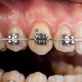
If significant occlusal discrepancies are identified, the dentist may recommend an orthodontic consultation. Modern orthodontics offers a variety of solutions, from traditional mechanics to advanced clear aligner therapy. Patients interested in understanding the biomechanics of tooth movement can explore how braces are made and the clinical engineering behind self-ligating systems. Correcting malocclusion is not merely an aesthetic endeavor; it is a functional necessity that improves the patient’s ability to maintain oral hygiene and distributes masticatory forces evenly across the dentition.
Clinical Case Study: The Value of Routine Diagnostics
A 34-year-old patient visited HCMC Dental Clinic in Ho Chi Minh City for a routine bi-annual examination, reporting no pain or sensitivity. During the visual inspection, the dentition appeared healthy. However, digital bitewing radiographs revealed a deep, asymptomatic carious lesion on the distal surface of the maxillary right first molar, encroaching on the pulp chamber. Because the decay was detected before it breached the pulp, Dr. Cuong was able to restore the tooth with a conservative composite restoration, sparing the patient from a complex root canal therapy and a subsequent crown. This case underscores the indispensable role of radiographic imaging in detecting hidden pathology during routine check-ups.
The Interconnection Between Oral Health and Systemic Conditions
Clinical evidence demonstrates a bidirectional relationship between periodontal health and systemic conditions, emphasizing the importance of routine dental examinations in overall medical management.
The oral cavity is not an isolated system; it is the primary gateway to the body, and its health is intricately linked to overall systemic well-being. A comprehensive dental examination serves as a critical screening tool for systemic diseases, as many medical conditions manifest early signs within the mucosal tissues and periodontium. Understanding this oral-systemic connection highlights why routine dental visits are a vital component of comprehensive healthcare.
One of the most extensively researched connections is the bidirectional relationship between periodontal disease and diabetes mellitus. Patients with uncontrolled diabetes are significantly more susceptible to severe periodontitis due to impaired immune function and altered collagen metabolism. Conversely, chronic periodontal inflammation can exacerbate insulin resistance, making it more difficult for diabetic patients to achieve glycemic control. Routine dental cleanings and periodontal maintenance are essential interventions that can help stabilize blood sugar levels in diabetic individuals[5].
Furthermore, there is a well-documented association between periodontal pathogens and cardiovascular disease. The chronic inflammation associated with periodontitis can lead to transient bacteremia—the entry of oral bacteria into the bloodstream. These pathogens can adhere to atheromatous plaques in the coronary arteries, potentially contributing to atherosclerosis and increasing the risk of myocardial infarction and stroke. By maintaining optimal oral hygiene and adhering to a schedule of professional prophylaxis, patients can actively reduce their systemic inflammatory burden.
When to See a Doctor for an Immediate Dental Check-Up
While routine bi-annual examinations are sufficient for maintaining oral health in most patients, certain clinical signs and symptoms necessitate immediate professional intervention. Delaying care when acute pathology is present can lead to the rapid progression of infection, irreversible tissue damage, and systemic complications. Patients should not wait for their scheduled check-up if they experience any of the following warning signs.
Important Clinical Considerations: Seek Immediate Evaluation If You Experience:
- Spontaneous or Throbbing Pain: Unprovoked dental pain, especially pain that wakes you up at night, is a classic symptom of irreversible pulpitis or an acute periapical abscess requiring immediate endodontic evaluation.
- Severe Gingival Bleeding or Swelling: While mild bleeding during flossing can indicate early gingivitis, profuse bleeding, purulent exudate (pus), or localized swelling of the gums suggests a severe periodontal infection.
- Facial Swelling or Lymphadenopathy: Swelling that extends to the face, jaw, or neck, accompanied by fever or difficulty swallowing, is a medical emergency indicating a spreading odontogenic infection that requires immediate antibiotic therapy and surgical drainage.
- Trauma or Tooth Mobility: Any physical trauma resulting in a fractured, luxated (displaced), or avulsed (knocked-out) tooth requires emergency dental care to maximize the chances of saving the dentition.

Dr. Cuong advises that any sudden changes in the oral cavity, including unexplained lesions, persistent bad breath (halitosis), or sudden sensitivity to hot and cold stimuli, should be evaluated promptly. Early diagnostic intervention is the key to conservative, successful treatment outcomes. For patients considering aesthetic enhancements after ensuring their foundational oral health is stable, a consultation regarding porcelain veneers or other restorative options can be integrated into the treatment plan.
To schedule your comprehensive dental examination and ensure your oral health is meticulously maintained, contact the clinical team at HCMC Dental Clinic today. Our specialists are dedicated to providing evidence-based diagnostics and personalized preventative care.
Frequently Asked Questions
How often should I have my teeth checked by a dentist?
Clinical guidelines generally recommend a comprehensive dental examination every six months. However, patients with a history of periodontal disease, high caries risk, or systemic conditions like diabetes may require evaluations every three to four months. Your dentist will determine the optimal frequency based on a personalized risk assessment and your specific clinical needs.
Does a routine dental check-up and cleaning hurt?
A routine dental examination and prophylaxis are generally painless. Patients with severe gingival inflammation or dentin hypersensitivity may experience mild, transient discomfort, which can be managed with topical anesthetics or desensitizing agents during the procedure. Modern ultrasonic scalers are designed to maximize efficiency while minimizing patient discomfort.
What is the difference between a routine check-up and a deep cleaning?
A routine check-up includes a standard prophylaxis for supragingival plaque removal. A deep cleaning, or scaling and root planing, is a therapeutic procedure targeting subgingival calculus and infected root surfaces in patients with active periodontal disease. Deep cleanings often require local anesthesia and are performed over multiple appointments to ensure thorough debridement.
Can a dentist detect other health issues during an exam?
Yes, a comprehensive oral examination can reveal signs of systemic conditions. Dentists frequently identify early indicators of diabetes, nutritional deficiencies, autoimmune disorders, and cardiovascular issues through the assessment of mucosal tissues and periodontal health. The oral cavity often mirrors the overall health of the body, making dentists crucial partners in systemic healthcare.
Are dental X-rays safe during a routine check-up?
Dental X-rays are highly safe and utilize minimal radiation, especially with modern digital radiography. The diagnostic benefits of detecting interproximal decay, bone loss, and pathology far outweigh the negligible risks associated with the low radiation exposure. Clinicians adhere to the ALARA (As Low As Reasonably Achievable) principle, utilizing lead aprons and thyroid collars for added protection.
References
- Journal of the American Dental Association. Clinical guidelines for the frequency of dental examinations and prophylaxis. (2021).
- Journal of Periodontology. The pathogenesis of periodontal disease and the efficacy of mechanical debridement. (2020).
- International Dental Journal. Diagnostic accuracy of digital radiography in detecting early carious lesions. (2019).
- Oral Surgery, Oral Medicine, Oral Pathology and Oral Radiology. Protocols for comprehensive oral cancer screening in clinical practice. (2022).
- Clinical Oral Investigations. The bidirectional relationship between periodontal health and systemic diseases. (2018).