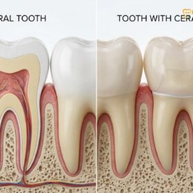
A crown after a root canal is clinically recommended to protect the treated tooth from fracturing and reinfection. Because endodontic therapy removes the living pulp, the remaining tooth structure becomes brittle. Placing a custom dental crown restores its structural integrity, bite force, and natural aesthetic appearance.
Clinical Summary:
Following root canal treatment, a tooth loses its blood supply and hydration, significantly increasing its susceptibility to structural fractures under chewing forces. A dental crown acts as a protective helmet, distributing occlusal stress evenly and sealing the access cavity against bacterial microleakage. While anterior teeth (incisors and canines) may sometimes only require a composite restoration if sufficient tooth structure remains, posterior teeth (premolars and molars) almost universally mandate full-coverage crowns due to the heavy masticatory loads they bear. Modern restorative dentistry utilizes high-strength materials like monolithic Zirconia and E.max porcelain to ensure long-term durability and seamless aesthetics. Timely placement—typically within two to four weeks post-endodontic therapy—is critical to prevent catastrophic vertical root fractures that could necessitate extraction.
Key Takeaways:
- Root-treated teeth become brittle and require structural reinforcement to withstand daily chewing forces.
- Posterior teeth (molars and premolars) almost always need a crown, while anterior teeth may occasionally be restored with fillings alone.
- Delaying crown placement increases the risk of irreparable vertical root fractures and bacterial reinfection.
- Zirconia and porcelain-fused-to-metal (PFM) are the most durable materials for restoring posterior teeth.
- Properly crowned root-canaled teeth can last 10 to 15 years or longer with excellent oral hygiene.
Why a Crown is Clinically Necessary After Root Canal Therapy
A crown provides essential structural reinforcement and a bacterial seal for a root-treated tooth, preventing catastrophic fractures and endodontic failure.
To fully comprehend why a custom dental crown is so frequently prescribed following endodontic therapy, one must understand the profound biomechanical changes that occur within the tooth during and after the procedure. Root canal treatment is a highly successful, tooth-saving intervention designed to eradicate infection from the pulpal chamber and root canals. However, the very nature of this procedure alters the physical properties of the tooth.
The Loss of Vitality and Hydration
The dental pulp is the living heart of the tooth, containing nerves, blood vessels, and connective tissue. It provides continuous hydration and nutrients to the surrounding dentin. When the pulp is irreversibly inflamed or infected, it must be completely extirpated (removed) during the root canal procedure. Once the pulp is gone, the tooth is rendered non-vital. Without its internal blood supply, the dentin gradually loses its moisture content, becoming significantly more brittle and prone to desiccation over time[1]. This brittleness means the tooth is less capable of absorbing and dissipating the heavy forces generated during mastication (chewing).
Alteration of Tooth Architecture
Beyond the loss of hydration, the physical architecture of the tooth is compromised. To access the infected pulp, the endodontist or general dentist must drill an access cavity through the occlusal (biting) surface of the tooth. This access preparation, combined with the removal of any pre-existing decay or old filling materials, significantly reduces the volume of healthy, intact tooth structure. The roof of the pulp chamber, which normally acts as a structural cross-tie holding the walls of the tooth together, is removed. Consequently, the remaining walls of the tooth are left unsupported and highly vulnerable to flexing and fracturing under occlusal loads.
“The primary cause of failure in root-canaled teeth is not endodontic reinfection, but rather structural fracture due to the lack of timely coronal coverage and biomechanical reinforcement.”
The Loss of Proprioception
Another critical, yet often overlooked, factor is the loss of proprioception. Vital teeth possess nerve endings in the periodontal ligament and the pulp that provide sensory feedback to the brain regarding how hard we are biting. When a tooth is root-canaled, its internal sensory mechanism is eliminated. Patients often unknowingly exert significantly more biting force on a non-vital tooth because they lack the protective reflex that prevents over-stressing the tooth. This increased force, applied to a weakened and brittle structure, creates a perfect storm for a catastrophic fracture if the tooth is not protected by a crown.
As Dr. Nguyen Van Cuong frequently advises patients, the success of endodontic therapy is intrinsically linked to the quality of the final coronal restoration. The root canal saves the foundation, but the crown saves the tooth itself.
Which Teeth Require a Crown After Endodontic Treatment?
Posterior teeth almost universally require crowns due to heavy chewing forces, whereas anterior teeth may only need a crown if significant tooth structure is lost.
While a crown is the gold standard for restoring most root-treated teeth, the absolute necessity depends heavily on the tooth’s anatomical location, its function in the dental arch, and the amount of residual healthy tooth structure[2].
Posterior Teeth: Molars and Premolars
The posterior teeth—the molars and premolars—are the workhorses of the mouth. They are responsible for the heavy lifting of grinding and crushing food. The occlusal forces exerted on these teeth can exceed 150 to 200 pounds of pressure per square inch. Because of their multi-cusp anatomy and their position near the temporomandibular joint (the hinge of the jaw), they absorb the maximum impact during mastication.
For molars and premolars, a crown after a root canal is almost universally mandatory. Attempting to restore a root-canaled posterior tooth with only a composite filling is highly risky. The cusps of the tooth can easily wedge apart under pressure, leading to a vertical root fracture—a devastating complication that almost always requires the complete extraction of the tooth.
Anterior Teeth: Incisors and Canines
The anterior teeth—the incisors and canines—serve a different function. They are designed for shearing and tearing food, rather than heavy grinding. Consequently, they are subjected to significantly lower occlusal forces. Furthermore, the access cavity for a root canal on an anterior tooth is typically made through the lingual (back) surface, leaving the entire facial (front) surface and the incisal edge intact.
If an anterior tooth has undergone a root canal but remains structurally robust—meaning it has not suffered extensive decay, large previous fillings, or trauma—it may sometimes be successfully restored with a bonded composite resin restoration rather than a full-coverage crown. However, if the anterior tooth is heavily discolored (a common side effect of pulpal necrosis), fractured, or has large existing restorations, a crown or Porcelain Veneers may be required to restore both strength and optimal aesthetics.
| Tooth Type | Primary Function | Occlusal Force | Crown Recommendation Post-Root Canal |
|---|---|---|---|
| Molars | Grinding and crushing | Very High | Highly Recommended / Mandatory. High risk of vertical fracture without full coverage. |
| Premolars | Tearing and initial grinding | High | Highly Recommended. Prone to cusp fractures due to anatomical shape. |
| Canines | Tearing and guiding the bite | Moderate | Case-Dependent. Crown needed if heavily decayed or if guiding function is compromised. |
| Incisors | Cutting and shearing | Low to Moderate | Case-Dependent. May be restored with composite if structural integrity is largely intact. |
Timing: How Long Should You Wait to Get a Crown?
Dentists typically recommend placing a permanent crown within two to four weeks after a root canal to minimize the risk of tooth fracture.
The timing of crown placement following endodontic therapy is a critical factor in the long-term survival of the tooth. Patients often wonder if they can delay the crown procedure, especially if the tooth feels fine and is no longer causing pain. However, clinical guidelines strongly advise against prolonged delays.
The Initial Healing Phase
Immediately following a root canal, the tooth is typically sealed with a temporary filling material. The surrounding periapical tissues (the bone and ligaments at the tip of the root) may be slightly inflamed from the infection and the instrumentation used during the procedure. Dentists generally recommend a brief observation period—usually two to four weeks—before proceeding with the permanent crown. This window allows the inflammation to subside, ensures that the tooth is asymptomatic, and confirms that the endodontic treatment was successful.
The Risks of Delayed Restoration
While a short waiting period is beneficial, extending this delay for months or years is highly detrimental. Temporary fillings are exactly that—temporary. They are not designed to withstand the harsh environment of the oral cavity over the long term. Over time, temporary materials can wear down, wash out, or develop micro-cracks.
Clinical Warning: Delaying crown placement beyond a few months leaves the temporary filling vulnerable to degradation, risking bacterial microleakage and vertical root fractures that often require complete tooth extraction.
If the temporary seal fails, saliva and oral bacteria can seep back into the root canal system, re-infecting the gutta-percha (the rubber-like material used to fill the canals). This phenomenon, known as coronal leakage, is a leading cause of endodontic failure and may necessitate a complex retreatment. Furthermore, every day the tooth remains unprotected by a crown is a day it is at risk of fracturing under normal chewing forces. For patients who travel frequently, understanding Temporary Dental Crown Care While Traveling: Clinical Guide is essential to prevent premature failure of the temporary seal before the final restoration can be placed.
Types of Dental Crowns for Root-Canaled Teeth
Material selection depends on the tooth’s location and aesthetic needs, with Zirconia offering maximum strength for molars and E.max providing superior aesthetics for front teeth.
Modern restorative dentistry offers a variety of advanced materials for fabricating dental crowns. The choice of material depends on several clinical factors, including the tooth’s location, the patient’s bite force, aesthetic requirements, and the presence of parafunctional habits like bruxism (teeth grinding)[3].
Monolithic Zirconia Crowns
Zirconia (zirconium dioxide) is a highly durable, biocompatible ceramic material that has revolutionized restorative dentistry. Monolithic zirconia crowns are milled from a single solid block of material, offering exceptional flexural strength (often exceeding 1000 MPa). This makes them virtually indestructible under normal chewing forces, rendering them the premier choice for restoring posterior molars that have undergone root canal therapy. Furthermore, modern zirconia can be shaded and characterized to blend seamlessly with natural teeth, overcoming the opaque appearance of older generations of the material.
Lithium Disilicate (E.max) Crowns
Lithium disilicate, commonly known by the brand name E.max, is a glass-ceramic material renowned for its unparalleled aesthetic properties. It mimics the natural translucency, opalescence, and light-reflecting characteristics of natural enamel better than almost any other material. While not quite as strong as monolithic zirconia, E.max is still highly durable and is the material of choice for anterior teeth (incisors and canines) and premolars where aesthetic demands are paramount.
Porcelain-Fused-to-Metal (PFM) Crowns
For decades, PFM crowns were the standard of care in restorative dentistry. They consist of a strong metal alloy substructure overlaid with tooth-colored porcelain. PFM crowns offer a reliable balance of strength and aesthetics. However, they have some drawbacks. The underlying metal can sometimes create a dark line at the gum margin, especially if gum recession occurs over time. Additionally, the porcelain overlay can occasionally chip or fracture under heavy occlusal loads. Patients often ask, Are Porcelain Fused to Metal Crowns Safe? Clinical Guide—the answer is yes, they remain a highly viable and safe option, particularly for patients requiring long-span bridges or those with specific occlusal challenges.
Gold and Metal Alloys
Although less popular today due to aesthetic concerns, cast gold or base metal alloy crowns remain the most conservative and durable restorations available. They require the least amount of tooth preparation, meaning more of the natural tooth structure is preserved. Gold is highly biocompatible, gentle on opposing teeth, and will never fracture. For patients who suffer from severe bruxism and do not mind a metallic appearance on their second or third molars, gold remains an outstanding clinical choice.
The Clinical Workflow: Placing a Crown After Root Canal
The restoration process involves a core build-up, precise tooth preparation, digital or physical impressions, and the final cementation of the custom crown.
The process of restoring a root-canaled tooth is a meticulous, multi-step clinical workflow designed to ensure maximum retention, structural integrity, and marginal adaptation. The procedure typically requires two appointments, though clinics equipped with CAD/CAM technology may offer same-day restorations.
Step 1: The Post and Core Build-Up
Because a root-canaled tooth has lost a significant amount of its internal structure, there is often not enough healthy dentin remaining to support and retain a crown. To solve this, the dentist must perform a “core build-up.” This involves using a strong composite resin material to replace the missing dentin and rebuild the foundation of the tooth.
In cases where the tooth structure is severely compromised, a dental post may be required. The dentist removes a small amount of gutta-percha from one of the treated root canals and cements a post (usually made of flexible fiberglass or cast metal) into the canal space. The post acts as an anchor, providing retention for the composite core material that is built up around it. It is important to note that a post does not strengthen the root; rather, it simply retains the core, which in turn retains the crown[4].
Clinical Case Study: A 45-year-old patient visited HCMC Dental Clinic in Ho Chi Minh City with a fractured upper molar following a delayed crown placement post-root canal. Dr. Nguyen Van Cuong performed a thorough evaluation using 3D CBCT imaging. Fortunately, the fracture had not reached the root bifurcation. A custom cast post and core were fabricated, followed by the placement of a monolithic Zirconia crown. The patient regained full masticatory function and reported no sensitivity, highlighting the importance of timely and precise restorative intervention.
Step 2: Margination and Tooth Preparation
Once the core is securely in place, the dentist prepares the tooth to receive the crown. This involves circumferentially reducing the enamel and dentin (or the core material) by 1.0 to 2.0 millimeters, depending on the chosen crown material. The goal is to create a precise margin (the ledge where the crown will sit) and to shape the tooth into a miniature, tapered version of itself. This specific geometry ensures that the crown can slide over the tooth and be retained by mechanical friction as well as dental cement.
Step 3: Impressions and Temporization
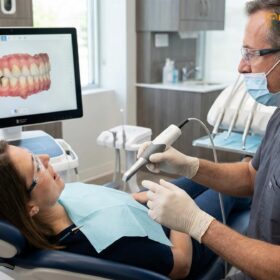
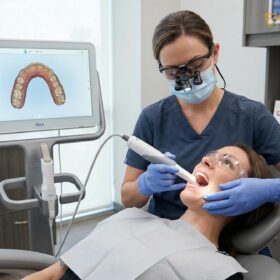
After the tooth is prepared, an accurate impression must be taken to capture the exact dimensions of the preparation and the surrounding teeth. Modern clinics increasingly use digital intraoral scanners to capture highly accurate 3D optical impressions, eliminating the need for messy traditional putty. The digital file is then sent to a dental laboratory where skilled technicians design and mill the final crown. While the permanent crown is being fabricated, a temporary acrylic or composite crown is cemented onto the tooth to protect it, maintain the space, and provide aesthetic continuity.
Step 4: Final Cementation and Occlusal Adjustment
At the final insertion appointment, the temporary crown is carefully removed, and the tooth is cleaned. The permanent crown is tried in to verify the marginal fit, interproximal contacts (how it touches neighboring teeth), and aesthetics. Once the fit is confirmed, the tooth is isolated, etched, and bonded. The crown is then permanently affixed using a high-strength dual-cure resin cement or a glass ionomer cement. Finally, the dentist meticulously checks the patient’s occlusion (bite) using articulating paper, making micro-adjustments to ensure the new crown harmonizes perfectly with the opposing teeth.
Longevity and Care for Your Restored Tooth
With meticulous oral hygiene and regular dental check-ups, a crowned root-canaled tooth can function optimally for 10 to 15 years or longer.
A root-canaled tooth restored with a high-quality crown is designed to be a long-term, highly durable solution. Clinical studies indicate that with proper care, these restorations boast survival rates exceeding 90% over a ten-year period. However, the longevity of the restoration is heavily dependent on the patient’s home care and routine professional maintenance[5]. For patients exploring international options for high-quality restorations, understanding Thailand Dental Crowns: Clinical Guide & SEA Tourism can provide valuable context on global standards of care.
Maintaining Marginal Integrity
The most vulnerable part of any dental crown is the margin—the microscopic junction where the artificial crown meets the natural tooth structure. While the ceramic or metal crown itself cannot decay, the exposed natural root surface below the margin is highly susceptible to bacterial acid attacks. If plaque is allowed to accumulate at the gum line, secondary caries (new decay) can develop underneath the crown, potentially destroying the remaining tooth structure and leading to the failure of the entire restoration.
“A root-canaled tooth is immune to hot and cold sensitivity, but the surrounding gingival tissues remain susceptible to periodontal disease if plaque is not meticulously managed.”
Best Practices for Post-Operative Care
To maximize the lifespan of a crowned, root-treated tooth, patients must adhere to strict oral hygiene protocols:
- Meticulous Brushing: Brush twice daily using a soft-bristled toothbrush and fluoridated toothpaste, paying special attention to the gum line around the crowned tooth.
- Interdental Cleaning: Floss daily or use interdental brushes to remove plaque and food debris from the tight spaces between the teeth where a toothbrush cannot reach.
- Avoid Destructive Habits: Refrain from using teeth as tools (e.g., opening packages, biting nails) and avoid chewing excessively hard items like ice, hard candies, or unpopped popcorn kernels, which can chip porcelain or fracture the underlying root.
- Night Guards for Bruxism: Patients who clench or grind their teeth at night should wear a custom-fitted occlusal splint (night guard) to protect the ceramic crown from excessive wear and to shield the root from stress fractures.
Occasionally, patients may experience transient discomfort after the final cementation. Understanding Tooth Crown Pain Chewing Sensitivity: Clinical Causes & Solutions can help differentiate between normal post-operative settling and issues that require clinical adjustment.
When to See a Doctor
While a crowned, root-canaled tooth should function seamlessly, certain symptoms warrant immediate clinical evaluation. You should schedule an appointment with your dentist or endodontist if you experience any of the following:
- Pain Upon Biting: If you feel a sharp, localized pain when applying pressure to the tooth, it could indicate that the crown’s bite is too high and requires adjustment, or more seriously, it could be a sign of a vertical root fracture.
- Swelling or Fistula: The appearance of swelling in the gums adjacent to the tooth, or a small pimple-like bump (a fistula) that drains pus, is a strong indicator of a recurrent periapical infection or a missed root canal anatomy.
- Mobility: If the crowned tooth feels loose, it may indicate a failure of the cement seal, a fracture of the post and core, or advanced periodontal bone loss.
- Foul Taste or Odor: A persistent bad taste or smell originating from the specific tooth can signal that the crown margin has leaked, allowing bacteria to ferment debris underneath the restoration.

Frequently Asked Questions
Can I just get a filling instead of a crown after a root canal?
While a filling may suffice for anterior teeth with minimal structural loss, posterior teeth almost always require a crown. Molars and premolars bear the brunt of chewing forces, and a simple filling cannot provide the necessary structural reinforcement to prevent a brittle, root-treated tooth from fracturing under pressure.
Does getting a crown after a root canal hurt?
The crown preparation and placement process is generally painless because the tooth no longer contains living nerve tissue following the root canal. However, the surrounding gum tissue may experience mild, temporary inflammation from the impression process or the placement of the retraction cord, which resolves quickly.
How long should I wait to get a crown after endodontic therapy?
Most dental professionals recommend placing the permanent crown within two to four weeks after the root canal procedure. This brief waiting period allows any periapical inflammation to subside and ensures the endodontic treatment is successful before permanently sealing the tooth with a custom restoration.
What happens if I wait too long to get a crown?
Delaying crown placement significantly increases the risk of catastrophic tooth fracture and bacterial reinfection. Temporary fillings are not designed for long-term wear; they can degrade, wash out, or leak, allowing oral bacteria to re-enter the root canal system and potentially necessitating a complete retreatment or extraction.
Can a tooth with a root canal and crown still get a cavity?
Yes, while the artificial crown itself cannot decay, the underlying natural tooth structure at the gum line remains vulnerable to plaque and bacteria. If oral hygiene is neglected, secondary caries can develop at the margin where the crown meets the tooth, potentially undermining the entire restoration.
References
- Journal of Endodontics. Biomechanical principles of restoring endodontically treated teeth. (2021).
- International Endodontic Journal. Survival rates of root-filled teeth restored with various coronal restorations. (2020).
- Journal of Prosthetic Dentistry. The ferrule effect and its importance in restorative dentistry. (2019).
- Clinical Oral Investigations. Fracture resistance of monolithic zirconia vs lithium disilicate crowns. (2022).
- Journal of the American Dental Association. Long-term outcomes of post and core build-ups in posterior teeth. (2018).