Porcelain veneers are ultra-thin ceramic shells bonded to the facial surfaces of teeth to correct discoloration, chipping, or misalignment. They provide a highly aesthetic, minimally invasive solution for smile makeovers, ensuring durable functional outcomes when supported by precise clinical diagnostics and proper bonding protocols.
Clinical Summary:
Porcelain veneers represent a cornerstone of modern aesthetic dentistry, offering predictable and conservative restoration of the anterior dentition. The clinical success of these restorations depends heavily on meticulous case selection, precise tooth preparation confined strictly to the enamel, and advanced adhesive bonding techniques. By utilizing high-strength ceramics like lithium disilicate or highly translucent feldspathic porcelain, clinicians can achieve exceptional biomimetic results. Comprehensive occlusal analysis and patient compliance with maintenance protocols are critical to ensuring the longevity of the restorations, preventing complications such as microleakage, debonding, or ceramic fractures over time.
Key Takeaways:
- Veneers require minimal enamel reduction, typically between 0.3mm and 0.5mm, preserving tooth structure.
- Lithium disilicate (E.max) offers superior fracture resistance for functional anterior restorations.
- Strict isolation using a rubber dam is mandatory during the adhesive bonding phase to prevent contamination.
- Patients with severe bruxism or insufficient enamel are generally contraindicated for delicate laminates.
- Properly maintained porcelain veneers can function aesthetically for over a decade without degradation.
Understanding Porcelain Veneers and Biomimetic Aesthetics
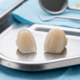
Porcelain veneers are custom-fabricated ceramic laminates designed to replace the buccal enamel, offering a conservative approach to modifying tooth shape, color, and alignment while preserving structural integrity.
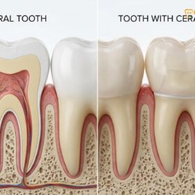
The evolution of aesthetic dentistry has shifted dramatically toward minimally invasive procedures that prioritize the preservation of natural tooth structure. Porcelain Veneers, also known as ceramic laminates, epitomize this biomimetic approach. Unlike traditional full-coverage restorations, which require circumferential reduction of the tooth, veneers are designed to replace only the visible facial enamel. This conservative preparation maintains the biomechanical integrity of the tooth while allowing the clinician to dramatically alter the patient’s aesthetic appearance. The primary goal is to mimic the optical properties of natural enamel, including its translucency, opalescence, and surface texture.
Patients often inquire about the potential for a long dental lifespan when investing in aesthetic restorations. The longevity of a porcelain veneer is intrinsically linked to the substrate to which it is bonded. Enamel provides a highly predictable, strong, and durable bond when treated with phosphoric acid and modern adhesive resins. When a veneer is bonded entirely to enamel, the risk of debonding or marginal leakage is virtually eliminated. Conversely, if the preparation extends deeply into the dentin, the bond strength decreases significantly due to the higher organic content and moisture present in dentinal tubules. Therefore, the clinical objective is always to remain within the enamel layer, which typically ranges from 0.5mm to 1.0mm in thickness on the facial aspect of anterior teeth.[1]
The optical integration of ceramic veneers is achieved through a complex interplay of material science and clinical artistry. Natural teeth exhibit a gradient of color and translucency, with the cervical third being more chromatic and opaque, while the incisal third is highly translucent. Modern dental ceramics, such as feldspathic porcelain and lithium disilicate, can be layered or monolithically milled to replicate this natural gradient. The clinician must carefully select the appropriate ceramic material based on the underlying tooth color, the desired final shade, and the functional demands of the patient’s occlusion.
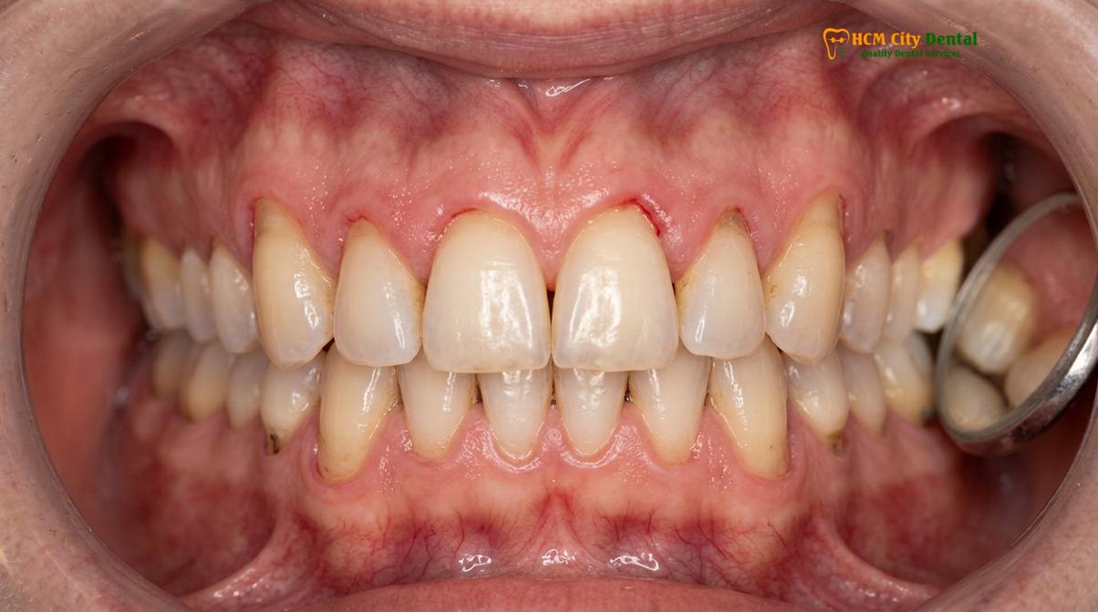
Furthermore, the success of these restorations is not solely dependent on the ceramic material itself, but also on the composite resin cement used for bonding. Light-cured resin cements offer excellent color stability and allow for extended working times, enabling the clinician to meticulously seat and clean the restorations before final polymerization. The combination of a high-quality ceramic, a precise enamel preparation, and a flawless bonding protocol ensures that the patient receives a restoration that is both functionally robust and aesthetically indistinguishable from natural dentition.
Clinical Indications for Aesthetic Veneers
Ideal candidates for veneers present with intrinsic staining, mild malocclusion, diastemas, or structural defects that do not compromise the overall biomechanical stability of the tooth.
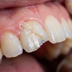
The decision to prescribe porcelain veneers requires a comprehensive clinical assessment of the patient’s aesthetic concerns, periodontal health, and occlusal dynamics. One of the most common indications for veneers is severe intrinsic discoloration that is unresponsive to traditional chemical bleaching protocols. Conditions such as tetracycline banding, severe fluorosis, or endodontic discoloration often require the masking capability of ceramic laminates. In these cases, the clinician must carefully balance the need for opacity to hide the underlying stain with the need for translucency to maintain a natural appearance. This often involves the use of specialized masking ceramics or deeper preparations to allow for adequate ceramic thickness.[2]
Another primary indication is the correction of morphological anomalies, such as microdontia (e.g., peg laterals), amelogenesis imperfecta, or generalized spacing (diastemas). Veneers provide an excellent modality for closing spaces and restoring ideal tooth proportions without the need for prolonged orthodontic therapy. However, it is crucial to recognize that veneers are not a substitute for orthodontics in cases of severe crowding or malocclusion. Attempting to correct significant misalignment with veneers alone can lead to excessive tooth reduction on the prominent teeth and excessively bulky restorations on the lingually displaced teeth, compromising both aesthetics and periodontal health.
Patients with a history of minor chipping or wear may also benefit from ceramic laminates, provided the underlying cause of the wear is identified and managed. If the wear is due to active parafunctional habits (bruxism), placing delicate ceramic veneers without addressing the occlusal forces will inevitably lead to catastrophic failure. In such scenarios, a comprehensive occlusal rehabilitation or the strict use of a protective occlusal guard is mandatory. The clinician must evaluate the anterior guidance and the envelope of function to ensure that the new restorations will not introduce occlusal interferences.
“Meticulous patient selection is the absolute foundation of aesthetic dentistry. We must evaluate not just the individual teeth, but the entire dynamic relationship of the lips, gingiva, and facial symmetry before ever touching a handpiece.”
Periodontal health is an absolute prerequisite for any restorative procedure, particularly those involving the gingival margins. The presence of active gingivitis or periodontitis will result in bleeding during the preparation and bonding phases, severely compromising the adhesive protocols. Furthermore, inflamed gingival tissues will recede following the resolution of the inflammation, potentially exposing the restorative margins and creating an aesthetic failure. Therefore, a thorough periodontal evaluation and necessary therapy must precede the initiation of any veneer case.
The Diagnostic and Preparation Workflow
The clinical workflow involves comprehensive digital smile design, conservative enamel preparation, precise elastomeric or digital impressions, and the fabrication of provisional restorations to guide the final outcome.
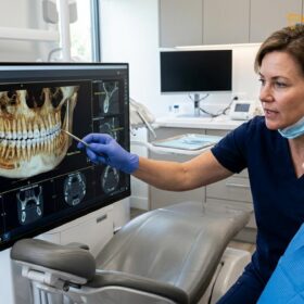
The contemporary workflow for aesthetic restorations begins long before the teeth are prepared. It starts with a comprehensive data collection phase, which includes high-resolution clinical photography, digital intraoral scanning, and often a facebow transfer to mount the diagnostic casts on a semi-adjustable articulator. This data is utilized to perform a Digital Smile Design (DSD), a process where the clinician digitally plans the new smile based on facial proportions, lip dynamics, and the golden ratio of tooth dimensions. The DSD allows the patient to visualize the proposed outcome and provides the dental laboratory with a precise blueprint for the diagnostic wax-up.
Once the diagnostic wax-up is completed, it is transferred to the patient’s mouth using a silicone matrix filled with bis-acrylic provisional material. This “mock-up” serves as a critical communication tool, allowing the patient to evaluate the aesthetics, phonetics, and function of the proposed restorations in real-time. Any necessary adjustments are made to the mock-up before proceeding. More importantly, the mock-up serves as a preparation guide. By preparing the teeth through the mock-up (the “shrink-wrap” technique), the clinician ensures that only the absolute minimum amount of enamel is removed to achieve the desired final volume, maximizing enamel preservation.
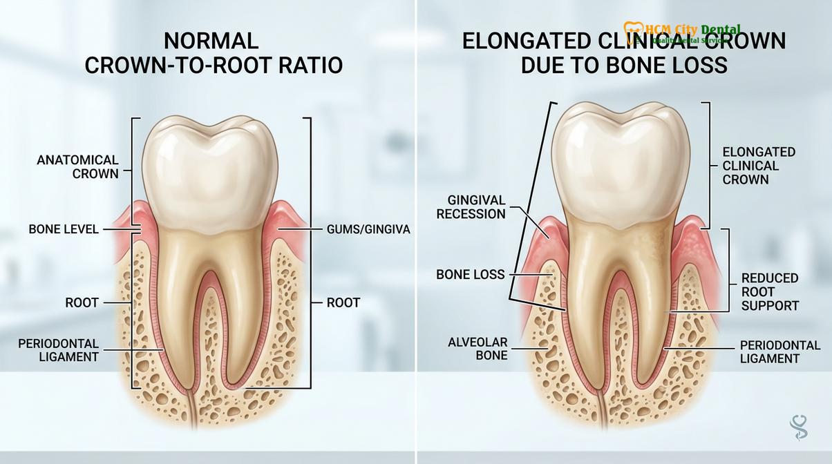
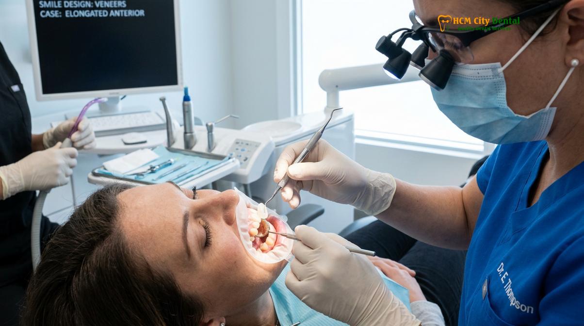
The actual tooth preparation for a ceramic veneer is a highly precise procedure that requires magnification and specialized diamond burs. The standard preparation involves a 0.3mm to 0.5mm reduction of the facial surface, a 1.5mm to 2.0mm incisal reduction to allow for ceramic layering and translucency, and a smooth, continuous chamfer margin placed at or slightly above the gingival crest. Understanding the difference between a dental crown and a veneer is crucial here, as crowns require significantly more aggressive circumferential reduction.
Following the preparation, precise impressions are captured using either traditional polyvinyl siloxane (PVS) materials or advanced digital intraoral scanners. Digital impressions offer the advantage of immediate evaluation of the preparation margins and occlusal clearance, reducing the likelihood of remakes. The final step in the preparation appointment is the fabrication of provisional restorations. These temporaries protect the prepared teeth, prevent sensitivity, and maintain the gingival architecture while the final ceramics are being fabricated by the laboratory. Patients must be instructed on how to care for temporary restorations, as they are inherently fragile and bonded with weak temporary cement.
Material Science: E.max, Zirconia, and Feldspathic Porcelain
Contemporary veneers primarily utilize feldspathic porcelain for unparalleled optical properties or lithium disilicate for enhanced flexural strength, depending on the patient’s functional requirements.
The selection of the appropriate ceramic material is a critical decision that dictates both the aesthetic outcome and the biomechanical durability of the restoration. Historically, feldspathic porcelain was the gold standard for veneers due to its exceptional optical properties. Feldspathic ceramics are created by meticulously layering glass-based powders and firing them in a porcelain furnace. This technique allows the master ceramist to incorporate internal characterizations, mamelons, and varying degrees of translucency, resulting in a restoration that perfectly mimics natural enamel. However, feldspathic porcelain has a relatively low flexural strength (approximately 90-120 MPa), making it highly susceptible to fracture if not bonded perfectly to a rigid enamel substrate.[3]
In recent years, lithium disilicate (commonly known by the brand name IPS e.max) has become the material of choice for the majority of anterior veneer cases. Lithium disilicate is a glass-ceramic that offers a remarkable balance of aesthetics and strength. It can be either pressed from an ingot or milled using CAD/CAM technology. With a flexural strength ranging from 360 to 400 MPa, lithium disilicate is significantly more robust than feldspathic porcelain, making it suitable for patients with higher functional demands or those requiring slightly more extensive restorations. While monolithic lithium disilicate is highly aesthetic, it can also be cut back and layered with fluorapatite veneering porcelain to achieve the ultimate optical vitality.
| Ceramic Material | Flexural Strength (MPa) | Optical Properties | Primary Clinical Indication |
|---|---|---|---|
| Feldspathic Porcelain | 90 – 120 MPa | Exceptional translucency and vitality | Highly aesthetic anterior cases with abundant enamel |
| Lithium Disilicate (E.max) | 360 – 400 MPa | Excellent aesthetics, versatile opacity | Standard anterior veneers, moderate functional risk |
| Zirconia (High Translucency) | 600 – 800 MPa | Good aesthetics, lower translucency | High bruxism risk, masking dark substrates |
Zirconia, a polycrystalline ceramic known for its immense strength, is generally not the first choice for pure aesthetic veneers. Zirconia lacks the glass phase necessary for traditional hydrofluoric acid etching and silanization, making adhesive bonding more challenging and less predictable than with silica-based ceramics. However, the development of highly translucent, cubic-phase zirconia has expanded its use in the anterior region. Zirconia may be indicated for patients with a history of severe bruxism or when attempting to mask extremely dark underlying tooth structure, though it is more commonly utilized for full-coverage smile makeovers rather than ultra-thin laminates.
Adhesive Bonding Protocols: The Key to Clinical Success
Successful integration of ceramic veneers relies on strict isolation, hydrofluoric acid etching of the intaglio surface, silanization, and the use of light-cured resin cements.
The adhesive bonding of porcelain veneers is arguably the most technique-sensitive procedure in restorative dentistry. The long-term success of the restoration is entirely dependent on the creation of a flawless, moisture-free bond between the ceramic, the resin cement, and the tooth structure. Any contamination by saliva, blood, or crevicular fluid during the bonding sequence will lead to immediate bond failure, subsequent microleakage, and eventual debonding of the veneer. Therefore, strict isolation using a rubber dam is considered the standard of care for this procedure.
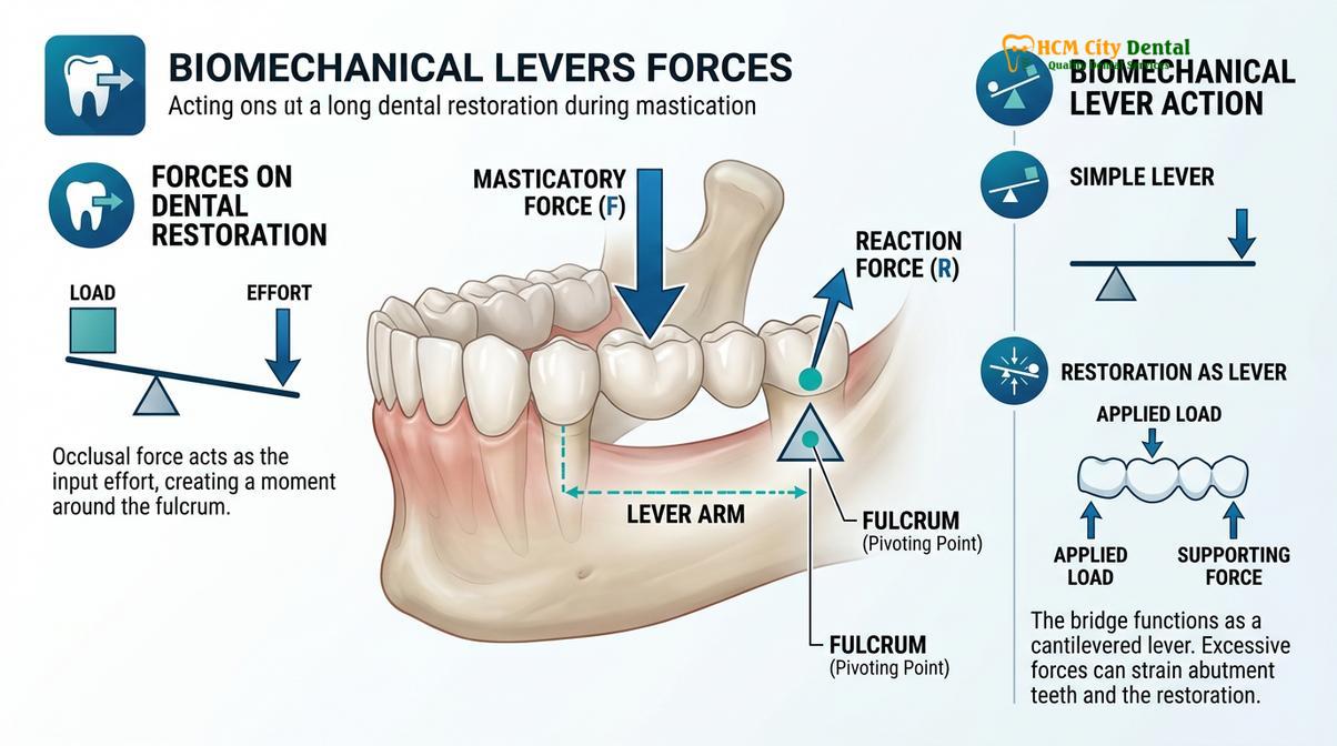
The bonding protocol begins with the meticulous preparation of the ceramic surface. For silica-based ceramics (feldspathic and lithium disilicate), the intaglio (internal) surface of the veneer is etched with hydrofluoric acid. This acid selectively dissolves the glass matrix, creating a micro-porous surface that provides mechanical retention for the resin cement. The etching time varies depending on the material; lithium disilicate typically requires 20 seconds of etching with 5% hydrofluoric acid, while feldspathic porcelain may require 90 seconds with a 9% concentration. Following etching, the ceramic is thoroughly rinsed, dried, and treated with a silane coupling agent. Silane acts as a chemical bridge, bonding the inorganic silica in the ceramic to the organic methacrylate groups in the resin cement.[4]
The tooth surface is simultaneously treated with phosphoric acid to etch the enamel, followed by the application of a dentin bonding agent. The veneer is then carefully seated using a light-cured or dual-cured resin cement. Excess cement is meticulously removed before final light polymerization. Proper marginal adaptation is crucial during this phase to prevent microleakage and ensure the long-term periodontal health of the restored tooth.[5]
Case Study: Aesthetic Rehabilitation in Ho Chi Minh City
A clinical case demonstrating the successful application of biomimetic principles and advanced ceramic materials to restore anterior aesthetics and function.
A recent case study at HCMC Dental Clinic in Ho Chi Minh City highlights the efficacy of this biomimetic approach. A patient presented with severe tetracycline staining and mild anterior spacing, seeking a comprehensive aesthetic rehabilitation. At HCMC Dental Clinic, Dr. Nguyen Van Cuong emphasizes the importance of biomimetic principles in restorative dentistry. Dr. Cuong meticulously evaluates each patient’s unique enamel structure to ensure that aesthetic enhancements do not compromise underlying tooth health. His approach integrates advanced digital smile design with conservative preparation techniques, delivering outcomes that are both beautiful and functionally sound.
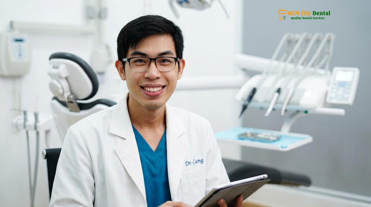
Following a detailed digital smile design, ultra-thin lithium disilicate veneers were fabricated. The conservative preparation allowed for optimal bonding entirely within the enamel layer, resulting in a vibrant, natural-looking smile that fully addressed the patient’s concerns while maintaining structural integrity. The patient’s successful treatment at HCMC Dental Clinic underscores the importance of combining high-quality materials with precise clinical execution.
When to Consult a Dentist: Important Clinical Considerations
Recognizing the signs of structural damage or aesthetic failure is critical for timely intervention and the preservation of natural dentition.
Patients experiencing structural damage, severe discoloration, or persistent tooth sensitivity should consult a qualified prosthodontist to explore their restorative options. According to guidelines aligned with the Vietnam Odonto-Stomatology Association, early intervention and proper case selection are vital for preventing further enamel degradation. It is important to note that not all patients are ideal candidates for delicate ceramic laminates; those with severe bruxism or insufficient remaining enamel may require alternative treatments to achieve a stable, long-lasting result.
“The longevity of any ceramic restoration is dictated not just by the material’s inherent strength, but by the precision of the adhesive protocol and the patient’s commitment to daily maintenance.”
For personalized advice on aesthetic restorations, contact HCMC Dental Clinic in Ho Chi Minh City to schedule a comprehensive evaluation and discover how modern biomimetic dentistry can transform your smile.
References
- Journal of Prosthetic Dentistry. Biomimetic restorative dentistry and enamel preservation techniques.
- International Journal of Periodontics and restorative dentistry. Long-term survival rates of lithium disilicate veneers.
- Dental Materials. Adhesion protocols for silica-based ceramics in anterior restorations.
- Journal of the American Dental Association. Clinical guidelines for aesthetic smile design and proportion.
- Clinical Oral Investigations. Marginal adaptation and microleakage in ceramic laminate veneers.