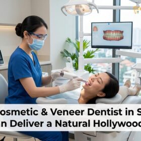
Replacing old veneers involves carefully removing degraded ceramic or composite material and bonding new, highly aesthetic restorations to preserve the underlying tooth structure. This procedure addresses marginal leakage, structural cracks, and aesthetic shifts, ensuring long-term functional stability and a renewed, natural-looking smile.
Clinical Summary:
The process of replacing old veneers is a highly precise clinical procedure designed to address failing restorations, aesthetic degradation, and underlying structural issues. Over time, older generation materials may suffer from marginal leakage, chipping, or discoloration, necessitating safe removal and replacement. Modern prosthodontic workflows utilize advanced digital scanning (such as the iTero Element 5D) and premium materials like E.max lithium disilicate or Zirconia HT to create ultra-thin, highly translucent replacements. By strictly adhering to biological width principles and employing minimal-prep techniques, clinicians can safely remove the old ceramic while preserving maximum natural enamel, resulting in a durable, lifelike, and biologically harmonious smile makeover.
Key Takeaways:
- Lifespan: Older porcelain restorations typically require replacement after 10-15 years due to marginal wear or aesthetic shifts.
- Safe Removal: Specialized magnification and precision instruments allow for the removal of old material without damaging the underlying enamel.
- Advanced Materials: Upgrading to modern E.max or Zirconia provides superior translucency, strength, and a more natural appearance.
- Digital Precision: Intraoral scanners eliminate messy impressions, ensuring a flawless marginal fit for the new restorations.
- Gum Health: Replacing ill-fitting restorations resolves dark lines and inflammation, restoring the natural biological width.
Clinical Indicators for Replacing Older Cosmetic Restorations
Recognizing the signs of failing restorations early prevents underlying tooth decay and structural damage. Clinical indicators include compromised margins, aesthetic degradation, and mechanical failure.
Dental restorations, while highly durable, are subject to the harsh biomechanical environment of the oral cavity. Over time, constant exposure to occlusal forces, thermal fluctuations, and acidic dietary components can compromise the integrity of older cosmetic work. Understanding the clinical indicators for replacing old veneers is crucial for maintaining both oral health and aesthetic harmony. The decision to pursue Porcelain Veneers as a replacement is typically driven by a combination of biological, mechanical, and aesthetic factors[1].
One of the primary reasons patients seek replacement is the natural aging of the materials. Older generation composites and early ceramics lacked the sophisticated optical properties and fracture toughness of modern materials. As these materials age, they may lose their surface glaze, becoming porous and susceptible to deep intrinsic staining that cannot be resolved through traditional whitening procedures. Furthermore, the underlying natural teeth may undergo physiological color changes as dentin naturally darkens with age, creating a mismatch between the restoration and the adjacent natural dentition.
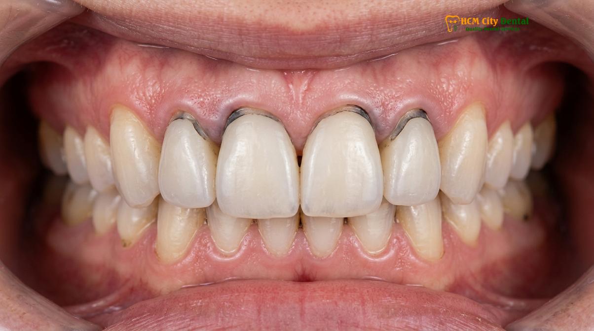
Dr. Nguyen Van Cuong, a leading specialist at HCMC Dental Clinic, emphasizes that the assessment of old restorations must go beyond mere aesthetics. “When evaluating a patient for replacing old ceramic, we must meticulously examine the marginal integrity, the health of the surrounding periodontium, and the structural soundness of the underlying abutment tooth,” notes Dr. Cuong. This comprehensive diagnostic approach ensures that the new restorations are placed on a healthy, stable foundation.
Another critical indicator is a change in the patient’s occlusal relationship. Over a decade or more, natural wear, minor tooth shifting, or the development of parafunctional habits like bruxism can alter the way the teeth come together. Restorations that were perfectly balanced when initially placed may begin to experience premature contacts or excessive shear forces, leading to micro-fractures or complete debonding. In such cases, replacing the restorations provides an opportunity to re-establish proper anterior guidance and occlusal harmony, often integrating a comprehensive Digital Smile Design (DSD) to optimize both function and form.
Marginal Leakage and Dark Lines Around the Gums
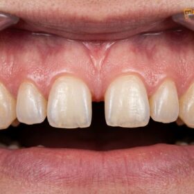
Dark lines at the gumline often indicate marginal leakage, where the adhesive bond has degraded, allowing bacteria to penetrate and stain the interface.
One of the most common and visually distressing signs of a failing restoration is the appearance of a dark line or shadow at the gingival margin. This phenomenon is frequently caused by marginal leakage, a process where the microscopic seal between the ceramic material and the natural tooth structure begins to break down. When this adhesive bond degrades, it creates a micro-gap that allows oral fluids, bacteria, and dietary pigments to infiltrate the space beneath the restoration[2].
The resulting marginal leakage stain is not merely an aesthetic issue; it is a significant biological concern. The bacteria that colonize this micro-gap produce acidic byproducts that can rapidly demineralize the vulnerable dentin or cementum, leading to secondary caries (decay) beneath the restoration. Because this decay is hidden beneath the opaque ceramic, it can progress silently, often remaining undetected until it causes significant structural damage or pulpal inflammation. Therefore, proactive margin decay prevention is a primary objective when planning the replacement of compromised restorations.
“Microleakage at the restoration-tooth interface remains the primary etiology for secondary caries and the premature failure of aesthetic bonded restorations. Achieving a flawless marginal seal through modern adhesive protocols is paramount for long-term clinical success.”
In older restorations, particularly those utilizing porcelain-fused-to-metal (PFM) techniques or early opaque ceramics, the dark line may also be attributed to the material itself. If the gingival tissue recedes even slightly, the opaque margin or the underlying metal substructure becomes visible. Modern replacement protocols eliminate this issue by utilizing highly translucent, all-ceramic materials like E.max (lithium disilicate) or Zirconia HT. These materials allow light to transmit naturally through the margin, creating a seamless, invisible transition between the restoration and the root surface, even if minor gingival changes occur in the future.
Addressing marginal leakage requires the meticulous and safe removal of the old material, followed by thorough caries excavation and the re-establishment of a pristine bonding surface. Clinicians utilize advanced isolation techniques, such as rubber dams, to ensure a completely dry and uncontaminated field during the final cementation protocol. This rigorous approach to adhesive dentistry ensures a durable, hermetic seal that protects the underlying tooth structure for decades.
Gum Recession: Exposing the Natural Root Edge
Gingival recession exposes the unveneered root surface, creating a visible aesthetic discrepancy and increasing the risk of root caries and dentinal hypersensitivity.
The relationship between a dental restoration and the surrounding gingival tissue is delicate and dynamic. Over time, patients may experience gum recession, a condition where the gingival margin migrates apically (towards the root), exposing the natural root surface. When this occurs around a veneered tooth, it creates a stark visual contrast between the highly polished ceramic and the darker, more yellow appearance of the natural root dentin. This gum recession root exposure is a frequent catalyst for patients seeking replacement procedures.
Gum recession can be multifactorial. It may result from aggressive brushing techniques, periodontal disease, anatomical predispositions (such as thin gingival biotypes), or even iatrogenic factors related to the original restorations. If the original restorations violated the biological width—the natural protective dimension of soft tissue attached to the tooth above the bone—the body responds with chronic inflammation and subsequent tissue recession[3]. Correcting this requires a deep understanding of periodontal architecture and precise gingival zenith calibration during the design of the new restorations.
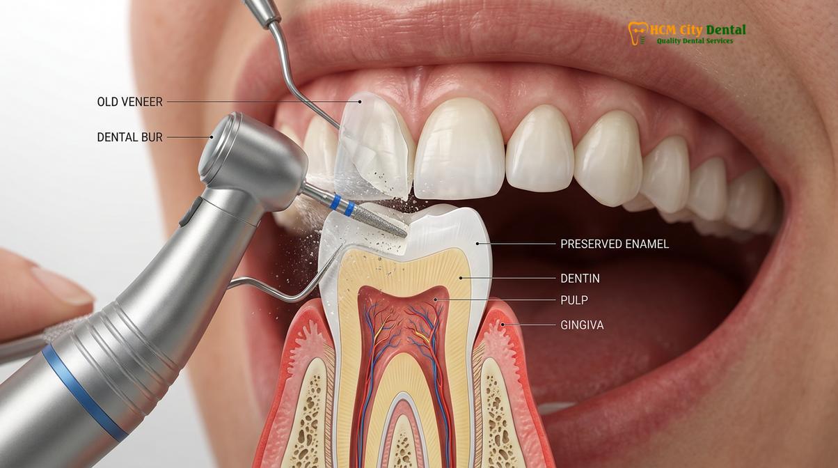
Clinical Warning: Ignoring exposed root surfaces can lead to severe dentinal hypersensitivity and rapid root caries. The root surface lacks the protective enamel layer, making it highly susceptible to acidic erosion and bacterial decay. Prompt clinical evaluation is essential to protect the structural integrity of the tooth.
When replacing restorations affected by recession, the clinician has several therapeutic options. If the recession is mild and the underlying periodontium is healthy, the new restorations can be designed with a slightly extended cervical margin to cover the exposed root, seamlessly blending into the new gingival contour. This approach requires meticulous minimal-prep techniques (often preserving 0.3mm to 0.7mm of enamel) to ensure the new margin is smooth, highly polished, and biologically compatible with the soft tissue.
In cases where the gingival architecture is highly irregular or asymmetrical, adjunctive procedures such as Laser Gum Contouring (using a soft-tissue diode laser) may be recommended prior to the final impressions. This procedure reshapes the gingival margin, creating a symmetrical and harmonious frame for the new smile. By addressing both the hard tissue (the tooth) and the soft tissue (the gums), clinicians can achieve a truly comprehensive and aesthetically superior result.
Structural Cracks, Chipping, and Failure of Adhesive Bond
Mechanical failures such as chipping or debonding occur due to excessive occlusal forces, trauma, or the natural degradation of older generation ceramic materials.
While dental ceramics are incredibly strong in compression, they can be vulnerable to tensile and shear forces. Structural cracks, chipping, or the complete failure of the adhesive bond are clear indications that a restoration has reached the end of its functional lifespan. These mechanical failures not only compromise the aesthetics of the smile but also create rough surfaces that harbor plaque and irritate the surrounding soft tissues.
The etiology of structural failure is often linked to occlusal dynamics. Patients who suffer from sleep bruxism (involuntary grinding or clenching of the teeth) place immense stress on their restorations. Over time, these repetitive forces can cause micro-fractures within the ceramic matrix, which eventually propagate and lead to visible chipping. To prevent this in the new restorations, comprehensive bite force protection is essential. This typically involves meticulous occlusal adjustment during the fitting phase and the fabrication of a custom night guard to protect the new ceramics during sleep.
Clinical Case Review: HCMC Dental Clinic
A 45-year-old patient presented to our facility in Ho Chi Minh City with multiple chipped composite restorations that were placed eight years prior. The patient exhibited signs of moderate nocturnal bruxism. The treatment plan involved the safe removal of the degraded composite, digital scanning with the iTero Element 5D, and the fabrication of six highly durable Zirconia HT restorations. Following cementation, a custom occlusal splint was provided to ensure long-term protection against parafunctional forces. The result was a restored, highly functional, and aesthetically flawless smile.
Debonding—where the restoration detaches entirely from the tooth—is another form of failure. This can occur if the original bonding protocol was compromised by moisture contamination, or if the restoration was bonded primarily to dentin rather than enamel. Enamel provides a significantly stronger and more predictable bond than dentin. When planning a replacement, the clinician must carefully evaluate the remaining tooth structure. If significant enamel has been lost, advanced adhesive protocols utilizing specific silane coupling agents and dual-cure resin cements are required to ensure the new restoration remains securely in place[4].
Upgrading to modern materials significantly reduces the risk of future structural failure. Lithium disilicate (E.max) offers exceptional flexural strength (approximately 400-500 MPa) while maintaining outstanding lifelike aesthetics. For patients with extremely heavy bites or those requiring restorations in high-stress areas, Zirconia HT (High Translucency) provides unparalleled durability (up to 1200 MPa) while still delivering excellent cosmetic results. The selection of the appropriate material is a critical step in planning your smile makeover.
The Veneer Replacement Process: How We Safely Remove Old Ceramic
Modern veneer replacement utilizes magnification and specialized burs to selectively remove old material without compromising the underlying natural enamel.
The prospect of removing old restorations can cause anxiety for some patients, but modern dental techniques have rendered the process highly safe, precise, and comfortable. The primary objective during removal is to eliminate all the old ceramic and degraded adhesive resin while strictly preserving the underlying natural tooth structure. This requires a delicate touch, high-powered magnification (dental loupes or a surgical microscope), and specialized instrumentation.
The safe veneer removal process begins with the administration of profound local anesthesia to ensure complete patient comfort. The clinician then uses specific diamond burs designed to cut through ceramic efficiently. A common technique involves creating depth grooves across the surface of the old restoration, carefully monitoring the transition between the ceramic, the adhesive layer, and the natural tooth. Because older ceramics and the underlying tooth can sometimes look similar in color, magnification and excellent illumination are critical to differentiate the layers.
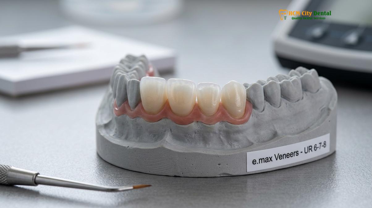
Once the bulk of the old material is removed, the clinician switches to finer, less abrasive instruments to gently polish away the remaining adhesive resin. This step is crucial for establishing a clean, pristine surface for the new bond. In some advanced clinics, erbium lasers may be utilized to degrade the adhesive bond, allowing the old restoration to be removed with minimal use of rotary instruments, further protecting the natural enamel.
| Clinical Aspect | Traditional Workflow | Modern Digital Workflow |
|---|---|---|
| Removal Technique | Standard rotary burs, higher risk of enamel loss | Magnification-guided selective removal, maximum enamel preservation |
| Impressions | Messy silicone putty, uncomfortable for patient | iTero Element 5D / Medit i700 Digital Scans (High precision, comfortable) |
| Material Selection | Opaque PFM or early generation composites | E.max Lithium Disilicate or Zirconia HT (High translucency) |
| Turnaround Time | 2-3 weeks, multiple temporary phases | Express 3-5 Day Turnaround via Direct Lab Partnership |
| Aesthetic Design | Manual wax-ups, limited patient preview | Digital Smile Design (DSD) 3D Mock-up for predictable results |
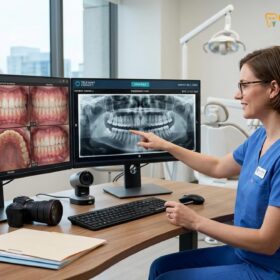
Following the complete removal of the old materials and the refinement of the tooth preparation, the digital workflow commences. State-of-the-art digital scanning technology, such as the iTero Element 5D or Medit i700 Intraoral Scanners, is used to capture highly accurate 3D images of the prepared teeth. This eliminates the need for uncomfortable traditional silicone impressions and provides the dental laboratory with flawless data to design the custom replacements. During the fabrication period, patients are fitted with aesthetic temporary restorations that protect the teeth and provide a preview of the final result.
Advanced Material Selection and Digital Workflow
Upgrading to advanced materials like lithium disilicate or high-translucency zirconia ensures superior aesthetics, enhanced durability, and precise marginal adaptation.
The success of a replacement procedure relies heavily on the quality of the materials utilized and the precision of the fabrication process. At HCMC Dental Clinic, we emphasize a Direct Lab Partnership with leading ceramists in Ho Chi Minh City. This direct collaboration between the prosthodontist and the laboratory enables complete quality control, allowing for intricate custom shading (such as matching a natural translucent BL1 or B1 shade) and precise morphological detailing.
Material Certification is a cornerstone of our practice. We utilize only authentic, globally certified materials from industry leaders in Germany and Liechtenstein. For patients prioritizing outstanding lifelike aesthetics and translucency, E.max (Ivoclar, Liechtenstein) lithium disilicate is the gold standard. For those requiring enhanced strength due to heavy occlusal forces, Zirconia HT (Cercon HT, Germany) provides an optimal balance of durability and aesthetics. Understanding the comparing porcelain and composite materials is essential for making an informed decision.
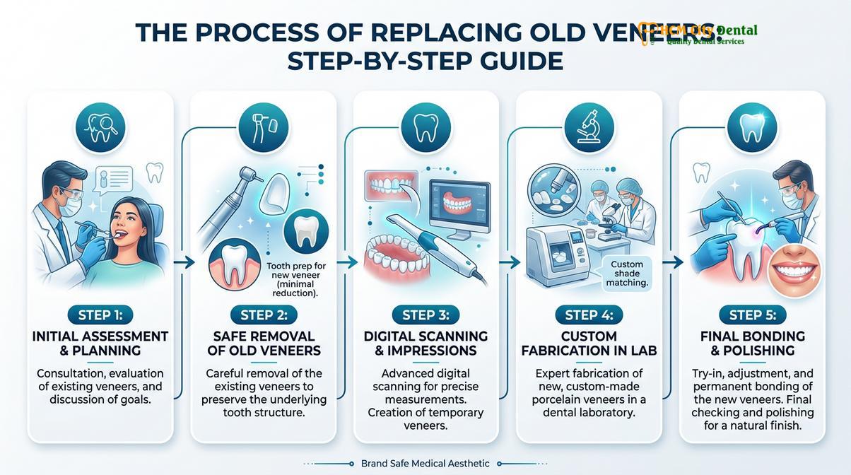
To accommodate the needs of international patients and busy professionals, we offer an Express 3-5 Day Turnaround. This streamlined workflow allows for complete smile makeovers to be fabricated, characterized, and permanently fitted within a single week, without compromising on quality or precision. Furthermore, our transparent Pricing Structure incorporates a significant 40% pre-arrival discount for WhatsApp bookings, making world-class cosmetic dentistry highly accessible:
- Composite Veneer (Chair-side): Walk-in: ~$80 | WhatsApp discount: From $48 (-40%).
- Zirconia Veneer (Cercon HT): Walk-in: ~$240 | WhatsApp discount: From $144 (-40%).
- E.max Porcelain Veneer (Ivoclar): Walk-in: ~$320 | WhatsApp discount: From $192 (-40%).
- Smile Design 3D Mock-up: Walk-in: ~$200 | WhatsApp discount: From $120 (Free if treatment is performed).
Every premium ceramic restoration is backed by Global manufacturer warranty cards, alongside a 3-year clinic bonding and fit guarantee for E.max and Zirconia. We also provide comprehensive remote follow-up support via WhatsApp, utilizing specific photo and video assessment protocols to monitor your new smile from anywhere in the world.
Long-Term Maintenance and Oral Hygiene Protocols
Maximizing the lifespan of new veneers requires strict adherence to specialized oral hygiene practices and routine professional maintenance.
Once your new restorations are permanently bonded, protecting your investment becomes the priority. While dental ceramics are impervious to biological decay, the underlying natural tooth structure and the surrounding gingival tissues remain vulnerable. A meticulous daily oral hygiene routine is essential to prevent marginal decay and maintain the health of the periodontium[5]. Understanding the lifespan of dental restorations helps set realistic expectations for long-term care.
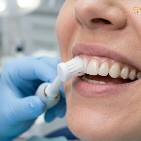
Effective plaque removal is critical. We strongly recommend the use of an electric soft toothbrush. The oscillating or sonic action of an electric brush removes plaque more efficiently than manual brushing, while the soft bristles ensure that the highly polished surface of the ceramic is not scratched or dulled over time. Abrasive whitening toothpastes should be strictly avoided, as they can degrade the surface glaze of the restorations.
“The longevity of aesthetic restorations is directly correlated with the patient’s commitment to meticulous oral hygiene and regular professional prophylaxis. Protecting the marginal seal from plaque accumulation is the most effective strategy for preventing secondary complications.”
Interdental cleaning is equally important. Mastering the correct flossing technique veneers is vital for removing bacteria from the tight spaces between the teeth and along the gumline, where the toothbrush cannot reach. Patients should use a smooth, unwaxed floss or a water flosser to gently clean these areas without snapping the floss aggressively against the margins. Additionally, incorporating a daily non alcoholic rinse helps reduce overall bacterial load without the drying effects of alcohol, which can irritate the gingival tissues and degrade composite bonding agents over time.
When to See a Doctor for Veneer Assessment
Prompt clinical evaluation is necessary if you experience pain, visible structural damage, or signs of marginal leakage around your restorations.
While high-quality restorations are designed for long-term stability, certain symptoms warrant immediate professional evaluation. You should schedule a clinical assessment if you experience any of the following:
- Persistent Sensitivity: Sharp pain when consuming hot, cold, or sweet foods may indicate that the adhesive bond has failed or that decay has developed beneath the restoration.
- Visible Cracks or Chipping: Even minor structural damage can compromise the integrity of the restoration and create rough edges that irritate the soft tissues.
- Gingival Inflammation: Red, swollen, or bleeding gums around specific teeth often point to marginal leakage, biological width violations, or plaque accumulation at the restoration margin.
- Mobility: If a restoration feels loose or shifts slightly when pressure is applied, the bonding agent has failed, and immediate recementation or replacement is required to prevent the restoration from detaching completely or being swallowed.
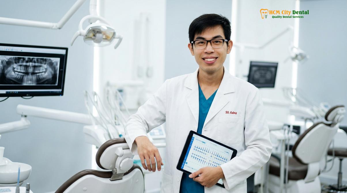
Regular professional check-ups, ideally every six months, allow your dentist to monitor the margins, assess the health of the surrounding tissues, and perform professional polishing to maintain the luster of your smile. If you are considering conservative cosmetic alternatives or need an assessment of your current restorations, our team is ready to assist.
Frequently Asked Questions
How do I know if my veneers need replacing?
You should consider replacing your veneers if you notice dark lines at the gumline, visible chipping, persistent sensitivity, or a dull, discolored appearance. A clinical examination is required to assess the integrity of the underlying tooth structure and the adhesive bond. Your dentist will use magnification and radiographs to determine if the restorations are failing mechanically or biologically.
Is it painful to remove and replace old veneers?
The procedure is generally highly comfortable, as it is performed under local anesthesia. Modern techniques utilize specialized magnification and precision instruments to gently remove the old material without causing trauma to the underlying natural enamel. Post-operative sensitivity is typically minimal and resolves quickly within a few days.
How long do older generation veneers last?
Older generation porcelain veneers typically last between 10 to 15 years, depending on the materials used and the patient’s oral hygiene habits. Composite resin veneers from older generations usually require replacement after 5 to 7 years due to staining and wear. Modern materials like E.max and Zirconia offer significantly extended lifespans when properly maintained.
Can I replace just one veneer, or do I need to replace all of them?
It is entirely possible to replace a single damaged veneer; however, achieving a perfect shade match with older, adjacent veneers can be clinically challenging. Dentists often recommend replacing them in pairs or sets to ensure a uniform, symmetrical smile, especially if the surrounding restorations are also showing signs of age or discoloration.
Will my new veneers look more natural than the old ones?
Yes, modern ceramic materials like lithium disilicate offer superior light transmission and translucency compared to older opaque porcelains. Advanced digital shade matching ensures the new restorations mimic the exact optical properties of natural enamel, resulting in a highly lifelike and vibrant appearance that blends seamlessly with your natural dentition.
References
- Journal of Esthetic and Restorative Dentistry. Translucency and optical properties of lithium disilicate. (2021).
- International Journal of Prosthodontics. Enamel preservation and bond strength in veneer replacement. (2020).
- The Journal of Prosthetic Dentistry. Flexural strength and fracture resistance of porcelain restorations. (2019).
- Clinical Oral Investigations. Diode laser gingival sculpting parameters for aesthetic dentistry. (2022).
- Journal of Clinical Periodontology. Biological width preservation around fixed restorations. (2018).