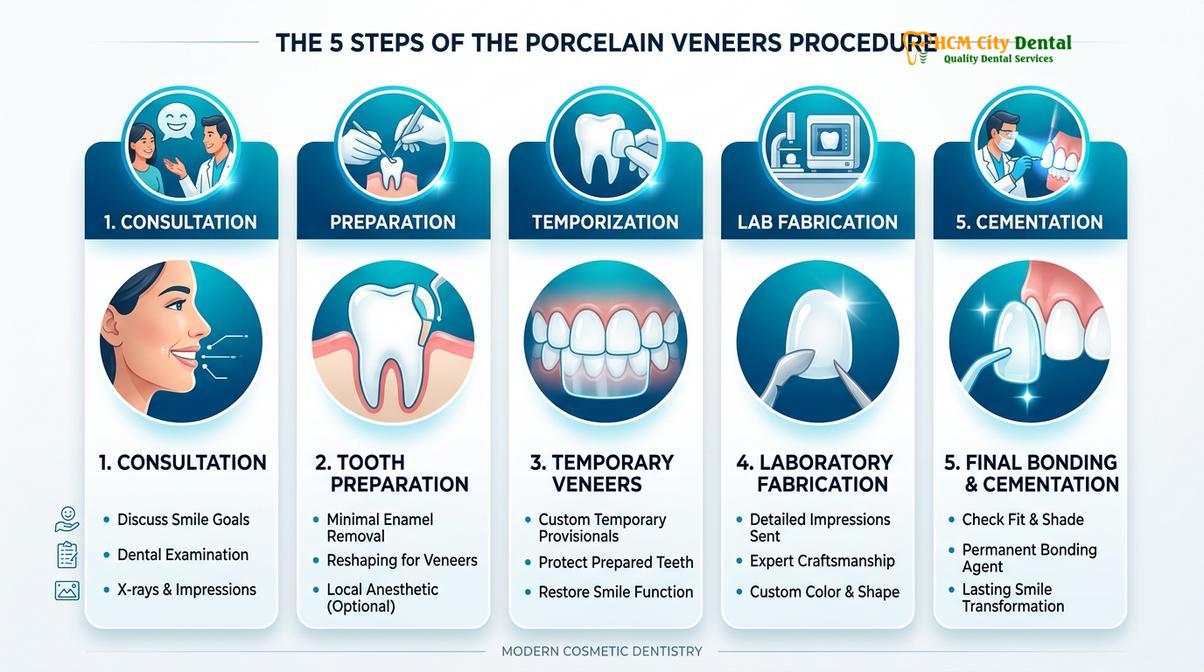
The porcelain veneers procedure is a highly precise cosmetic dental treatment that involves bonding ultra-thin ceramic shells to the facial surfaces of teeth. This minimally invasive workflow corrects discoloration, structural defects, and minor misalignments, restoring optimal dental aesthetics and biomechanical function with lifelike, durable materials.
Clinical Summary:
The clinical workflow for porcelain veneers encompasses comprehensive digital smile design, conservative enamel reduction, and advanced adhesive cementation. Utilizing state-of-the-art intraoral scanners, prosthodontists capture exact 3D topographies to fabricate custom restorations. Materials such as E.max (lithium disilicate) and Zirconia HT are selected based on occlusal stress and aesthetic demands. The procedure strictly adheres to biological width preservation and requires meticulous isolation protocols to ensure maximum bond strength at the dentin-enamel junction, resulting in a highly durable and natural-looking smile makeover.
Key Takeaways:
- Minimally Invasive: Tooth preparation typically requires only 0.3mm to 0.7mm of enamel reduction, preserving structural integrity.
- Advanced Materials: Certified E.max and Zirconia HT provide exceptional flexural strength and natural translucency.
- Digital Precision: Mess-free digital scanning ensures perfect marginal fit and optimal gingival health.
- Rapid Turnaround: Direct lab partnerships allow for complete smile makeovers within an express timeframe.
- Long-Term Durability: Strict adhesive bonding protocols yield restorations that can last over a decade with proper care.
Phase 1: Comprehensive Aesthetic Consultation and Digital Imaging
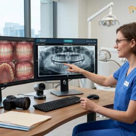
The initial consultation involves a detailed clinical assessment, digital smile design (DSD), and 3D intraoral scanning to establish a precise blueprint for your custom ceramic restorations.
The foundation of a successful porcelain veneers treatment lies in meticulous planning and comprehensive diagnostics. During the initial consultation, the prosthodontist evaluates the patient’s facial aesthetics, lip dynamics, phonetic patterns, and occlusal harmony. This holistic approach ensures that the final restorations not only look natural but also function seamlessly within the stomatognathic system.
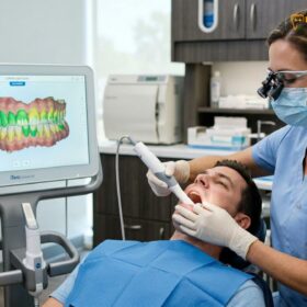
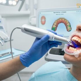
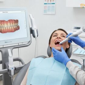
At HCMC Dental Clinic in Ho Chi Minh City, the diagnostic phase heavily relies on advanced digital technology. Traditional, uncomfortable silicone impressions have been entirely replaced by state-of-the-art digital scanning technology. These devices capture high-resolution 3D topographies of the dental arches in real-time, providing maximum comfort and unparalleled precision. The digital data is then integrated into Digital Smile Design (DSD) software, allowing the clinician to calibrate the gingival zenith, establish the ideal facial midline, and determine the optimal length and width ratios for each tooth.
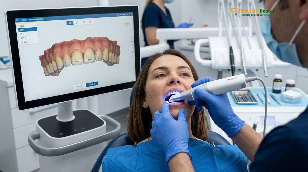
Patient communication during this phase is critical. By utilizing a 3D mock-up, individuals can visually preview their anticipated results before any irreversible treatment begins. This is also the stage where shade mapping occurs. The clinician works closely with the patient to select the ideal hue, value, and chroma, often utilizing the VITA shade guide to choose between natural translucent shades. If the patient presents with uneven gingival margins, a diode laser gum contouring procedure may be recommended to establish a symmetrical biological width prior to veneer preparation[4].
Understanding how many veneers you need is essential, as the required number depends on the width of your smile and the specific aesthetic issues being addressed. Whether correcting a single discolored tooth or performing a comprehensive eight-to-ten tooth anterior reconstruction, the digital imaging phase dictates the exact parameters for the subsequent clinical steps.
Phase 2: Tooth Preparation and Conservative Enamel Shaping
Tooth preparation involves the conservative removal of a microscopic layer of enamel to create space for the ceramic material, ensuring a flush marginal fit and optimal bond strength.
The preparation phase is arguably the most technique-sensitive aspect of the porcelain veneers procedure. The primary objective is to create sufficient restorative space for the ceramic material while preserving as much natural tooth structure as possible. To achieve this, the clinician utilizes an enamel depth-cutter bur, which creates precise horizontal grooves across the facial surface of the tooth. These depth grooves act as a strict guide, ensuring that the reduction is uniform and strictly controlled.
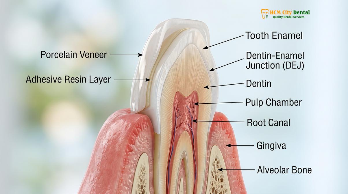
A minimally invasive preparation protocol typically requires the removal of only 0.3mm to 0.7mm of enamel. Staying within the enamel layer is paramount for the long-term success of the restoration. Enamel provides a highly mineralized, predictable substrate for adhesive bonding. If the preparation extends too deeply and breaches the dentin-enamel junction (DEJ), the bond strength decreases significantly due to the higher organic content and tubular fluid present in dentin[2]. Therefore, conservative preparation is not just an aesthetic preference; it is a biomechanical necessity.
“The longevity of a ceramic veneer is directly proportional to the volume of enamel preserved during preparation. Maximizing enamel retention at the margins ensures superior marginal seal, reduces microleakage, and virtually eliminates post-operative sensitivity.”
In certain clinical scenarios involving microdontia (undersized teeth) or teeth that are positioned slightly lingually, the clinician may opt for a prep-less approach. These ultra-thin restorations require minimal space and involve virtually no enamel removal. However, case selection for prep-less veneers must be rigorous, as adding material without adequate reduction can lead to bulky, over-contoured teeth that trap plaque and induce gingival inflammation.
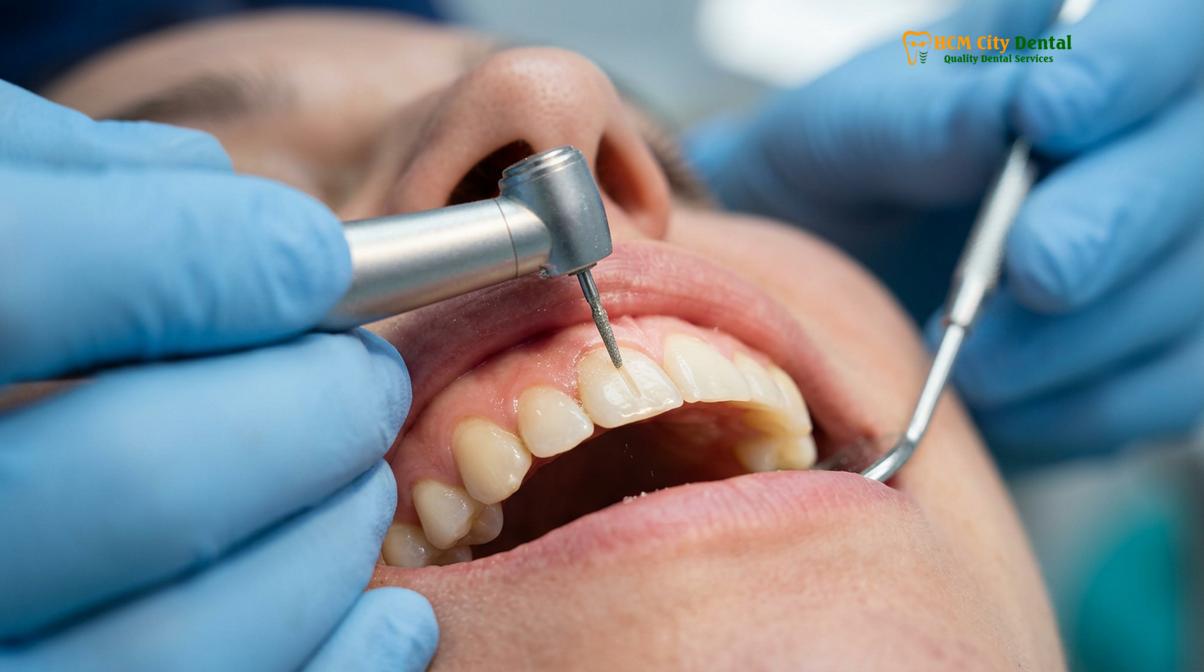
During the preparation phase, local anesthesia is administered to ensure complete patient comfort. The clinician carefully manages the gingival tissues, often placing a retraction cord into the sulcus to displace the gums slightly. This allows the preparation margin to be placed precisely at or slightly below the gingival crest, ensuring a seamless transition between the ceramic and the natural root structure, which is vital for both aesthetics and periodontal health.
Phase 3: Digital Scan (iTero/Medit) and Temporary Veneer Placement
Following preparation, a final digital scan is captured, and provisional restorations are fabricated to protect the teeth and provide an aesthetic preview during the lab fabrication phase.
Once the teeth have been meticulously prepared and the margins refined, the clinician captures the final digital scanning impression using advanced systems like iTero or Medit. This scan records the exact geometry of the prepared teeth, the surrounding soft tissues, and the patient’s bite registration. The digital file is instantly transmitted to the dental laboratory, eliminating the dimensional distortion and shipping delays associated with traditional physical impressions.
Because the prepared teeth have had a thin layer of enamel removed, they can be susceptible to thermal sensitivity and bacterial ingress. To protect the underlying dentin and maintain the patient’s appearance, provisional restorations are immediately fabricated chairside. These temporaries are typically made from a bis-acrylic composite resin material, utilizing a silicone matrix derived from the initial 3D diagnostic mock-up.
The placement of composite temporaries serves multiple critical functions. First, they provide immediate protection against thermal stimuli. Second, they stabilize the occlusal relationship and prevent the prepared teeth from shifting. Third, and perhaps most importantly, they act as a functional and aesthetic prototype. Patients wear these temporaries for a few days, allowing them to evaluate the proposed length, shape, and phonetics of their new smile in real-world conditions.
If the patient desires any modifications—such as softening the incisal angles or adjusting the length—these changes can be easily made to the temporaries and communicated to the ceramist. This ensures that the final porcelain restorations are perfectly aligned with the patient’s expectations. When comparing porcelain and composite veneers, it is important to note that while composite temporaries are excellent for short-term use, they lack the stain resistance and flexural strength of the final laboratory-milled ceramics.
Phase 4: Precision CAD/CAM Lab Fabrication
The dental laboratory utilizes CAD/CAM technology to mill and hand-characterize the final veneers from premium certified materials like E.max or Zirconia HT.
The fabrication of porcelain veneers represents the intersection of advanced digital engineering and meticulous artisanal craftsmanship. Upon receiving the digital scan, the laboratory technicians use Computer-Aided Design (CAD) software to virtually model each veneer. The design parameters are strictly controlled to ensure proper emergence profiles, optimal contact points, and precise marginal adaptation. Once the design is finalized, the data is sent to a Computer-Aided Manufacturing (CAM) milling unit, which carves the restorations from solid blocks of premium ceramic material.
Material selection is a critical determinant of the final outcome. The clinic utilizes only authentic, certified materials from industry-leading manufacturers. The two primary materials used are:
- E.max (Lithium Disilicate): Renowned for its outstanding lifelike aesthetics, high translucency, and opalescence. E.max is the gold standard for anterior (front) teeth where cosmetic demands are highest. It mimics the light-reflecting properties of natural enamel flawlessly[1].
- Zirconia HT: High-translucency zirconia offers exceptional flexural strength, making it the preferred choice for patients with heavy bite forces, a history of bruxism, or for restorations extending into the premolar regions[3].
After milling, the ceramist hand-finishes each veneer. This involves applying custom stains, creating surface micro-texture to mimic natural enamel anatomy, and glazing the restoration in a ceramic furnace. This artisanal touch ensures that the veneers do not look like opaque, artificial blocks, but rather vibrant, natural teeth.
Understanding the financial investment is an important part of the treatment planning process. The clinic offers transparent pricing with significant advantages for patients who book in advance. Below is the current pricing structure, highlighting the exclusive 40% pre-arrival discount available via WhatsApp booking:
| Treatment / Material | Standard Walk-in Price (Est.) | WhatsApp Pre-Booking (-40%) | Warranty & Support |
|---|---|---|---|
| Composite Veneer (Chair-side) | ~$80 (2.0M VND) | From $48 (1.2M VND) | 1-Year Guarantee |
| Zirconia Veneer (Cercon HT, Germany) | ~$240 (6.0M VND) | From $144 (3.6M VND) | 3-Year Clinic + Global Mfg |
| E.max Porcelain Veneer (Ivoclar) | ~$320 (8.0M VND) | From $192 (4.8M VND) | 3-Year Clinic + Global Mfg |
| Smile Design 3D Mock-up | ~$200 (5.0M VND) | From $120 (3.0M VND)* | *Free if treatment performed |
| Laser Gum Contouring (Diode) | ~$160 (4.0M VND) | From $96 (2.4M VND) / arch | Clinical Assessment Required |
To further assist patients, the clinic offers flexible payment options, making high-end cosmetic dentistry accessible without compromising on material quality or clinical expertise. When evaluating how long porcelain veneers last, investing in certified materials like E.max and Zirconia ensures decades of reliable function and aesthetic stability.
Phase 5: Try-In, Shade Verification, and Adhesive Cementation
The final phase involves the meticulous try-in of the ceramics followed by a strict adhesive bonding protocol to permanently fuse the veneers to the natural tooth structure.
The delivery appointment is the culmination of the entire porcelain veneers procedure. The temporary restorations are carefully removed, and the prepared teeth are thoroughly cleansed of any residual temporary cement using a pumice slurry. The permanent ceramic veneers are then placed onto the teeth using a water-soluble try-in paste. This critical step allows both the clinician and the patient to verify the marginal fit, interproximal contacts, and overall aesthetic integration.
During the try-in, the shade is meticulously evaluated under various lighting conditions. The color of the underlying tooth, the opacity of the ceramic, and the shade of the luting cement all interact to produce the final optical result. If the patient and the prosthodontist are completely satisfied with the aesthetics, the procedure moves to the permanent cementation phase.
The adhesive bonding protocol is a highly sensitive chemical process that requires absolute moisture control. To achieve this, the clinician utilizes rubber dam isolation or advanced cheek retractors to prevent salivary contamination, which would instantly compromise the bond strength. The internal (intaglio) surface of the porcelain veneer is treated with hydrofluoric acid to create micro-porosities, followed by the application of a silane coupling agent. Silane acts as a chemical bridge between the inorganic silica in the porcelain and the organic resin in the cement.
“The success of a ceramic restoration relies entirely on the integrity of the adhesive interface. Meticulous isolation and strict adherence to the chemical bonding protocol ensure a monolithic integration between the porcelain and the natural enamel, preventing microleakage and secondary decay.”
Simultaneously, the natural tooth structure is conditioned. A phosphoric acid etchant is applied to the enamel to create a micro-retentive surface. A bonding agent (primer and adhesive) is then applied and gently air-thinned. The veneer is loaded with a light-cure or dual-cure resin cement and gently seated onto the tooth. Excess cement is meticulously removed from the margins before a high-intensity curing light is used to polymerize the resin, permanently fusing the ceramic to the tooth. Strict adherence to national restorative and infection control protocols ensures patient safety throughout the procedure[5].
After curing, the margins are polished to a high gloss to prevent plaque accumulation, and the patient’s occlusion (bite) is carefully checked and adjusted. Proper occlusal calibration is vital to prevent premature contacts that could lead to ceramic fracture. For patients deciding between different restorative options, understanding the structural differences is essential. In some complex cases, a hybrid approach may be discussed, such as combining veneers and crowns to achieve the optimal functional and aesthetic outcome.
When to Consult a Prosthodontist & Important Clinical Notes
Understanding the indications and contraindications for porcelain veneers is crucial for ensuring a safe and successful cosmetic dental treatment.
Important Clinical Note: Porcelain veneers are an excellent solution for cosmetic enhancement, but they require a healthy foundation. Patients presenting with active periodontal disease, untreated dental caries, or severe malocclusion must have these underlying conditions fully resolved before proceeding with any aesthetic restorations.
While porcelain veneers offer transformative aesthetic benefits, they are not suitable for every patient. It is highly recommended to consult a qualified prosthodontist if you experience severe tooth discoloration that does not respond to professional whitening, noticeable enamel erosion, or minor structural damage such as chips and cracks. A thorough clinical evaluation is necessary to determine if your underlying dental health can support ceramic restorations. If veneers are not viable, your dentist may suggest an alternative for veneers to safely achieve your desired smile.
Case Study: A Smile Transformation Journey
Recently, a patient visited HCMC Dental Clinic in Ho Chi Minh City seeking a complete smile makeover to address severe enamel wear and intrinsic discoloration. After a comprehensive digital assessment, the clinical team determined that a combination of conservative preparation and E.max lithium disilicate restorations would provide the most durable and lifelike result.
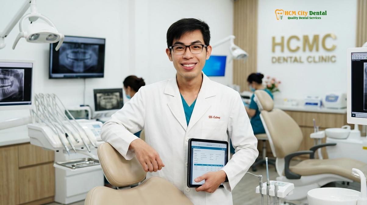
Dr. Nguyen Van Cuong, a leading specialist in restorative dentistry, meticulously planned her treatment. Dr. Cuong emphasizes that conservative preparation is the cornerstone of long-lasting aesthetics, a philosophy he details extensively when discussing dental crown vs veneer options with his patients. By utilizing digital smile design and precise depth-cutting techniques, the team was able to preserve maximum enamel while delivering a flawless, radiant smile.
The patient’s journey highlights the importance of choosing a clinic equipped with advanced CAD/CAM technology and experienced prosthodontists. The seamless integration of digital scanning, expert lab fabrication, and meticulous adhesive bonding resulted in a stunning transformation that restored both her confidence and her dental function.
References
- Journal of Esthetic and Restorative Dentistry. Translucency of lithium disilicate.
- International Journal of Prosthodontics. Enamel preservation and bond strength.
- The Journal of Prosthetic Dentistry. Flexural strength of porcelain restorations.
- Clinical Oral Investigations. Diode laser gingival sculpting parameters.
- Vietnam Odonto-Stomatology Association (VOSA). Guidelines on restorative dentistry and infection control protocols.
Ready to transform your smile with precision and care? Contact HCMC Dental Clinic in Ho Chi Minh City today to schedule your comprehensive consultation and discover how the porcelain veneers procedure can elevate your confidence.