Porcelain veneers offer a highly effective, permanent solution for deeply stained teeth that resist traditional whitening. By bonding custom-crafted ceramic shells over the affected enamel, prosthodontists can completely mask intrinsic discoloration, restoring a natural, vibrant, and structurally sound smile with minimal tooth preparation.
Clinical Summary:
Intrinsic tooth discoloration, such as tetracycline staining, originates deep within the dentin and cannot be resolved with chemical bleaching. Advanced ceramic restorations, specifically lithium disilicate (E.max) and zirconia, provide the necessary opacity to block underlying dark tones while maintaining lifelike translucency. Successful outcomes rely on precise enamel preparation, digital smile design, and strict adhesive bonding protocols to ensure long-term aesthetic and functional stability. International patients frequently seek these advanced treatments to achieve predictable, long-lasting smile transformations.
Key Takeaways:
- Intrinsic stains from medications or trauma require physical masking rather than chemical bleaching.
- Modern ceramic materials can block dark underlying dentin without appearing unnaturally opaque.
- Minimally invasive preparation preserves the enamel layer for optimal bond strength and longevity.
- Digital intraoral scanning ensures precise marginal fit and predictable aesthetic outcomes.
- Strict isolation protocols during cementation are critical for the long-term success of the restoration.
The Challenge of Deep Internal Tetracycline Discoloration
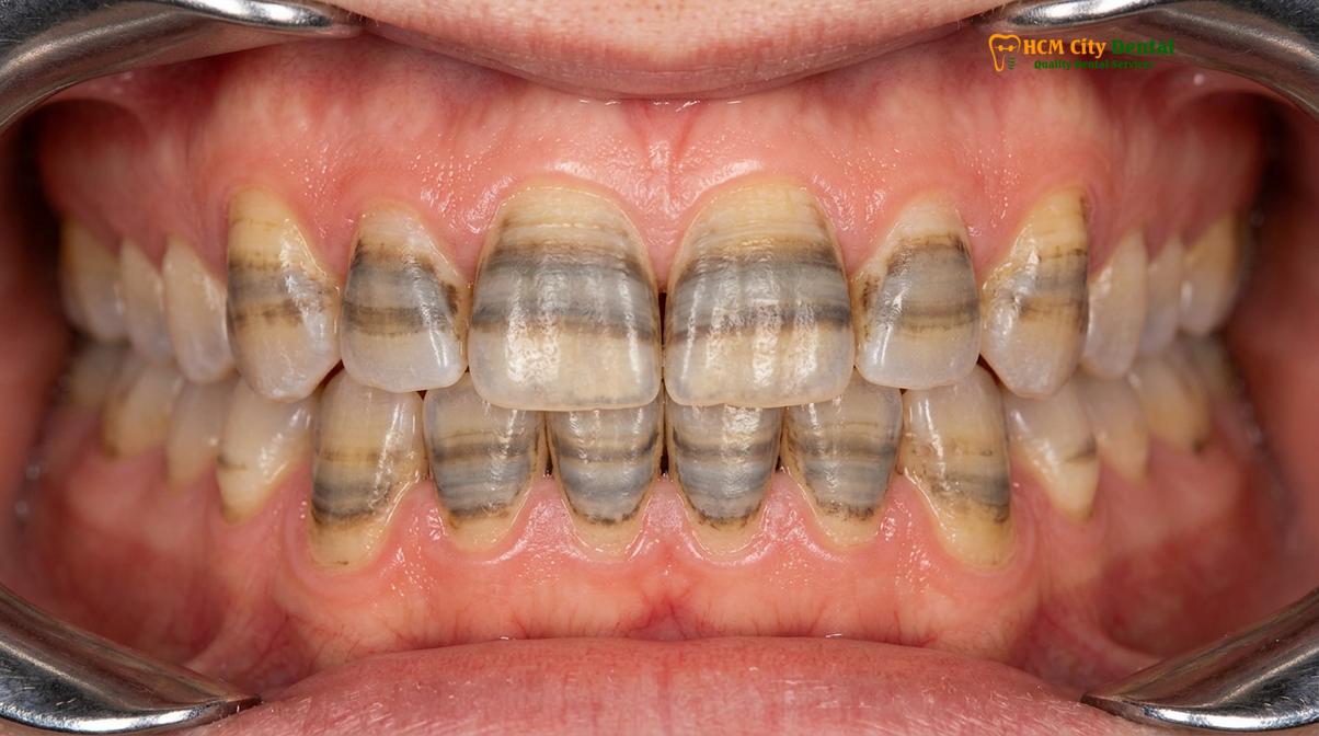
Tetracycline discoloration presents a unique clinical challenge because the staining is incorporated directly into the tooth’s crystalline structure during development, requiring advanced restorative techniques to mask effectively.
Tooth discoloration is broadly categorized into two types: extrinsic and intrinsic. Extrinsic stains occur on the surface of the enamel and are typically the result of lifestyle factors such as the consumption of coffee, tea, red wine, or tobacco use. These surface-level stains are generally responsive to routine dental prophylaxis and standard whitening procedures. However, intrinsic stains represent a fundamentally different clinical scenario. Intrinsic discoloration occurs deep within the inner structure of the tooth, specifically within the dentin, and is visible through the translucent outer enamel layer.
One of the most notoriously difficult forms of intrinsic staining is tetracycline discoloration. When the antibiotic tetracycline is administered during the critical periods of tooth development—typically in utero or during early childhood—the medication binds to the calcium orthophosphate in the developing teeth. This binding process creates a tetracycline-calcium orthophosphate complex that becomes permanently incorporated into the dentin and enamel [1]. Upon eruption and subsequent exposure to sunlight, this complex undergoes a photochemical reaction, oxidizing and turning into a dark gray, brown, or even bluish-purple hue.
The severity of tetracycline staining is often classified into different degrees, ranging from mild, uniform discoloration to severe, dark horizontal banding across the clinical crown. Because these stains are located deep near the dentin-enamel junction (DEJ), they are structurally embedded within the tooth. The depth and chemical nature of these stains make them highly resistant to conservative cosmetic treatments, necessitating a physical masking approach to achieve a bright, uniform aesthetic.
Why Chemical Bleaching and Whitening Fail for Internal Stains
Traditional peroxide-based whitening agents only oxidize surface-level organic pigments and cannot penetrate deeply enough to alter the structural discoloration embedded within the dentin.
Patients presenting with severe intrinsic discoloration often report a history of tooth bleaching failure. Traditional professional teeth whitening utilizes active ingredients such as hydrogen peroxide or carbamide peroxide. These chemical agents work by penetrating the porous enamel and breaking down the double carbon bonds of organic chromophores (stain molecules) into smaller, less pigmented compounds. While this oxidative process is highly effective for extrinsic stains and mild age-related yellowing, it is fundamentally limited when applied to structural intrinsic stains.
The primary limitation lies in the depth of penetration and the chemical nature of the stain itself. The tetracycline-calcium complex is an inorganic structural alteration, not a simple organic pigment. Even with prolonged exposure to high-concentration bleaching agents, the peroxide molecules cannot effectively dismantle these deep-seated complexes [2]. Furthermore, attempting to aggressively bleach severely stained teeth often leads to significant adverse effects. Prolonged bleaching dehydrates the tooth structure and can cause severe dentinal hypersensitivity without yielding any clinically acceptable improvement in shade.
“While external stains respond well to oxidation, attempting to bleach severe intrinsic banding often results in heightened sensitivity without meaningful color improvement, making ceramic coverage the definitive standard of care for these complex cases.”
In cases of severe intrinsic discoloration, the enamel may temporarily appear lighter immediately following a bleaching session due to dehydration. However, as the tooth rehydrates over the following days, the dark underlying dentin becomes visible once again—a phenomenon known as color rebound. Recognizing the limitations of chemical bleaching is the first step in guiding patients toward more predictable and permanent solutions, such as custom ceramic restorations.
Clinical Preparation Depth Required to Mask Deep Discoloration
Masking severe intrinsic stains requires a precise balance of tooth reduction, typically between 0.5mm and 0.7mm, to provide the ceramist enough restorative space to block out dark tones.
The success of a ceramic restoration in masking dark teeth relies heavily on the physical principles of light transmission and reflection. Dental ceramics are inherently translucent, designed to mimic the optical properties of natural enamel. If a ceramic shell is too thin, the dark color of the underlying dentin will simply show through the restoration, resulting in a dull, grayish appearance. Therefore, the concept of ultra-thin, prep-less restorations (often referred to as contact lens veneers, which are typically around 0.2mm thick) is generally contraindicated for severely discolored teeth.
To effectively block dark underlying tones, the clinician must create adequate restorative space. This is achieved through a highly controlled, minimally invasive preparation of the facial enamel. Using a specialized enamel depth-cutter, the prosthodontist carefully reduces the facial surface of the tooth by approximately 0.5mm to 0.7mm. This specific depth is critical: it provides the dental laboratory with enough room to layer opaque masking materials and translucent surface ceramics, while still remaining within the enamel layer [3].
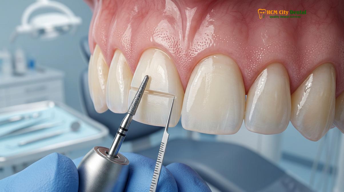
Preserving the enamel is paramount for the long-term success of the restoration. The bond strength between ceramic and enamel is significantly higher and more predictable than the bond to exposed dentin. By meticulously controlling the preparation depth, the clinician ensures that the restoration will be securely anchored, minimizing the risk of debonding or microleakage over time.
Clinical Warning: Insufficient preparation on severely dark teeth forces the laboratory to over-contour the restoration to achieve the desired shade. This leads to bulky, unnatural margins that violate the biological width, creating plaque traps that promote chronic gingival inflammation and periodontal disease.
Material Selection: Opaque Ingot Substructures vs. Translucent Layering
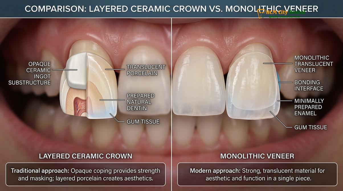
The choice between lithium disilicate and zirconia depends on the severity of the underlying stain, utilizing opaque substructures layered with translucent ceramics to achieve a natural appearance.
When addressing deeply stained teeth, material selection is arguably the most critical decision in the treatment planning phase. The chosen material must possess the dual capability of blocking the dark underlying tooth structure while simultaneously reflecting light in a manner indistinguishable from natural enamel. Modern cosmetic dentistry relies primarily on two advanced materials for this purpose: lithium disilicate (commonly known by the brand name E.max) and high-translucency zirconia.
For moderate to severe discoloration, a monolithic (single-layer) translucent ceramic will fail to hide the stain. Instead, the laboratory utilizes a bi-layered approach. The foundation of the restoration is an opaque ceramic ingot. This substructure acts as a light-blocking shield, neutralizing the dark gray or brown tones of the prepared tooth. Once the opaque core is milled or pressed, a master ceramist meticulously hand-layers highly translucent, opalescent porcelain over the facial surface [4]. This layering technique creates depth, internal characterization, and a lifelike surface texture.
Lithium disilicate (E.max, Ivoclar, Liechtenstein) is highly regarded for its exceptional aesthetic properties and strong adhesive bonding capabilities. It is available in various levels of opacity (such as High Opacity or Medium Opacity ingots), allowing the ceramist to customize the masking ability based on the specific needs of the tooth. Zirconia (such as Cercon HT, Germany), on the other hand, is inherently more opaque and possesses significantly higher flexural strength. Zirconia is often the material of choice for patients who exhibit severe bruxism (teeth grinding) or when masking the darkest of tetracycline stains, though it requires specialized bonding protocols.
| Material Type | Masking Ability | Aesthetic Quality | Best Clinical Indication |
|---|---|---|---|
| Lithium Disilicate (E.max) | Moderate to High (using HO/MO ingots) | Exceptional, highly lifelike translucency | Anterior teeth, moderate stains, high aesthetic demands |
| High-Translucency Zirconia | Very High (inherently opaque core) | Excellent, though slightly less translucent than E.max | Severe tetracycline stains, heavy grinders, posterior teeth |
| Composite Resin | Low to Moderate | Good initially, but prone to dulling over time | Temporary solutions, minor localized stains, budget constraints |
Designing Veneers to Block Gray/Brown Tones Without Looking Chalky
Achieving a lifelike result on dark teeth requires sophisticated shade mapping and layering techniques to neutralize gray or brown undertones without resulting in a flat, artificial appearance.
One of the greatest risks when masking grey teeth is creating a restoration that appears overly opaque, flat, or “chalky.” Natural teeth are not monochromatic; they exhibit a gradient of color, with higher chroma (intensity) at the gingival margin and higher value (brightness) and translucency at the incisal edge. When a ceramist attempts to block a dark stain using only highly opaque materials, the resulting tooth can look artificial, resembling a white chiclet rather than a natural biological structure.
To prevent this, advanced cosmetic clinics employ Digital Smile Design (DSD) and comprehensive shade mapping. Before any preparation begins, high-resolution clinical photographs are taken under color-corrected lighting. The clinician evaluates the value, hue, and chroma of the underlying stain. If the patient desires a bright, Hollywood-style smile (such as a VITA BL1 shade), the ceramist must calculate the exact thickness of the opaque core needed to raise the value of the dark tooth, while leaving enough space for the translucent enamel layer.
The illusion of natural vitality is created in the final millimeters of the porcelain. By incorporating subtle internal effects—such as mamelons, halo effects at the incisal edge, and varying degrees of opalescence—the ceramist ensures that light scatters naturally across the surface. This meticulous attention to detail is what separates a standard dental crown from a bespoke cosmetic restoration.
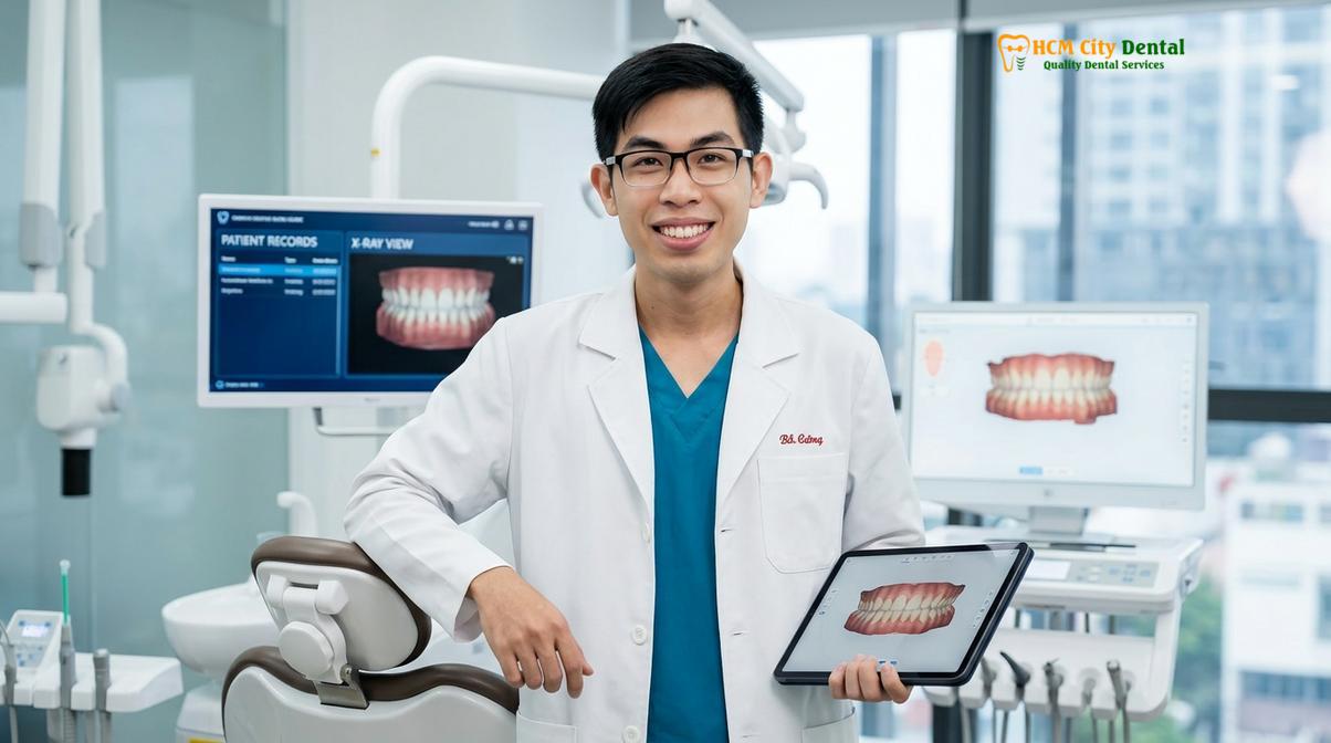
Clinical Case Study: A 34-year-old patient visited HCMC Dental Clinic in Ho Chi Minh City presenting with severe, dark-banded tetracycline discoloration that had failed multiple bleaching attempts. Under the clinical direction of Dr. Nguyen Van Cuong, a comprehensive DSD protocol was initiated. The treatment involved a precise 0.6mm facial preparation to preserve the enamel, followed by the fabrication of custom E.max restorations utilizing Medium Opacity (MO) ingots layered with highly translucent facial porcelain. The final result successfully masked the intrinsic banding while delivering a vibrant, natural BL2 shade, dramatically improving the patient’s aesthetic confidence.
Advanced Clinical Workflows: From Scanning to Cementation
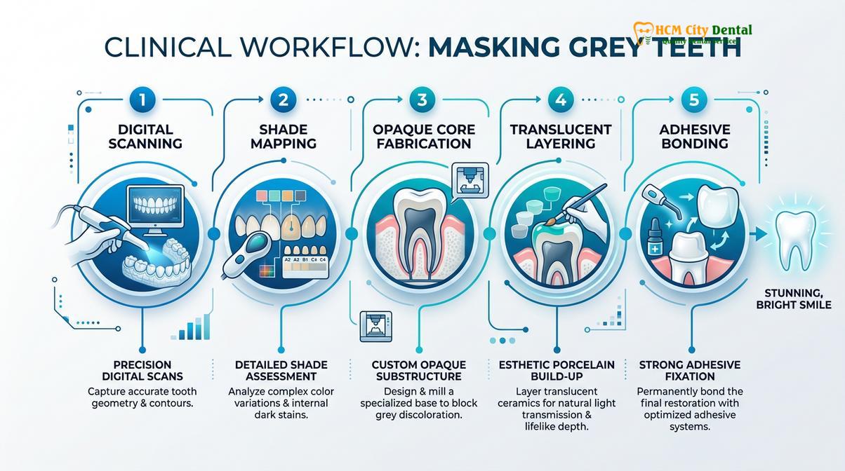
Modern veneer fabrication relies on digital precision and meticulous clinical execution, from 3D intraoral scanning to moisture-controlled adhesive bonding.
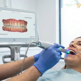
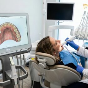
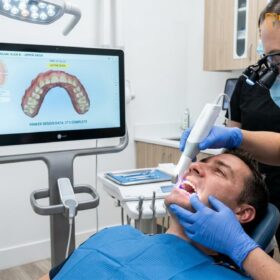
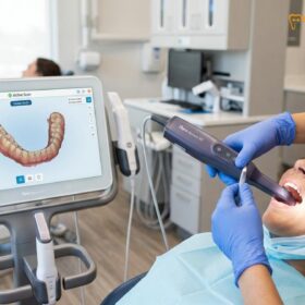
The clinical workflow for restoring deeply stained teeth demands absolute precision at every stage. The process begins with state-of-the-art digital scanning technology. Utilizing devices such as the iTero Element 5D or the Medit i700 Intraoral Scanners, the clinician captures a highly accurate 3D topographical map of the prepared teeth. This digital approach eliminates the need for messy, uncomfortable traditional silicone impressions, providing maximum comfort for the patient and eliminating the risk of dimensional distortion.
Following the preparation and scanning, the patient is fitted with custom provisional restorations. These temporaries serve multiple critical functions: they protect the prepared enamel, prevent dentinal sensitivity, maintain the health of the gingival tissues, and provide a “test drive” for the proposed new smile. The patient can evaluate the shape, length, and phonetics of the temporaries, allowing the clinician to communicate any necessary adjustments to the laboratory before the final ceramics are milled.
The final, and arguably most critical, phase is the cementation process. Ceramic restorations are not merely glued into place; they are chemically fused to the tooth structure through a complex adhesive bonding protocol. This protocol requires absolute moisture control, which is typically achieved through rubber dam isolation. The presence of even microscopic amounts of saliva or crevicular fluid can severely compromise the bond strength.
“The long-term success of a ceramic restoration is dictated not just by the material, but by the rigor of the adhesive bonding protocol under strict moisture control. A compromised bond leads to microleakage, recurrent decay, and ultimate failure of the aesthetic result.”
During bonding, the internal surface of the ceramic is treated with hydrofluoric acid to create micro-porosities, followed by the application of a silane coupling agent. Simultaneously, the tooth enamel is etched with phosphoric acid and treated with a dentin bonding agent. A specialized light-cured resin cement is then used to unite the two surfaces, creating a seamless, monolithic structure that is highly resistant to fracture and bacterial infiltration.
Treatment Timeline, Pricing, and Warranty in Ho Chi Minh City
International patients can complete a full smile makeover within a single week, benefiting from direct laboratory partnerships, transparent pricing, and comprehensive global warranties.
For patients seeking high-end cosmetic dentistry, Ho Chi Minh City has emerged as a premier destination, offering world-class clinical standards at a fraction of the cost found in Western countries. By maintaining a Direct Lab Partnership with leading ceramic laboratories in HCMC, clinics can ensure complete quality control and direct collaboration between the prosthodontist and the master ceramist.
This streamlined workflow allows for an Express 3-5 Day Turnaround. Tailored specifically for dental tourists and expatriates, this accelerated timeline means that complete smile makeovers—including the fabrication, characterization, and final fitting of multiple restorations—can be completed within a single week without compromising clinical excellence.
Transparency in financial planning is a cornerstone of ethical patient care. According to the latest clinic fee schedule, the pricing structure (incorporating a 40% pre-arrival discount for WhatsApp bookings) is highly competitive:
- Composite Veneer (Chair-side): Walk-in: ~$80 (2.0M VND) | WhatsApp discount: From $48 (1.2M VND) (-40%).
- Zirconia Veneer (Cercon HT): Walk-in: ~$240 (6.0M VND) | WhatsApp discount: From $144 (3.6M VND) (-40%).
- E.max Porcelain Veneer (Ivoclar): Walk-in: ~$320 (8.0M VND) | WhatsApp discount: From $192 (4.8M VND) (-40%).
- Smile Design 3D Mock-up: Walk-in: ~$200 (5.0M VND) | WhatsApp discount: From $120 (3.0M VND) (Free if treatment is performed).
- Laser Gum Contouring (Diode): Walk-in: ~$160 (4.0M VND) | WhatsApp discount: From $96 (2.4M VND) per arch.
- In-Office Teeth Whitening (Laser): Walk-in: ~$140 (3.5M VND) | WhatsApp discount: From $84 (2.1M VND).
To ensure peace of mind for international patients, treatments are backed by robust guarantees. Patients receive global manufacturer warranty cards, alongside a 3-year clinic bonding and fit guarantee for E.max and Zirconia restorations, and a 1-year guarantee for Composite work. Furthermore, Dr. Cuong emphasizes that remote follow-up support via WhatsApp—utilizing specific photo and video assessment protocols—allows the clinical team to monitor the health and stability of the restorations long after the patient has returned home. For those requiring flexible financials, 0% interest monthly credit card installment plans are also available.
When to See a Doctor
While ceramic restorations offer a transformative solution for stained teeth, thorough clinical evaluation is necessary to determine candidacy. Patients should schedule a comprehensive consultation if they experience severe intrinsic discoloration that has not responded to professional whitening, or if they notice structural defects such as chipping or enamel wear accompanying the discoloration. For patients exploring their options, comparing ceramic and resin options with a qualified professional is highly recommended.
During the examination, the clinician will assess the biological width and calibrate the gingival zenith to ensure that the proposed restorations will harmonize with the surrounding soft tissues [5]. It is important to note that active periodontal disease, severe untreated malocclusion, or insufficient remaining enamel are contraindications for this procedure and must be addressed prior to cosmetic intervention. Additionally, patients with a history of sleep bruxism will require the fabrication of a custom nightguard to protect their new investment from nocturnal grinding forces. Proper planning of your smile architecture ensures both aesthetic brilliance and long-term functional stability.
Frequently Asked Questions
Can veneers hide dark grey tetracycline stains?
Yes, custom ceramic restorations can effectively hide dark grey tetracycline stains. By utilizing opaque substructures layered with translucent porcelain, prosthodontists can block the underlying dark tones while maintaining a natural, lifelike appearance without looking artificially chalky. The success of this masking relies heavily on the skill of the ceramist and the specific opacity of the chosen material.
Do I need thicker veneers for severely discolored teeth?
Severely discolored teeth generally require slightly thicker restorations, typically between 0.5mm and 0.7mm. This additional thickness provides the ceramist with the necessary restorative space to incorporate opaque masking layers while preserving the surface translucency required for natural aesthetics. Ultra-thin, prep-less options are usually insufficient for blocking deep intrinsic stains.
Will the dark color show through my veneers over time?
High-quality porcelain and zirconia are highly stable and will not allow dark colors to show through over time. Unlike composite resin, which can absorb fluids, stain, and degrade, premium ceramics maintain their opacity and structural integrity for decades when properly bonded. The color achieved on the day of cementation is permanent.
Is the preparation process painful for sensitive stained teeth?
The preparation process is highly controlled and performed under local anesthesia, ensuring a comfortable experience. Because the procedure focuses on the outermost enamel layer, post-operative sensitivity is typically minimal. Any temporary sensitivity is managed effectively with high-quality provisional restorations worn between the preparation and final cementation appointments.
How do I maintain my new smile after the procedure?
Maintenance involves standard oral hygiene, including brushing with non-abrasive toothpaste, daily flossing, and routine professional cleanings. Additionally, wearing a custom nightguard is highly recommended to protect the ceramics from nocturnal bruxism and ensure long-term structural stability. Avoiding using your teeth as tools will also prevent accidental chipping.
References
- Journal of Esthetic and Restorative Dentistry. Translucency of lithium disilicate and masking ability. (2021).
- Operative Dentistry. Efficacy and limitations of vital tooth bleaching. (2019).
- International Journal of Prosthodontics. Enamel preservation and bond strength in ceramic restorations. (2020).
- The Journal of Prosthetic Dentistry. Flexural strength and optical properties of zirconia and lithium disilicate. (2022).
- Journal of Clinical Periodontology. Biological width around restorations and gingival health. (2018).