Are veneers safe? Yes, when performed by a qualified prosthodontist using conservative preparation techniques, dental veneers are a highly safe and predictable cosmetic treatment. The procedure relies on preserving natural tooth enamel to ensure long-term structural integrity and prevent underlying nerve damage.
Clinical Summary:
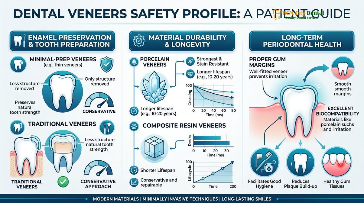
The medical safety of dental veneers depends entirely on the clinical protocols utilized during tooth preparation and bonding. Modern cosmetic dentistry prioritizes minimally invasive techniques, typically removing only 0.3mm to 0.7mm of enamel to accommodate the ceramic shell. This conservative approach preserves the tooth’s structural integrity, minimizes the risk of dentin exposure, and drastically reduces the likelihood of pulpitis or nerve damage. Utilizing advanced materials like authentic E.max lithium disilicate or Zirconia HT, combined with precision digital scanning (such as the iTero Element 5D), ensures optimal marginal fit and biocompatibility. While complications like debonding or transient sensitivity can occur, adhering to strict diagnostic criteria, respecting the biological width, and utilizing custom nightguards for bruxism make porcelain veneers a highly safe, long-term restorative option.
Key Takeaways:
- Enamel Preservation: Safe veneer placement requires keeping the preparation strictly within the enamel layer (0.3mm – 0.7mm) to ensure maximum bond strength and protect the tooth nerve.
- Material Biocompatibility: Certified materials like E.max and Zirconia are highly biocompatible, resisting plaque accumulation and supporting healthy gingival tissues.
- Digital Precision: Intraoral scanners eliminate traditional impression errors, ensuring a seamless marginal fit that prevents microleakage and secondary decay.
- Risk Factors: Aggressive over-preparation by inexperienced practitioners is the primary cause of severe complications, including irreversible pulp necrosis.
- Strict Contraindications: Patients with severe untreated sleep bruxism, active periodontal disease, or insufficient enamel should explore alternative restorative treatments.
The Medical Safety Profile of Modern Porcelain Veneers
Modern porcelain veneers boast an exceptional safety profile, primarily due to advancements in biocompatible ceramics and digital scanning technologies that ensure precise marginal fit and periodontal health.
When evaluating the question of whether cosmetic dental procedures are medically sound, the safety profile of Porcelain Veneers stands out as one of the most extensively researched topics in prosthodontics. The fundamental safety of this treatment is rooted in the evolution of dental materials and the shift toward tissue-preserving protocols. Historically, older techniques sometimes required aggressive tooth reduction, which compromised the vitality of the tooth. Today, the paradigm has shifted entirely toward biomimetic dentistry—a philosophy that aims to mimic the natural biomechanics of the tooth while preserving as much healthy tissue as possible.
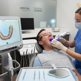
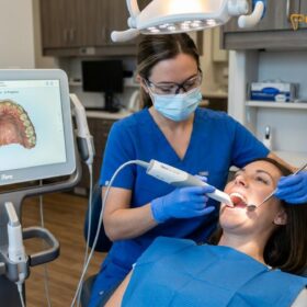
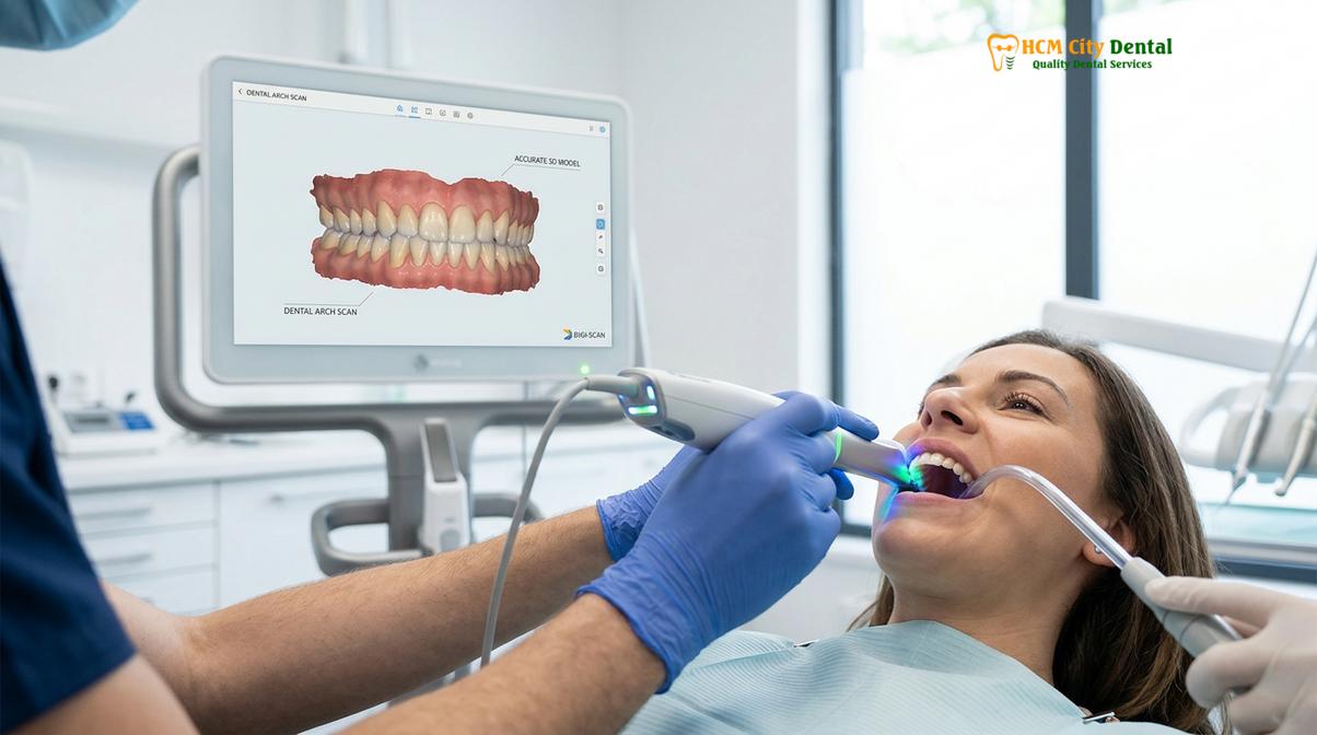
The cornerstone of this safety profile is the utilization of state-of-the-art digital scanning technology. At leading facilities, the integration of the iTero Element 5D & Medit i700 Intraoral Scanners has revolutionized the diagnostic and preparatory phases. These advanced optical devices eliminate the need for messy, uncomfortable traditional silicone impressions. Instead, they capture thousands of frames per second to create a hyper-accurate 3D model of the patient’s dentition. This maximum comfort and 3D precision allow the prosthodontist to design custom veneers with microscopic accuracy, ensuring that the final ceramic restoration fits seamlessly against the natural tooth structure without creating bulky margins that trap plaque.
Furthermore, the safety and longevity of the restoration are heavily dependent on material certification. Reputable clinics use only authentic, certified materials from industry-leading manufacturers in Germany and Liechtenstein. For instance, Zirconia HT (Cercon HT, Germany) is often selected for high-stress areas or for patients with a history of mild grinding, due to its exceptional flexural strength. Conversely, E.max (Ivoclar, Liechtenstein) lithium disilicate is the gold standard for outstanding lifelike aesthetics in the aesthetic zone. E.max provides unparalleled translucency matching and shade mapping (such as achieving a natural VITA BL1 or B1 shade) while maintaining robust mechanical properties [1]. To ensure patient safety and material authenticity, providing a genuine emax identity card post-treatment is a standard clinical practice that verifies the origin and quality of the ceramic used.
“The predictability and safety of ceramic veneers are directly correlated to the precision of the marginal adaptation. Digital workflows have reduced marginal discrepancies to the micron level, virtually eliminating the risk of cement washout and secondary caries.”
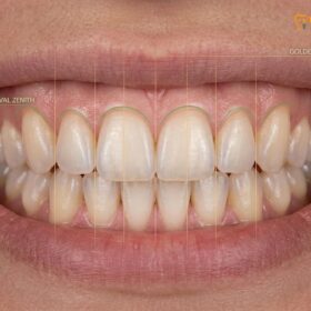
The clinical approach to aesthetic choices also factors into the overall safety. By utilizing Digital Smile Design (DSD), the clinician can plan the exact dimensions of the veneers before a single tooth is touched. This pre-visualization ensures that the proposed changes are functionally sound and harmonize with the patient’s dynamic facial expressions, preventing occlusal interferences that could lead to premature ceramic fracture.
Understanding the Biological Cost: Enamel Removal and Dentin Exposure
The primary biological cost of veneers is the irreversible removal of a microscopic layer of enamel, typically ranging from 0.3mm to 0.7mm, which is necessary to accommodate the ceramic shell.
To fully grasp the medical implications of veneer placement, patients must understand the concept of the “biological cost.” Every restorative dental procedure requires some alteration of the natural anatomy. For veneers, this involves the strategic reduction of the tooth’s outermost layer: the enamel. Enamel is the hardest substance in the human body, composed primarily of hydroxyapatite crystals. It serves as the ultimate protective barrier for the sensitive dentin and the vital dental pulp (nerve) beneath it.
The goal of a minimally invasive preparation is to keep the entire reduction strictly within the enamel layer. Standard porcelain veneers typically require a preparation depth of 0.3mm to 0.7mm. In cases where prep-less or ultra-thin options like Lumineers are indicated, the reduction may be as minimal as 0.2mm, or sometimes no preparation is required at all, depending on the tooth’s original volume and alignment. Preserving the enamel is not merely a conservative ideal; it is a strict biomechanical requirement for the long-term success of the restoration [2].
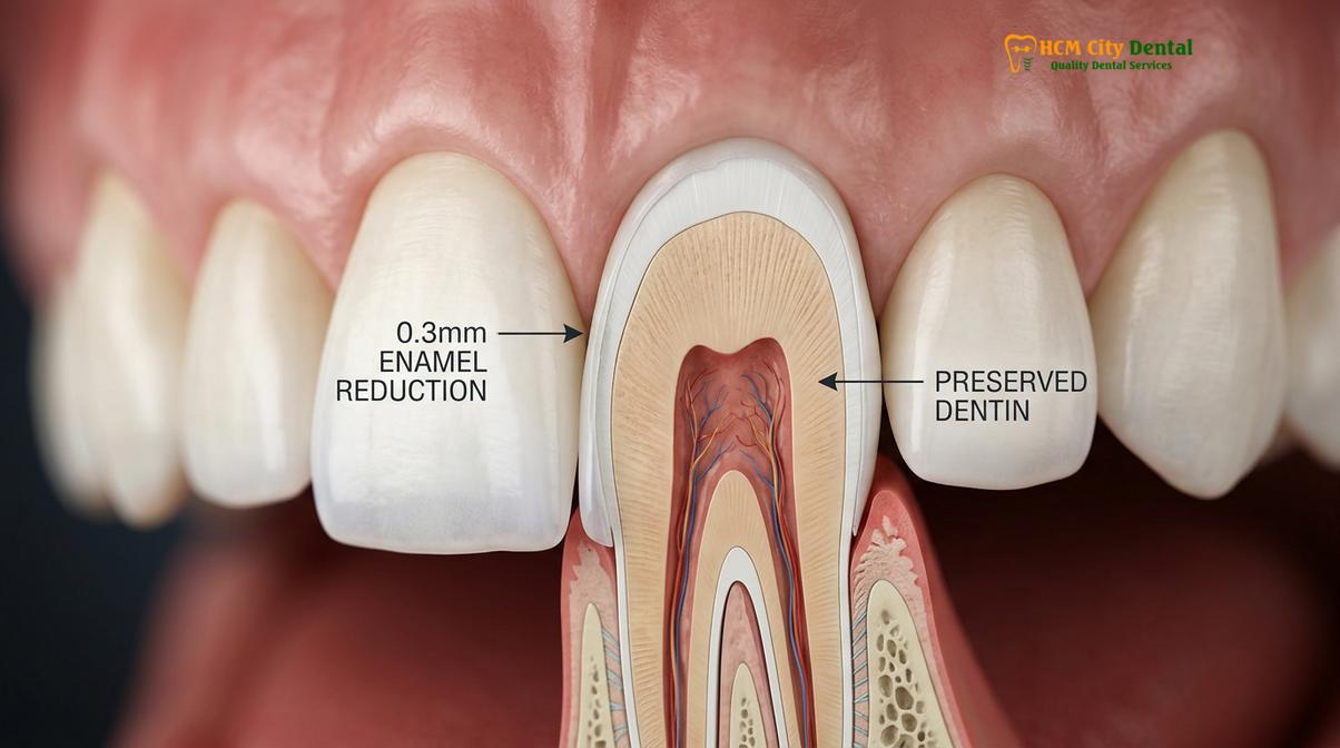
Dental adhesives bond significantly better to the highly mineralized structure of enamel than they do to the organic, fluid-filled tubules of dentin. When a veneer is bonded entirely to enamel, the shear bond strength is maximized, creating a virtually impenetrable seal that mimics the natural enamel-dentin junction. This robust bond prevents microleakage—the seepage of bacteria and oral fluids beneath the restoration—which is the primary cause of secondary decay.
Conversely, if the preparation is too aggressive and exposes large areas of dentin, the biological cost increases exponentially. Dentin bonding is inherently less predictable over time. Furthermore, cutting into dentin opens the dentinal tubules, creating a direct pathway to the dental pulp. This exposure is the primary mechanism behind post-operative sensitivity and increases the risk of bacterial ingress. Therefore, the safety of the procedure is entirely dependent on the clinician’s skill in executing precise enamel preservation techniques, ensuring that the biological cost remains as low as possible.
Common Complications and Their True Frequency (Clinical Data)
While generally safe, potential complications such as transient sensitivity, microleakage, or debonding can occur, though their frequency is drastically reduced with proper clinical protocols.
No medical or dental procedure is entirely devoid of risk. However, when discussing veneer complications, it is crucial to differentiate between transient, expected side effects and true pathological failures. Clinical data indicates that when strict case selection and proper bonding protocols are followed, the survival rate of porcelain veneers exceeds 90% over a 10-year period [3]. Nevertheless, patients must be fully informed of the potential risks to make an educated decision.
The most frequently reported complication is post-operative thermal sensitivity. This occurs because the removal of enamel, even microscopically, temporarily alters the thermal insulation of the tooth. In most cases, this sensitivity is transient, resolving within a few days to a couple of weeks as the pulp naturally produces secondary dentin to insulate itself. If sensitivity persists for months, it may indicate that the dentin was heavily exposed or that the bonding interface is compromised.
Another potential issue is debonding, where the veneer detaches from the tooth. This is relatively rare when bonding to enamel but becomes a significant risk if the veneer is bonded primarily to dentin or if the patient subjects the teeth to excessive shear forces (such as tearing packages open with their teeth). Microleakage is a more insidious complication; it occurs when the marginal seal degrades over time, allowing bacteria to infiltrate and cause decay beneath the veneer. This highlights the absolute necessity of precise marginal adaptation during the fabrication process.
To provide a clear clinical perspective, the following table outlines the most common complications, their estimated clinical frequency, and the preventive protocols employed by top-tier prosthodontists:
| Clinical Complication | Estimated Frequency | Primary Clinical Cause | Preventive Protocol |
|---|---|---|---|
| Transient Thermal Sensitivity | 15% – 20% | Mild dentin exposure during prep | Immediate dentin sealing (IDS) and desensitizing agents |
| Ceramic Chipping / Fracture | 4% – 8% | Occlusal trauma or bruxism | Custom nightguard fabrication and occlusal equilibration |
| Veneer Debonding | 2% – 5% | Bonding to dentin instead of enamel | Strict enamel preservation and proper isolation during cementation |
| Marginal Microleakage | 1% – 3% | Poor marginal fit or cement degradation | High-resolution digital scanning (iTero/Medit) and precise milling |
| Gingival Inflammation | 3% – 6% | Violation of biological width or bulky margins | Careful gingival zenith calibration and highly polished ceramic margins |
Understanding these risks allows patients to appreciate the importance of selecting a highly qualified clinician. The safety of the procedure is not inherent to the ceramic material itself, but rather to the meticulous execution of the clinical workflow.
Nerve Damage, Pulp Necrosis — When Veneers Can Go Wrong
Aggressive tooth preparation that penetrates too deeply into the dentin can traumatize the dental pulp, potentially leading to irreversible pulpitis or nerve necrosis.
While the aesthetic benefits of a smile makeover are highly desirable, the most severe risk associated with cosmetic dentistry is dental nerve damage. This catastrophic complication is almost exclusively the result of iatrogenic error—specifically, aggressive over-preparation of the tooth structure by an inexperienced or overly aggressive practitioner.
The dental pulp, located in the center of the tooth, contains the nerves and blood vessels that keep the tooth vital. It is highly sensitive to thermal and mechanical trauma. During the preparation phase, the friction from the high-speed dental bur generates significant heat. If adequate water coolant is not used, or if the clinician cuts too deeply into the dentin, the heat and vibration can cause severe inflammation of the pulp tissue, a condition known as pulpitis.
Clinical Warning: If a patient experiences severe, spontaneous, throbbing pain that wakes them up at night following veneer placement, it is a strong indicator of irreversible pulpitis. This condition requires immediate endodontic intervention (root canal therapy) to remove the dying nerve tissue and prevent the formation of a periapical abscess.
In cases of extreme over-preparation, the pulp may undergo necrosis (death) silently over several months or years, eventually presenting as a dark discoloration of the underlying tooth or a localized infection at the root apex. This is why the distinction between a veneer and a crown is so critical. A veneer should only replace the facial enamel. If a tooth requires 360-degree reduction or massive structural removal, a Dental Crown vs Veneer assessment must be carefully conducted, as forcing a veneer onto a heavily compromised tooth is a violation of biomechanical principles.
To mitigate these risks, expert prosthodontists utilize depth-limiting burs and magnification loupes or dental microscopes during preparation. This ensures that the reduction is strictly controlled and uniform, safeguarding the vital pulp and ensuring the long-term safety of the tooth.
Contraindications: Who Should NOT Get Veneers
Veneers are contraindicated for patients with severe untreated bruxism, insufficient remaining enamel, active periodontal disease, or profound structural tooth damage.
A critical component of medical safety is proper patient selection. Not every individual is a suitable candidate for ceramic restorations. Identifying veneer contraindications during the initial comprehensive examination is essential to prevent premature failure and safeguard the patient’s oral health.
The most significant biomechanical contraindication is severe, untreated sleep bruxism (chronic teeth grinding and clenching). The sheer force exerted by the masticatory muscles during nocturnal bruxism can easily exceed the flexural strength of even the most robust ceramics, leading to catastrophic fracturing or debonding of the veneers. While mild to moderate grinders can often be treated successfully using high-strength Zirconia HT and the mandatory nightly use of a custom nightguard, severe cases may require orthodontic intervention or full-coverage crowns instead.
Another absolute contraindication is a lack of sufficient healthy enamel. Patients who have suffered from severe acid erosion (due to GERD or bulimia), aggressive over-brushing, or previous excessive dental treatments may not have enough enamel left to achieve a predictable bond. Attempting to bond veneers primarily to dentin in these cases will almost certainly result in failure. In such scenarios, alternative restorative strategies must be employed.
Furthermore, active periodontal disease or rampant dental caries must be fully resolved before any cosmetic work is initiated. Placing veneers over inflamed, bleeding gums or decaying tooth structure is a severe violation of clinical standards. The foundation must be perfectly healthy; otherwise, the restorations will fail, and the underlying disease will progress rapidly beneath the ceramics.
How Conservative Preparation Minimizes All Known Risks
Adhering to conservative preparation guidelines and respecting the gingival architecture ensures optimal bonding strength, prevents periodontal inflammation, and guarantees long-term success.
The ultimate safety of a smile makeover lies in the meticulous execution of conservative clinical protocols. By respecting the natural anatomy and utilizing advanced digital workflows, the risks associated with cosmetic dentistry are reduced to near zero. A critical aspect of this is maintaining the biological width attachment—the natural dimension of the soft tissue attached to the portion of the tooth above the alveolar bone. If a veneer margin is placed too deeply beneath the gum line, it violates this biological width, triggering chronic gingival inflammation, bleeding, and eventual bone loss [4].
To ensure perfect harmony between the ceramic and the soft tissue, clinicians often employ Diode Laser Gum Contouring prior to scanning. This minimally invasive laser sculpting recalibrates the gingival zenith (the highest point of the gum contour), creating a symmetrical frame for the new teeth without causing bleeding or requiring sutures. Following this, the cementation protocol is executed with absolute precision. The enamel is etched, a silane coupling agent is applied to the internal surface of the ceramic, and a high-quality bonding resin is used to fuse the veneer to the tooth under strict moisture control.
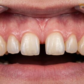
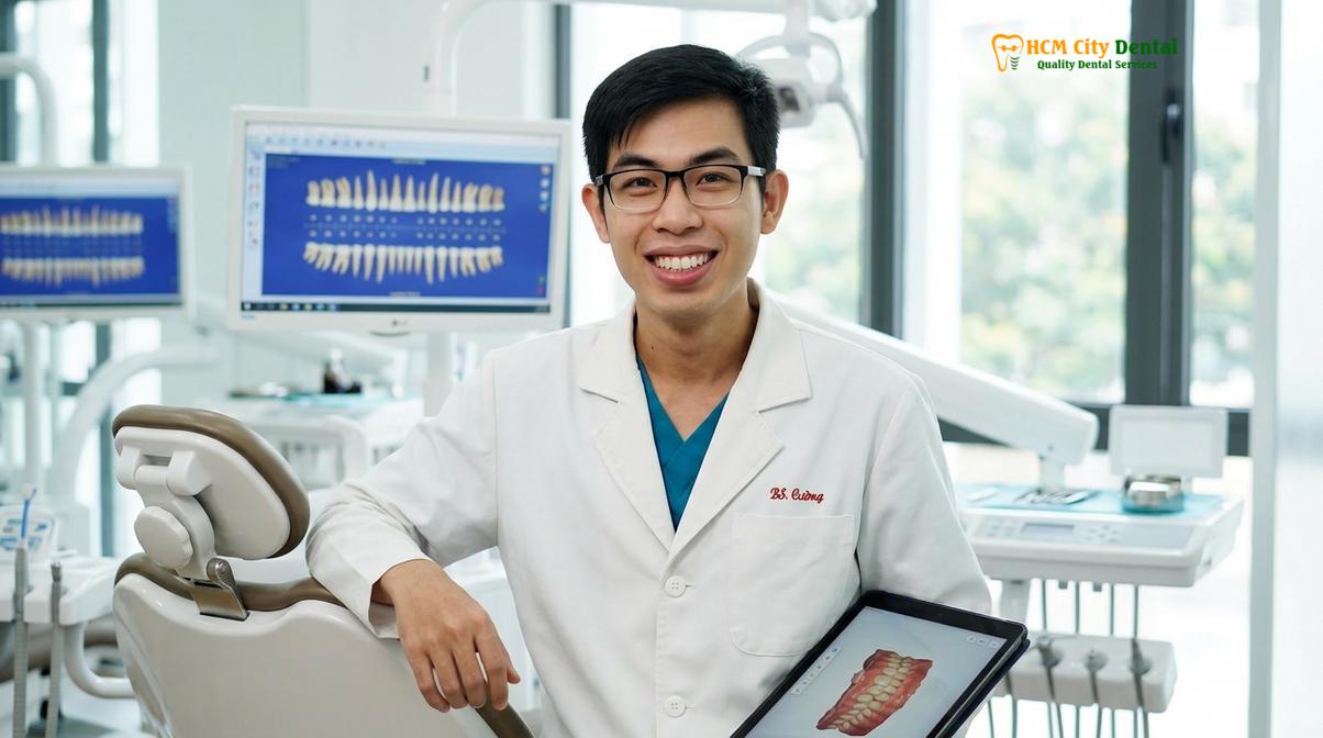
Clinical Case Review: A 34-year-old patient presented to HCMC Dental Clinic in Ho Chi Minh City with severe tetracycline staining and mild diastemas (gaps). Dr. Nguyen Van Cuong utilized a comprehensive smile photo guide and DSD to plan the case. By employing a minimal-prep approach (0.5mm reduction) and utilizing highly opaque yet natural-looking E.max BL1 ceramics, the stains were completely masked. The preservation of enamel ensured zero post-operative sensitivity, and the precise marginal fit resulted in perfectly healthy, stippled gingiva at the one-year follow-up.
For international patients seeking treatment, adhering to a strict safety checklist dental abroad is paramount. This includes verifying the clinic’s sterilization protocols, material certifications, and the credentials of the prosthodontist. At HCMC Dental Clinic, we partner directly with a leading laboratory in Ho Chi Minh City, enabling complete quality control and direct collaboration between the prosthodontist and the master ceramist. This Direct Lab Partnership facilitates an Express 3-5 Day Turnaround, tailored specifically for dental tourists and expats, allowing complete smile makeovers to be fabricated, characterized, and fitted within a single week.
Understanding the financial parameters and warranty structures is also a key part of a safe and transparent patient experience. According to the latest clinic fee schedule, we offer a highly structured pricing model, incorporating a 40% pre-arrival discount for WhatsApp bookings to make world-class care accessible:
- Composite Veneer (Chair-side): Walk-in: ~$80 (2.0M VND) | WhatsApp discount: From $48 (1.2M VND) (-40%).
- Zirconia Veneer (Cercon HT): Walk-in: ~$240 (6.0M VND) | WhatsApp discount: From $144 (3.6M VND) (-40%).
- E.max Porcelain Veneer (Ivoclar): Walk-in: ~$320 (8.0M VND) | WhatsApp discount: From $192 (4.8M VND) (-40%).
- Smile Design 3D Mock-up: Walk-in: ~$200 (5.0M VND) | WhatsApp discount: From $120 (3.0M VND) (Free if treatment is performed).
- Laser Gum Contouring (Diode): Walk-in: ~$160 (4.0M VND) | WhatsApp discount: From $96 (2.4M VND) per arch.
- In-Office Teeth Whitening (Laser): Walk-in: ~$140 (3.5M VND) | WhatsApp discount: From $84 (2.1M VND).
To further guarantee patient safety and peace of mind, we provide global manufacturer warranty cards alongside a 3-year clinic bonding and fit guarantee for E.max and Zirconia restorations, and a 1-year guarantee for Composite. We also offer remote follow-up support via WhatsApp, utilizing strict photo and video assessment protocols to monitor your healing and oral health from anywhere in the world. Financial flexibility is supported through 0% interest monthly credit card installment plans. If you are planning your smile makeover, a comprehensive clinical evaluation is the first step toward a safe, stunning result.
“The true art of cosmetic dentistry lies not just in creating a beautiful smile, but in engineering a restoration that respects the biology of the human body, ensuring safety, function, and longevity for decades to come.”
When to See a Doctor
While the safety profile of modern ceramic restorations is exceptionally high, patients must remain vigilant regarding their oral health. You should schedule an immediate clinical evaluation with your prosthodontist if you experience any of the following symptoms after your treatment:
- Prolonged or Severe Sensitivity: While mild thermal sensitivity is normal for a few days, sharp, lingering pain when consuming hot or cold foods that lasts for weeks may indicate nerve inflammation or a compromised bonding margin.
- Bite Imbalance or High Spots: If your teeth do not come together smoothly, or if you feel a “high spot” when chewing, the ceramic may need minor occlusal adjustment to prevent fracturing.
- Gingival Bleeding or Swelling: Healthy gums around ceramics should be firm, pink, and stippled. Localized redness, swelling, or bleeding when flossing indicates periodontal inflammation, potentially due to retained cement or a violation of the biological width [5].
- Visible Cracks or Debonding: Any structural damage to the ceramic shell or a feeling that the restoration is loose requires immediate professional attention to prevent bacterial leakage and decay.
Frequently Asked Questions
Do veneers damage your teeth permanently?
No, veneers do not inherently damage teeth, but the procedure is irreversible because a microscopic layer of enamel must be removed. When performed by a skilled prosthodontist using minimal-prep techniques, the underlying tooth structure remains healthy, vital, and structurally sound for a lifetime.
Can veneers cause nerve damage?
Yes, nerve damage is a rare but possible complication if the tooth is over-prepared. Aggressive drilling that penetrates deep into the dentin can traumatize the dental pulp, potentially leading to irreversible pulpitis or necrosis, which is why conservative enamel preservation is mandatory.
Who should not get dental veneers?
Patients with severe untreated sleep bruxism, active periodontal disease, extensive tooth decay, or insufficient natural enamel are not ideal candidates. These conditions compromise the bonding strength and longevity of the ceramic restorations, making alternative treatments like full crowns or orthodontics more appropriate.
Are veneers safe long term?
Yes, porcelain veneers are highly safe for long-term wear when properly maintained. With excellent oral hygiene, routine professional cleanings, and the use of a custom nightguard to protect against nocturnal grinding, high-quality ceramic veneers can safely last 10 to 15 years or more.
How do I know if my veneers are failing?
Signs of failing veneers include persistent thermal sensitivity, visible microleakage (dark lines at the gum margins), chipped ceramic, or localized gingival inflammation. If you experience any of these symptoms, a clinical evaluation is necessary to assess the integrity of the bond and the health of the underlying tooth.
References
- Journal of Esthetic and Restorative Dentistry. Translucency and mechanical properties of lithium disilicate. (2021).
- International Journal of Prosthodontics. Enamel preservation and shear bond strength in ceramic restorations. (2020).
- The Journal of Prosthetic Dentistry. Flexural strength and marginal adaptation of porcelain veneers. (2019).
- Clinical Oral Investigations. Diode laser gingival sculpting parameters for aesthetic dentistry. (2022).
- Journal of Clinical Periodontology. Biological width around restorations and periodontal health. (2018).