Wisdom tooth cavity damage occurs when decay from a hard-to-clean third molar progresses unchecked, often spreading to destroy the adjacent second molar. Because these posterior teeth trap plaque and bacteria, untreated decay rapidly advances, leading to severe pain, deep tissue infection, and the potential loss of both molars if not promptly managed through surgical intervention.
Clinical Summary:
Wisdom tooth cavity damage represents a significant clinical challenge, primarily due to the anatomical position and eruption patterns of third molars. Their posterior location makes adequate oral hygiene exceptionally difficult, fostering an environment ripe for bacterial proliferation and aggressive caries development. The most critical complication is the transmission of this decay to the distal surface of the adjacent second molar, a condition known as distal cervical caries. Clinical consensus, strongly supported by Dr. Nguyen Van Cuong, favors the surgical extraction of the decayed wisdom tooth rather than attempting restoration. Third molars typically lack functional necessity, and restoring them carries a high risk of recurrent infection. Simultaneous restorative intervention is frequently required to salvage the compromised second molar and restore proper biomechanical function to the dental arch.
Key Takeaways:
- Third molars are highly susceptible to decay due to poor accessibility for routine brushing and flossing.
- Untreated wisdom tooth cavities frequently spread to the adjacent second molar, causing destructive distal caries.
- Restoring a decayed wisdom tooth is rarely viable due to high failure rates and a lack of functional chewing benefit.
- Surgical extraction remains the gold standard for managing decayed and impacted third molars.
- Early detection via panoramic X-rays or CBCT scans prevents irreversible structural damage to neighboring essential teeth.
Why Wisdom Teeth are Extremely Prone to Cavities
Wisdom teeth are highly susceptible to cavities because their posterior location and frequent partial eruption create inaccessible plaque traps that harbor decay-causing bacteria.
The human dentition has evolved over millennia, but modern jaw sizes have generally decreased, leaving insufficient space for the third molars, commonly known as wisdom teeth, to erupt properly. This lack of space is the primary anatomical reason why these teeth are so vulnerable to structural compromise. When a wisdom tooth attempts to emerge into a crowded dental arch, it often becomes impacted or only partially erupts through the gingival tissue. This partial eruption creates a flap of gum tissue known as an operculum, which partially covers the biting surface of the tooth. The space beneath this operculum is virtually impossible to clean with a standard toothbrush or dental floss, creating an ideal, anaerobic incubator for pathogenic bacteria.
Even when wisdom teeth fully erupt, their location at the very back of the oral cavity makes them notoriously difficult to maintain. The standard toothbrush head often struggles to reach the distal (back) surfaces of these teeth without triggering a gag reflex. Consequently, plaque—a sticky biofilm composed of saliva, food debris, and bacteria—accumulates rapidly. The dominant bacteria in this biofilm, particularly Streptococcus mutans and Lactobacilli, metabolize dietary carbohydrates and excrete lactic acid as a byproduct. This localized acidic environment initiates the demineralization of the tooth’s enamel, marking the genesis of a cavity in back tooth wisdom structures. Because the enamel on third molars can sometimes be less mineralized or feature deep, irregular developmental grooves, the acidic attack penetrates the tooth structure with alarming speed.
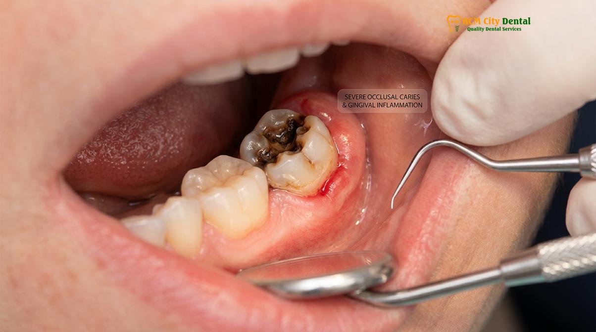
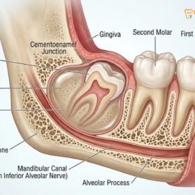
Furthermore, the angulation of the wisdom tooth plays a critical role in its susceptibility to decay. Mesioangular impactions, where the wisdom tooth tilts forward toward the front of the mouth, are particularly problematic. This angulation creates a deep, V-shaped crevice between the wisdom tooth and the adjacent second molar. This crevice acts as a mechanical trap for food particles. Every time a patient chews, fibrous foods and carbohydrates are packed tightly into this space—a phenomenon known clinically as food impaction. The constant presence of fermentable carbohydrates in this stagnant zone provides an endless fuel source for cariogenic bacteria, accelerating the decay process exponentially. [1]
The histological progression of a wisdom tooth cavity follows the same pathological pathway as any other tooth, but often goes unnoticed for longer periods. The decay begins in the outer enamel layer, which is devoid of nerve endings, meaning the patient experiences no pain. As the demineralization breaches the dentinoenamel junction (DEJ) and enters the dentin, the decay spreads laterally and progresses more rapidly due to the tubular structure of dentin. It is only when the bacterial toxins approach the dental pulp—the innermost chamber containing nerves and blood vessels—that the patient begins to experience thermal sensitivity or spontaneous throbbing pain. By the time symptoms manifest, the structural damage to the wisdom tooth is usually extensive and irreversible.
How a Cavity Spreads from Wisdom Tooth to Second Molar
Decay spreads from a wisdom tooth to the second molar through a shared contact point where trapped food and bacterial acids continuously erode the adjacent enamel.
One of the most devastating consequences of an untreated third molar cavity is its ability to compromise the adjacent, highly functional second molar. This phenomenon, often referred to as wisdom tooth decay next tooth involvement, is a classic example of how localized dental pathology can cascade into a broader regional issue within the mouth. The mechanism of this spread is not a direct biological infection from one tooth’s internal structure to another, but rather an environmental contamination caused by the physical proximity of the two teeth.
When a wisdom tooth is impacted or poorly positioned, it often rests directly against the distal (back) surface of the second molar. In a healthy dental arch, adjacent teeth touch at a small, convex area called the contact point, with a triangular space below it (the embrasure) filled by healthy gum tissue (the interdental papilla). However, the abnormal angulation of a wisdom tooth destroys this natural, self-cleansing architecture. Instead of a pinpoint contact, there is often a broad, flat area of contact, or worse, a deep periodontal pocket where the wisdom tooth crown abuts the second molar’s root surface.
“The interproximal space between a mesioangular wisdom tooth and a second molar is a highly stagnant zone. The continuous accumulation of plaque in this inaccessible area guarantees that the acidic byproducts of bacterial metabolism will simultaneously attack the enamel of both teeth, leading to dual-tooth structural failure if left unmanaged.”
As the cavity on the wisdom tooth grows, it creates a rough, cavitated surface that harbors even more plaque. The bacterial load in this specific interproximal space skyrockets. The acidic byproducts produced by these bacteria do not discriminate; they attack the enamel of the wisdom tooth and the adjacent second molar with equal ferocity. Because the space is impossible to clean, the acid remains in constant contact with the second molar’s distal surface. This continuous acid challenge overwhelms the natural remineralizing capacity of the patient’s saliva, leading to the breakdown of the second molar’s enamel. [2]
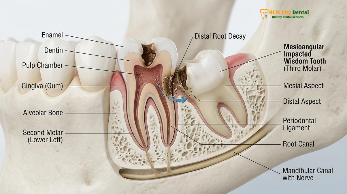
The physical dynamics of chewing exacerbate this problem. The occlusal forces generated during mastication can cause slight micromovements of the teeth. When food is wedged between the second and third molars, these micromovements grind the abrasive food particles and acidic plaque directly into the enamel of the second molar. This combination of chemical dissolution and mechanical abrasion accelerates the formation of a cavity on the second molar. Furthermore, the inflammatory response of the surrounding gum tissue (gingivitis or periodontitis) causes the gums to bleed and swell, providing a nutrient-rich exudate that further fuels bacterial growth and shifts the local microbiome toward more aggressive, tissue-destroying pathogens.
The Threat of Distal Caries on the Second Molar
Distal caries on the second molar is a severe threat because the decay often occurs below the gumline, rapidly approaching the dental pulp and complicating restoration.
When a patient develops a second molar cavity wisdom tooth complication, the clinical scenario shifts from a routine extraction to a complex restorative challenge. The decay that forms on the back of the second molar—clinically termed distal cervical caries—is uniquely dangerous due to its specific anatomical location. Unlike cavities that form on the chewing surfaces of teeth, which are easily visible and surrounded by thick enamel, distal cervical caries forms at or below the cementoenamel junction (CEJ). This is the area where the crown of the tooth meets the root.
The enamel at the CEJ is extremely thin, and in some cases, the root surface (composed of cementum and dentin) is directly exposed to the oral environment. Cementum and dentin are significantly softer and less mineralized than enamel, meaning that once the acidic bacterial attack reaches this area, the decay progresses toward the tooth’s nerve at an alarming rate. Because this decay is hidden deep between the teeth and often below the gumline, it is entirely invisible to the naked eye during a standard visual examination. It is typically only discovered through routine bitewing or panoramic radiographs, or when the patient presents with acute pain.
The proximity of this decay to the dental pulp is a major clinical concern. The pulp chamber in a second molar is relatively large, and the distance from the distal root surface to the nerve is short. If the decay penetrates the pulp, the patient will require root canal therapy to save the tooth, adding significant complexity, time, and expense to the treatment plan. In severe cases where the decay has destroyed too much of the root structure, the second molar may become non-restorable and require extraction alongside the wisdom tooth, resulting in a significant loss of chewing function.
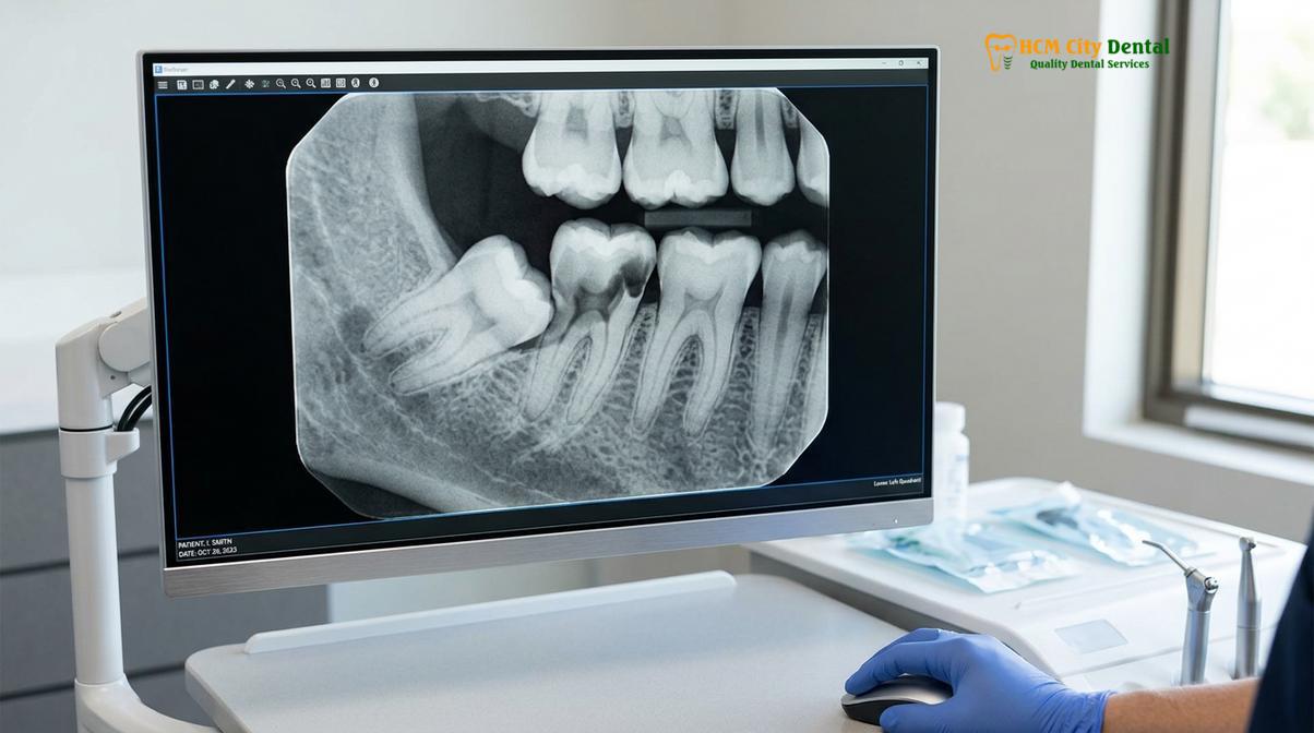
Restoring a second molar with distal cervical caries is one of the most technically demanding procedures in restorative dentistry. To properly repair a cavity, a dentist must remove all the decayed tissue and place a filling material in a completely dry, isolated environment. However, because the decay is located far back in the mouth, below the gumline, and adjacent to a bleeding, inflamed periodontal pocket, achieving this isolation is incredibly difficult. Blood and saliva easily contaminate the area, which can compromise the chemical bond of modern composite resin filling materials. [3]
| Clinical Parameter | Isolated Wisdom Tooth Decay | Combined Decay (Involving Second Molar) |
|---|---|---|
| Primary Symptom | Localized dull ache, occasional swelling | Severe thermal sensitivity, sharp radiating pain |
| Treatment Complexity | Straightforward surgical extraction | Extraction + complex subgingival restoration |
| Risk to Chewing Function | Minimal (third molars are non-essential) | High (second molars are critical for mastication) |
| Endodontic Risk | None (tooth is removed) | High risk of root canal therapy for second molar |
| Long-term Prognosis | Excellent following extraction | Guarded, depends on the depth of the distal caries |
Furthermore, the physical presence of the wisdom tooth itself often blocks the dentist’s instruments from reaching the decay on the second molar. In almost all cases of distal cervical caries, the wisdom tooth must be surgically extracted first, simply to gain the physical access necessary to clean and restore the second molar. The timing of this restoration is critical; it is often performed immediately after the extraction while the patient is still numb, or delayed slightly to allow the extraction socket to heal, depending on the severity of the bleeding and the depth of the cavity.
Why Restoring a Wisdom Tooth Cavity is Rarely Recommended
Dentists rarely restore wisdom teeth because they offer little chewing value, and fillings in this inaccessible area have an exceptionally high rate of recurrent decay.
When patients experience decayed wisdom tooth pain, their first instinct is often to ask if the tooth can simply be filled, just like any other cavity. While technically possible in a small minority of cases where the wisdom tooth is perfectly straight, fully erupted, and easily accessible, the overwhelming consensus in modern dentistry is that restoring a decayed wisdom tooth is a poor clinical investment. The rationale behind this recommendation is multifaceted, involving considerations of biomechanics, long-term prognosis, and overall oral health.
First and foremost is the issue of functional utility. In the modern human diet, third molars provide virtually no significant contribution to mastication (chewing). The first and second molars bear the brunt of the occlusal load required to break down food. Therefore, expending time, effort, and financial resources to save a tooth that does not contribute to oral function is generally counterproductive. Retaining a compromised wisdom tooth simply maintains a high-risk area in the mouth without offering any functional reward.
Clinical Case Review: The Futility of Third Molar Restoration
A 26-year-old patient visited HCMC Dental Clinic in Ho Chi Minh City complaining of severe pain in the lower left jaw. Two years prior, at a different facility, the patient had opted for a large composite filling on a partially erupted wisdom tooth to avoid extraction. Upon examination, Dr. Nguyen Van Cuong noted that the filling had failed entirely due to recurrent decay at the gingival margin, a common result of poor moisture control during the initial procedure. The decay had now progressed into the nerve, causing an acute abscess. The tooth was surgically extracted, highlighting why attempting to restore inaccessible third molars often merely delays the inevitable need for removal while increasing the risk of severe infection.
The technical challenges of placing a successful restoration in a wisdom tooth cannot be overstated. Dental materials, particularly tooth-colored composite resins, require a pristine, dry environment to bond correctly to the tooth structure. The posterior location of wisdom teeth makes isolation extremely difficult. The patient’s tongue, cheek muscles, and copious salivary flow constantly threaten to contaminate the working field. If even a microscopic amount of moisture contaminates the tooth during the bonding process, the filling will eventually leak. This microleakage allows bacteria to seep underneath the filling, leading to recurrent decay that destroys the tooth from the inside out. [4]
Furthermore, even if a filling is placed perfectly, the underlying anatomical and environmental factors that caused the cavity in the first place remain unchanged. The tooth is still located in a hard-to-clean area, and the patient will likely continue to struggle with plaque removal. Consequently, the rate of secondary caries (new decay forming around the edges of the filling) is exceptionally high in third molars. Dr. Nguyen Van Cuong frequently advises patients that filling a wisdom tooth is often just a temporary stopgap that delays the inevitable need for extraction, while simultaneously increasing the risk of the decay spreading to the vital second molar during the interim period.
Treatment Options: Extracting Wisdom Tooth and Restoring Second Molar
The standard treatment protocol involves surgically extracting the decayed wisdom tooth and simultaneously restoring the damaged second molar to preserve chewing function.
When a patient presents with combined wisdom tooth cavity damage affecting the adjacent teeth, a comprehensive, two-phased treatment approach is required. The primary objective is to eliminate the source of the infection (the wisdom tooth) and rehabilitate the functional tooth (the second molar). This workflow requires precise surgical and restorative skills to ensure a predictable, long-term outcome.
The first phase of treatment is the surgical removal of the decayed third molar. Depending on the degree of impaction and the extent of the decay, this procedure is typically performed under local anesthesia, though sedation options are available for anxious patients. The oral surgeon or specialized dentist will carefully elevate the gingival tissue to expose the bone and the tooth. Because decayed teeth are structurally weak and prone to fracturing under pressure, the surgeon will often use a specialized handpiece or an ultrasonic piezotome to section the tooth into smaller pieces. This technique, known as odontotomy, allows the tooth to be removed gently without applying excessive force to the surrounding jawbone or the adjacent second molar. For more details on this surgical protocol, patients can review our comprehensive guide on Tooth Extraction.
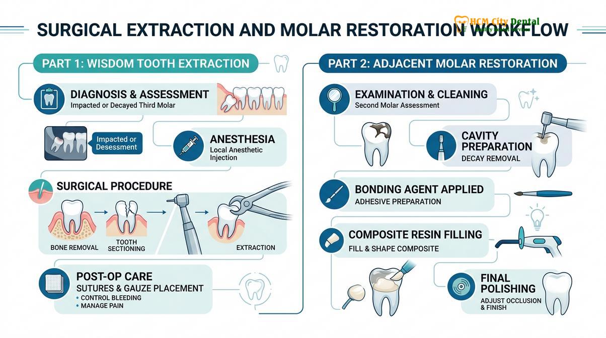
Once the wisdom tooth is successfully removed, the surgical site is thoroughly irrigated with sterile saline or an antimicrobial solution to flush out any remaining debris, infected tissue, or bacterial colonies. At this critical juncture, the dentist finally has unobstructed visual and physical access to the distal surface of the second molar. The second phase of treatment—restoring the second molar—can now commence. If the cavity on the second molar is moderate, the dentist will carefully excavate the decayed dentin using slow-speed instruments to avoid irritating the underlying pulp.
Because the cavity is often located below the gumline, achieving proper isolation is paramount. The dentist will utilize specialized matrix bands and wedges to create a temporary wall around the tooth, ensuring that the restorative material is contoured correctly and does not overhang into the healing extraction socket. A high-quality, moisture-tolerant bonding agent is applied, followed by the incremental placement of a bulk-fill composite resin or a glass ionomer cement. Glass ionomer is sometimes preferred in these deep cervical areas because it chemically bonds to dentin and releases fluoride, which helps remineralize the surrounding tooth structure and prevent future decay.
“The simultaneous management of third molar extraction and second molar restoration requires meticulous moisture control and precise matrix adaptation. Failure to properly seal the distal margin of the second molar will inevitably lead to recurrent caries and eventual endodontic failure.”
In severe cases where the decay has already breached the pulp chamber of the second molar, a root canal treatment will be necessary. This involves removing the infected nerve tissue, disinfecting the internal canals, and sealing them with a biocompatible material called gutta-percha. Following a root canal, the second molar will be significantly weakened and will require a full-coverage dental crown to protect it from fracturing under the heavy forces of mastication. The complexity of this combined treatment underscores the critical importance of addressing wisdom tooth issues before they compromise adjacent structures.
Preventive Care and Early Detection in Saigon
Proactive radiographic screening and rigorous oral hygiene are essential to detect hidden wisdom tooth decay before it inflicts irreversible damage on neighboring teeth.
Preventing wisdom tooth cavity damage requires a proactive approach, combining advanced diagnostic imaging with meticulous home care. Because the most destructive decay often occurs below the gumline and between the teeth, relying solely on visual inspections or waiting for pain to occur is a dangerous strategy. By the time symptoms arise, the damage is usually extensive. Therefore, early detection is the cornerstone of preserving posterior dental health.
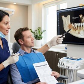
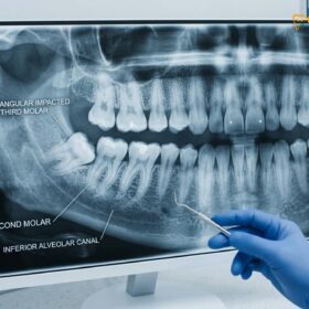
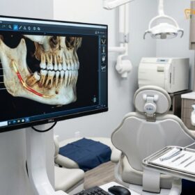
At HCMC Dental Clinic in Ho Chi Minh City, the standard of care for evaluating third molars involves comprehensive radiographic imaging. While traditional panoramic X-rays provide a good two-dimensional overview of the jaw and tooth positioning, they can sometimes obscure the fine details of early distal cervical caries due to the overlapping shadows of the teeth and bone. For a definitive diagnosis, especially when a second molar is suspected to be compromised, a Cone Beam Computed Tomography (CBCT) scan is highly recommended. A CBCT scan generates a high-resolution, 3D image of the teeth and surrounding bone, allowing the dentist to view the exact relationship between the wisdom tooth and the second molar from any angle. This imaging capability is invaluable for detecting hidden cavities, assessing the proximity of the tooth roots to the inferior alveolar nerve, and planning a safe, minimally invasive extraction. [5]
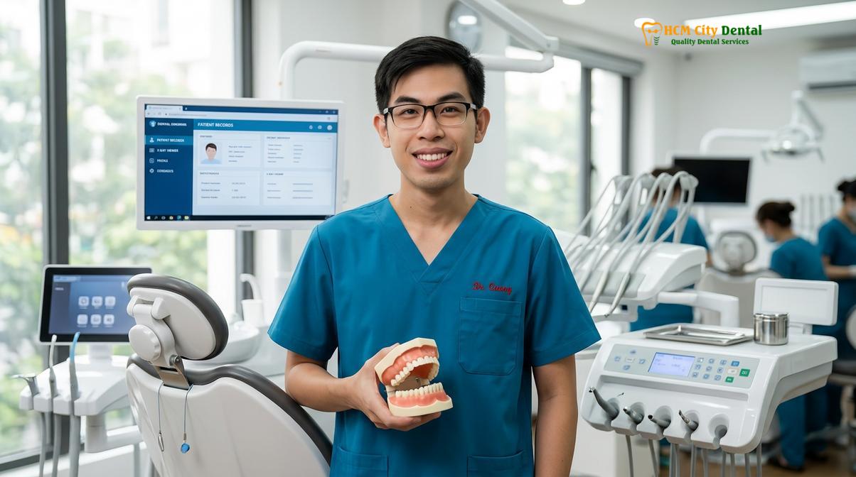
In addition to professional diagnostics, patients must optimize their daily oral hygiene routines to mitigate the risk of decay in the posterior regions of the mouth. Standard brushing is often insufficient for cleaning around partially erupted wisdom teeth. Patients are advised to use a single-tufted brush (an end-tuft brush), which has a small, angled head designed specifically to reach behind the last molars and sweep away plaque from the operculum and distal surfaces. Furthermore, incorporating a water flosser into the daily routine is highly effective. The pressurized stream of water can flush out trapped food particles and disrupt the bacterial biofilm in the deep crevices between the second and third molars, areas where traditional string floss often struggles to reach effectively.
Regular professional cleanings and checkups are non-negotiable for patients who retain their wisdom teeth. During these visits, dental hygienists use specialized ultrasonic scalers to remove hardened calculus (tartar) from the posterior teeth, reducing the bacterial load and minimizing gingival inflammation. If you are experiencing any discomfort in the back of your mouth or have not had your wisdom teeth evaluated recently, scheduling a consultation at HCMC Dental Clinic is a vital step in protecting your long-term oral health and preventing complex restorative procedures.
When to See a Doctor
Important Clinical Considerations & Warning Signs
Wisdom tooth decay can rapidly escalate from a minor localized issue to a severe systemic infection. You should seek immediate clinical evaluation if you experience any of the following symptoms:
- Spontaneous, throbbing pain in the back of the jaw that radiates to the ear or neck.
- Visible swelling of the gums, cheek, or jawline, indicating a spreading abscess.
- A persistent bad taste in the mouth or foul breath (halitosis) caused by purulent discharge (pus).
- Difficulty opening your mouth fully (trismus) or pain when swallowing.
- Extreme sensitivity to hot or cold temperatures in the second molar region.
Do not rely on over-the-counter pain medications to mask these symptoms. A personalized clinical examination and radiographic assessment are required to determine the appropriate surgical or restorative intervention.
Frequently Asked Questions
Can a dentist fill a cavity on a wisdom tooth?
Yes, a dentist can technically fill a cavity on a wisdom tooth if it is fully erupted and functional. However, extraction is almost always recommended instead, as fillings in this inaccessible area have a high failure rate and the tooth offers little chewing value. The difficulty in maintaining a dry environment during the filling process often leads to poor bonding and recurrent decay.
What happens if a decayed wisdom tooth is not removed?
If a decayed wisdom tooth is not removed, the decay will penetrate the dental pulp, causing severe infection, abscesses, and excruciating pain. Furthermore, the bacterial infection will likely spread to the adjacent second molar and surrounding jawbone. This can lead to localized bone loss, systemic infection, and the eventual need to extract multiple teeth.
How does a wisdom tooth damage the molar next to it?
A wisdom tooth damages the adjacent molar by creating a tight crevice where food and plaque become trapped. This stagnant bacterial environment produces acids that rapidly erode the distal surface of the second molar, leading to deep cavities. The physical pressure of an impacted wisdom tooth can also cause root resorption on the adjacent tooth.
Is a cavity on a second molar serious?
Yes, a cavity on a second molar is highly serious, especially when caused by an adjacent wisdom tooth. These cavities often form below the gumline near the tooth’s nerve, making them difficult to restore and increasing the risk of root canal therapy or tooth loss. The second molar is critical for chewing, so preserving it is a top clinical priority.
How much does it cost to treat a wisdom tooth cavity and second molar in Vietnam?
Currently, the cost varies based on the complexity of the extraction and the type of restoration required for the second molar. Patients can expect a highly competitive fee schedule compared to Western countries, though an exact quote requires a clinical and radiographic evaluation. Simple extractions cost less than complex surgical removals involving bone impaction.
References
- Journal of Oral and Maxillofacial Surgery. Pathological conditions associated with impacted third molars. (2021).
- International Journal of Dentistry. Distal cervical caries in the second molar associated with impacted third molars. (2020).
- British Dental Journal. Restorative challenges of the mandibular second molar following third molar extraction. (2019).
- Journal of the American Dental Association. Prophylactic removal of third molars: A clinical review. (2022).
- Clinical Oral Investigations. Microbiological profile of pericoronitis and third molar caries. (2018).