Upper wisdom tooth sinus pressure occurs when the roots of the upper third molars compress against or penetrate the delicate floor of the maxillary sinus cavity. This close anatomical relationship means that dental impactions, localized inflammation, or infections can directly trigger sinus pain, congestion, and referred headaches.
Clinical Summary:
The maxillary sinus cavities rest directly above the upper jaw, often sharing a microscopic bony boundary with the roots of the upper wisdom teeth. When these third molars become impacted, erupt at abnormal angles, or develop periodontal infections, the resulting inflammation can easily transfer across the sinus floor. This phenomenon, known as odontogenic sinusitis, frequently causes unilateral facial pressure, nasal congestion, and headaches that mimic traditional sinus infections. Accurate diagnosis relies on 3D Cone Beam Computed Tomography (CBCT) to map the root anatomy. Surgical extraction of the offending tooth typically resolves the sinus symptoms, provided strict post-operative sinus precautions are followed to prevent oroantral communication.
Key Takeaways:
- Upper wisdom teeth roots often sit millimeters away from, or even protrude into, the maxillary sinus cavity.
- Dental infections can spread to the sinus lining, causing odontogenic sinusitis (sinusitis of dental origin).
- Symptoms often include unilateral facial pressure, localized gum pain, and referred headaches.
- 3D CBCT imaging is a critical diagnostic tool to assess the risk of sinus exposure prior to extraction.
- Following surgery, strict sinus precautions (no nose blowing, no straws) are mandatory to ensure the sinus floor heals safely.
The Close Relationship Between Upper Wisdom Teeth and Sinus Cavities
The upper wisdom teeth and the maxillary sinuses share a highly intimate anatomical boundary, separated only by a thin layer of bone and a delicate mucosal membrane.
To understand why a dental issue can feel exactly like a respiratory or allergy problem, one must first examine the complex anatomy of the human maxilla (upper jaw). The maxillary sinuses are the largest of the paranasal sinuses—hollow, air-filled spaces located behind the cheekbones, extending from just below the eyes down to the roots of the upper posterior teeth. As the human skull develops and matures, these sinus cavities undergo a process called pneumatization, where they expand downward into the alveolar bone of the upper jaw. This expansion frequently brings the sinus floor into direct contact with the roots of the upper molars, particularly the second and third molars (wisdom teeth)[1].
In many patients, the separation between the upper wisdom teeth sinus roots and the sinus cavity is incredibly thin—sometimes measuring less than a millimeter of cortical bone. In certain anatomical variations, there is no bony separation at all; the roots of the wisdom teeth actually protrude directly into the sinus cavity, covered only by the Schneiderian membrane, which is the delicate mucosal lining of the sinus. This microscopic proximity creates a direct pathway for mechanical pressure and bacterial transmission.
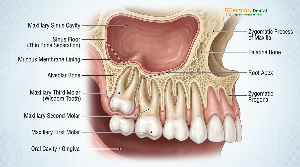
When an upper wisdom tooth begins to erupt, it requires adequate space in the posterior maxilla. However, modern human jaws frequently lack the necessary arch length to accommodate these late-erupting third molars. As the tooth attempts to push through the gum tissue, it may become impacted—trapped within the jawbone or angled improperly against the adjacent second molar. The physical force of this impaction, combined with the natural growth trajectory of the tooth, can exert upward pressure directly against the sinus floor. This mechanical compression alone is often enough to trigger a sensation of fullness, pressure, and dull aching in the cheekbones, even in the absence of an active bacterial infection.
Furthermore, the periodontal ligament—the fibrous connective tissue that anchors the tooth to the surrounding bone—is rich in nerve endings and blood vessels. When an upper wisdom tooth is under stress from impaction, this ligament becomes inflamed. Because the vascular and neural networks of the posterior maxilla are intimately shared with the maxillary sinus, localized dental inflammation can cause a sympathetic inflammatory response in the sinus lining. The Schneiderian membrane may become engorged and edematous, reducing the volume of the sinus cavity and impairing its natural ability to drain mucus through the ostium (the sinus drainage pathway into the nasal cavity). This cascade of events perfectly illustrates the profound dental sinus connection that complicates the diagnosis of facial pain.
How Upper Wisdom Teeth Cause Sinus Pressure and Headaches
Impacted or infected upper wisdom teeth trigger localized inflammation that compresses the sinus floor and irritates the trigeminal nerve, leading to referred pain and headaches.
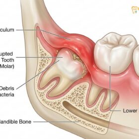
The sensation of sinus pressure and the development of headaches related to wisdom teeth are primarily driven by two distinct but overlapping mechanisms: mechanical obstruction leading to mucosal inflammation, and complex neural pathways causing referred pain. When an upper wisdom tooth is partially erupted, it creates a deep periodontal pocket—a flap of gum tissue known as an operculum—that is notoriously difficult to keep clean. Food debris and oral bacteria inevitably accumulate beneath this flap, leading to a localized infection called pericoronitis. As the infection takes hold, the body’s immune response triggers a massive release of inflammatory mediators, including cytokines and prostaglandins, into the surrounding tissues.
Because the upper wisdom teeth sinus roots are situated so close to the sinus floor, this inflammatory soup easily diffuses upward. The sinus membrane reacts by swelling and increasing mucus production. If the swelling is severe enough to block the sinus ostium, the mucus becomes trapped. The trapped fluid creates a buildup of hydrostatic pressure within the rigid bony walls of the maxillary sinus. Patients experience this internal pressure as a deep, throbbing ache in the mid-face, often describing it as a classic sinus pain wisdom teeth scenario. The pressure worsens when bending over, lying down, or experiencing changes in barometric pressure, which are hallmark signs of sinus congestion[2].
“The anatomical convergence of the maxillary dentition and the paranasal sinuses means that odontogenic pathology must always be ruled out when a patient presents with unilateral sinus pressure that is refractory to standard decongestant therapy.”
Beyond the physical pressure of trapped mucus, the neurological aspect of this condition is equally significant. The entire facial region, including the upper teeth, the periodontal ligaments, and the maxillary sinus lining, is innervated by the trigeminal nerve (Cranial Nerve V). Specifically, the maxillary division (V2) of the trigeminal nerve supplies sensory information from both the upper molars and the sinus cavity to the brain. When an impacted wisdom tooth causes severe localized pain, the intense barrage of pain signals traveling along the V2 pathway can overwhelm the central nervous system’s ability to pinpoint the exact source of the irritation.
This phenomenon is known as referred pain. The brain misinterprets the distress signals originating from the periodontal ligament of the wisdom tooth as coming from the broader region supplied by the nerve, including the sinus cavity, the temples, and the forehead. Consequently, a patient may suffer from a debilitating wisdom tooth sinus headache without ever realizing that the root cause lies in their dental arch. This referred pain is often described as a continuous, dull ache that radiates from the back of the upper jaw up through the cheekbone and into the temporal region, making it easily confusable with a tension headache or a severe sinus migraine.
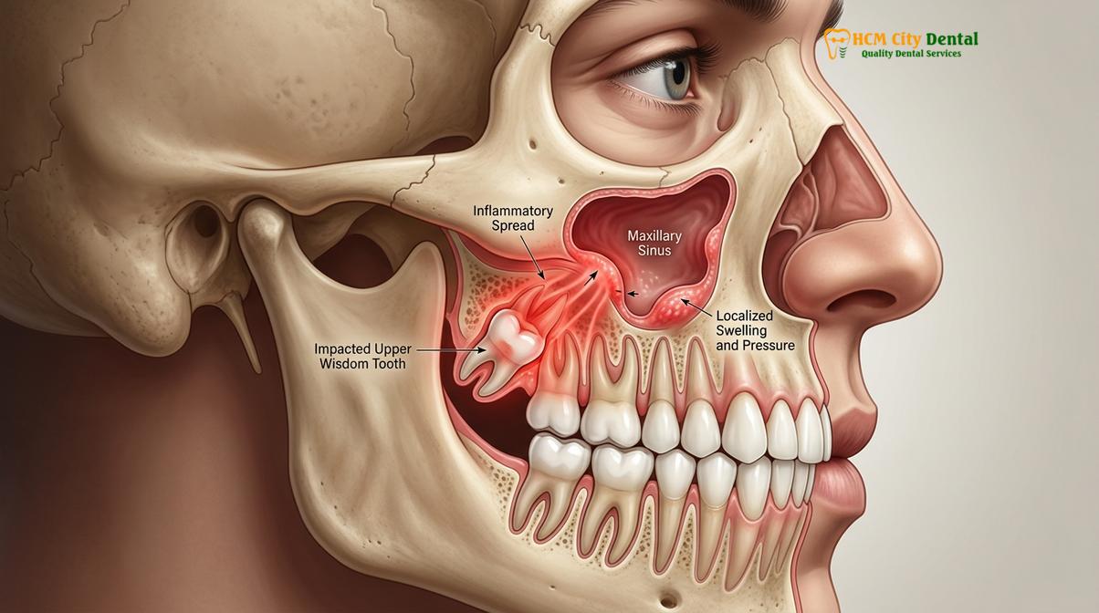
It is also crucial to consider the role of chronic, low-grade infections. Unlike acute pericoronitis, which presents with obvious swelling and severe pain, a chronic apical abscess (an infection at the very tip of the tooth root) may remain largely asymptomatic in the mouth. The bacteria slowly erode the thin bone separating the tooth from the sinus, eventually breaching the sinus floor. Once the bacteria enter the sinus cavity, they establish a secondary infection. This is a classic presentation of odontogenic sinusitis, where the primary source of the sinus infection is entirely dental. In these cases, treating the sinus infection with traditional antibiotics or nasal sprays will only provide temporary relief; the sinus pressure will inevitably return until the offending wisdom tooth is professionally addressed through clinical extraction procedures.
Symptoms: Sinusitis vs Wisdom Tooth Infection
Distinguishing between a primary sinus infection and sinusitis of dental origin requires evaluating specific clinical signs, such as unilateral symptoms and localized dental tenderness.
For both patients and healthcare providers, differentiating between rhinogenic sinusitis (a standard sinus infection caused by a virus, allergy, or airborne bacteria) and odontogenic sinusitis (a sinus infection caused by a dental issue) is a critical diagnostic challenge. Because the symptoms overlap so heavily, misdiagnosis is common. Patients often endure multiple rounds of broad-spectrum antibiotics prescribed by general physicians, only to experience a rapid recurrence of symptoms once the medication is finished. This cycle continues until the underlying dental pathology is identified and treated.
The most definitive hallmark of odontogenic sinusitis is the unilateral nature of the symptoms. Standard respiratory infections and seasonal allergies typically affect the upper respiratory tract symmetrically; therefore, rhinogenic sinusitis usually presents with bilateral congestion, meaning both the left and right sinus cavities are blocked and painful. In stark contrast, a sinus infection originating from an upper wisdom tooth is almost always unilateral. The patient will experience intense pressure, pain, and nasal discharge strictly on the side of the face where the problematic tooth is located. If you are experiencing severe sinus pressure in your right cheekbone while your left cheekbone feels completely normal, there is a high clinical probability of a dental etiology[3].
Another crucial differentiating factor is the nature of the nasal discharge. While viral sinus infections often produce clear or mildly yellow mucus, odontogenic sinusitis is frequently characterized by thick, purulent (pus-filled), and foul-smelling nasal discharge. The bacteria responsible for dental infections are typically anaerobic (thriving in environments without oxygen), such as *Peptostreptococcus*, *Fusobacterium*, and *Prevotella*. When these anaerobic bacteria colonize the sinus cavity, they produce sulfur compounds that result in a distinctively bad odor and a foul taste in the back of the throat, often described as post-nasal drip with a metallic or putrid flavor.
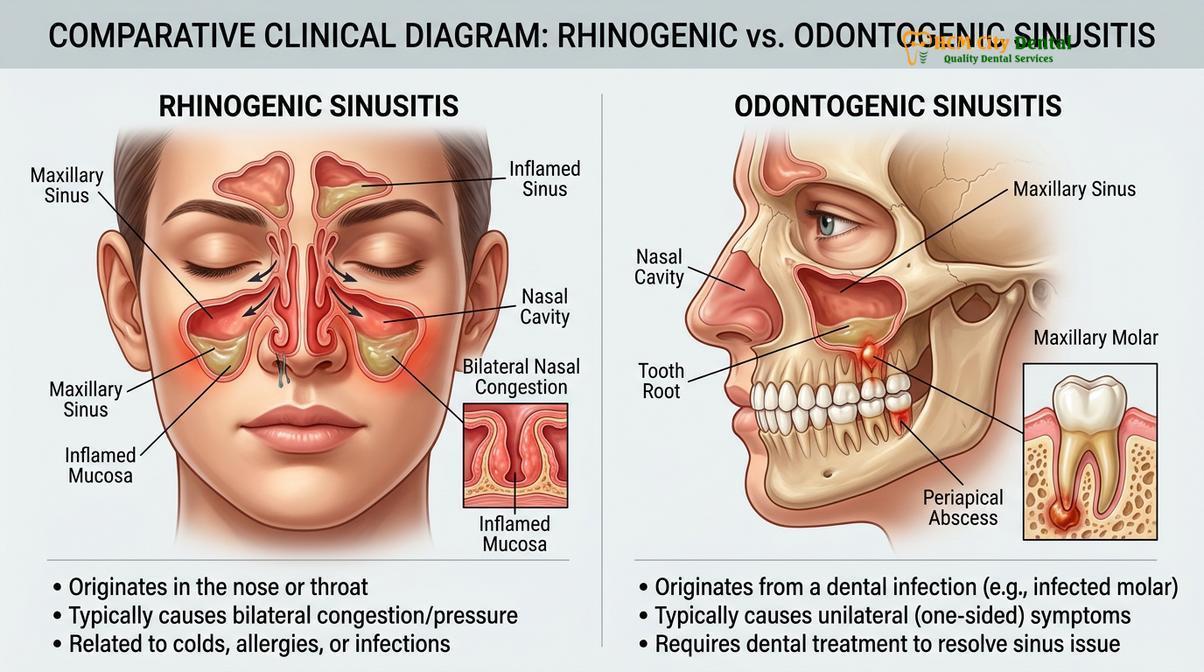
To provide a clear clinical framework, the following table outlines the primary diagnostic differences between these two conditions:
| Clinical Symptom | Rhinogenic Sinusitis (Standard) | Odontogenic Sinusitis (Dental Origin) |
|---|---|---|
| Symmetry of Symptoms | Usually bilateral (both sides of the face) | Almost always unilateral (one side of the face) |
| Nasal Discharge | Clear, white, or light yellow mucus | Thick, purulent, foul-smelling/foul-tasting discharge |
| Dental Symptoms | Generalized mild aching in all upper teeth | Severe, localized pain in one specific back tooth |
| Response to Decongestants | Often provides significant temporary relief | Minimal to no relief; pressure remains constant |
| History of Dental Issues | Not applicable | Recent toothache, gum swelling, or deep cavities |
Furthermore, localized dental symptoms are a primary indicator. While a severe standard sinus infection can cause generalized aching across all the upper teeth due to the swollen sinus floor pressing down on the entire maxillary nerve plexus, an infected wisdom tooth will present with highly localized signs. The patient may notice that the gum tissue specifically behind the last visible molar is red, swollen, and exquisitely tender to the touch. Biting down on that specific side may elicit a sharp, shooting pain, indicating inflammation of the periodontal ligament. If these localized dental signs accompany unilateral sinus pressure, a comprehensive dental evaluation is imperative to prevent the infection from spreading further into the maxillofacial spaces.
Understanding Sinus Communication Risk During Extraction
Removing upper wisdom teeth carries a risk of creating an oroantral communication, a temporary opening between the mouth and sinus that requires careful management.
When conservative management fails and an upper wisdom tooth is deemed the source of chronic sinus pressure or infection, surgical extraction is the definitive treatment. However, the very anatomy that causes the problem—the microscopic distance between the tooth roots and the sinus floor—also introduces a specific surgical risk known as an oroantral communication (OAC). An OAC is an unnatural opening or pathway that connects the oral cavity directly to the maxillary sinus cavity. This occurs when the thin plate of bone separating the two spaces is removed along with the tooth, or when the roots of the tooth were already protruding through the bone and into the sinus lining prior to extraction.
During the extraction of an upper third molar, the oral surgeon applies controlled, precise forces to elevate and deliver the tooth from its socket. If the upper wisdom teeth sinus roots are deeply embedded in the sinus floor, their removal leaves behind a void. In many cases, the Schneiderian membrane remains intact, and the socket heals normally. However, if the membrane is exceptionally thin, inflamed, or firmly attached to the tooth roots, it may tear during the extraction process. This tear creates a direct communication between the mouth and the sinus. The surgeon will typically test for this immediately after the tooth is removed by asking the patient to gently pinch their nose and blow out (a gentle Valsalva maneuver). If air bubbles appear in the empty tooth socket, an OAC is confirmed[4].
Clinical Warning: If an oroantral communication occurs during extraction, it is vital to avoid any actions that create negative pressure in the mouth or positive pressure in the nose. Failure to follow strict sinus precautions can prevent the opening from closing, leading to a chronic oroantral fistula that requires complex surgical repair.
The management of an oroantral communication depends entirely on its size. Small communications (less than 2 millimeters in diameter) frequently heal spontaneously without additional surgical intervention, provided the patient strictly adheres to post-operative sinus precautions to allow a stable blood clot to form and organize. The blood clot acts as a natural biological scaffold, sealing the opening while new tissue regenerates across the gap. For moderate communications (2 to 6 millimeters), the surgeon may place a specialized collagen sponge or a platelet-rich fibrin (PRF) membrane into the socket and secure it with figure-eight sutures to stabilize the clot and encourage rapid soft tissue closure.
In cases where a large communication occurs (greater than 6 millimeters), or if a small communication fails to heal and epithelializes into a permanent tunnel known as an oroantral fistula (OAF), more advanced surgical closure is required. Techniques such as a buccal advancement flap or the mobilization of the buccal fat pad are utilized to physically stretch healthy tissue over the extraction site, creating a watertight seal. To minimize these risks, highly experienced clinicians prioritize meticulous pre-operative planning and utilize advanced imaging technologies to map the dental sinus connection long before the first incision is made.
Pre-operative 3D CBCT Scanning for Root Mapping
Cone Beam Computed Tomography (CBCT) provides a precise 3D map of the tooth roots and sinus floor, allowing surgeons to plan extractions with maximum safety.
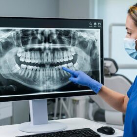
The cornerstone of safe and predictable upper wisdom tooth extraction, particularly when sinus involvement is suspected, is advanced diagnostic imaging. Historically, dentists relied on 2D panoramic X-rays (OPG) to evaluate the position of wisdom teeth. While a panoramic X-ray provides a helpful general overview of the jaws, it suffers from a significant limitation: it compresses three-dimensional anatomical structures into a flat, two-dimensional image. On a standard X-ray, the roots of an upper wisdom tooth may appear to be deeply embedded inside the sinus cavity, when in reality, they might be situated safely to the side of the sinus wall (buccally or palatally). This superimposition makes it impossible to accurately gauge the true risk of sinus perforation.
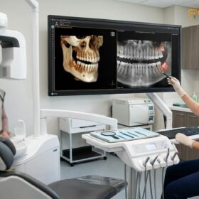
To eliminate this dangerous guesswork, modern oral surgery relies on Cone Beam Computed Tomography (CBCT). A CBCT scanner rotates around the patient’s head, capturing hundreds of high-resolution images from different angles. Advanced software then reconstructs these images into a highly detailed, distortion-free 3D model of the patient’s maxillofacial anatomy. This technology allows the surgeon to virtually slice through the jawbone in any direction (axial, coronal, and sagittal planes) to examine the exact spatial relationship between the tooth roots and the sinus floor down to the fraction of a millimeter.
At HCMC Dental Clinic in Ho Chi Minh City, the utilization of 3D CBCT imaging is a mandatory protocol for complex upper wisdom tooth impactions. By analyzing the CBCT scan, the surgical team can determine the exact thickness of the bone separating the tooth from the sinus, identify the presence of any pre-existing sinus pathology (such as mucosal thickening or polyps), and map the precise curvature of the roots. If the scan reveals that the roots are highly divergent or hooked around the sinus floor, the surgeon can plan a specialized approach, such as carefully sectioning the tooth into smaller pieces to remove it without pulling on the delicate sinus membrane[5].
Clinical Case Review: A 26-year-old patient presented to HCMC Dental Clinic in Saigon complaining of chronic right-sided sinus pressure and a persistent wisdom tooth sinus headache. A standard 2D X-ray was inconclusive. Dr. Nguyen Van Cuong ordered a 3D CBCT scan, which clearly revealed that the mesial root of the upper right third molar had breached the sinus floor, causing localized mucosal thickening. Based on this precise 3D mapping, Dr. Cuong performed a minimally invasive extraction with immediate socket grafting. The patient’s sinus symptoms resolved completely within two weeks.
Furthermore, CBCT imaging is invaluable for patient communication and informed consent. When a patient can visually see the exact position of their tooth in relation to their sinus cavity on a computer monitor, they gain a much deeper understanding of the surgical complexities and the absolute necessity of following post-operative care instructions. This level of diagnostic precision not only drastically reduces the incidence of unexpected surgical complications but also ensures that patients receive the most conservative, tissue-sparing treatment possible.
Sinus Precautions After Upper Wisdom Tooth Removal
Strict adherence to post-operative sinus precautions is mandatory to protect the blood clot and ensure the delicate sinus floor heals without complications.
Following the successful removal of an upper wisdom tooth, the immediate post-operative period is critical for ensuring that the extraction socket heals properly and that the integrity of the maxillary sinus is maintained. Even if a formal oroantral communication was not detected during the surgery, the bone separating the socket from the sinus may be paper-thin. The primary goal of recovery is to protect the fragile blood clot that forms in the socket. This clot is the foundation of the healing process; if it is dislodged, the patient risks developing a painful condition known as dry socket, or worse, creating a delayed opening into the sinus cavity.
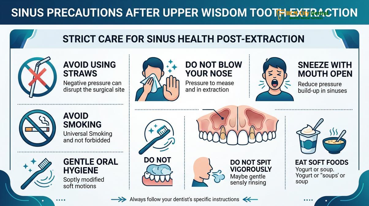
To protect this delicate healing environment, patients must strictly adhere to a set of guidelines known as “sinus precautions” for a minimum of 10 to 14 days following the procedure. The fundamental principle behind these precautions is the avoidance of any activity that creates significant pressure changes—either positive or negative—within the oral and nasal cavities. The most critical rule is to absolutely avoid blowing the nose. If a patient feels congested, they must gently wipe their nose rather than blowing forcefully. The sudden spike in intranasal pressure from blowing can easily rupture the thin membrane separating the sinus from the mouth, blowing the blood clot entirely out of the socket.
“The success of an upper third molar extraction is heavily dependent on patient compliance during the first two weeks of recovery. Protecting the sinus floor from barometric trauma is just as important as the surgical technique itself.”
Similarly, patients must alter how they sneeze. If a sneeze is unavoidable, it must be done with the mouth wide open. Sneezing with a closed mouth forces the explosive air pressure upward into the sinus cavities, which can cause severe damage to the healing surgical site. Furthermore, creating negative pressure in the mouth is equally dangerous. Patients must strictly avoid using drinking straws, smoking cigarettes, or forcefully spitting. The suction created by these actions acts like a vacuum, easily pulling the stabilizing blood clot out of the extraction site. For comprehensive guidance on managing the recovery timeline, patients should review detailed clinical recovery protocols.
In addition to behavioral modifications, pharmacological support plays a vital role in post-operative sinus management. If the surgeon notes a high risk of sinus involvement or confirms a small communication, they will typically prescribe a specific regimen of medications. This often includes a broad-spectrum antibiotic (such as Amoxicillin-Clavulanate) to prevent secondary bacterial colonization of the sinus, and an oral decongestant (such as Pseudoephedrine) to keep the sinus ostium open and prevent fluid buildup. Saline nasal sprays may also be recommended to keep the nasal passages moist without causing irritation. Patients who plan to travel shortly after surgery must also be aware of the risks associated with cabin pressure changes and should consult guidelines regarding flight safety after extractions.
When to See a Doctor
Immediate clinical evaluation is required if you experience severe unilateral facial pain, foul-tasting nasal drainage, or a fever following an extraction.
While mild swelling and discomfort are normal parts of the healing process after any dental surgery, certain symptoms indicate that a complication, such as an infection or an unhealed sinus communication, has occurred. It is crucial to distinguish between normal post-operative recovery and signs that require immediate medical intervention. Delaying treatment for an advancing maxillofacial infection can lead to severe systemic complications.
You should seek immediate evaluation from an oral surgeon or specialized dental professional if you experience any of the following red flag symptoms:
- Foul-Tasting Drainage: A persistent salty, metallic, or putrid taste in your mouth, or foul-smelling yellow/green discharge from your nose, strongly indicates an active bacterial infection in the sinus cavity.
- Air Passing Through the Socket: If you notice air whistling through the extraction site when you speak or breathe, or if liquids you drink leak out through your nose, you likely have an oroantral fistula that requires surgical closure.
- Severe, Unrelenting Pain: Pain that worsens after the first 72 hours, radiates intensely across the cheekbone and eye, and does not respond to prescribed pain medication.
- Systemic Symptoms: The development of a high fever, chills, or significant lethargy suggests that a localized dental infection is spreading systemically.
- Visual Disturbances: Any swelling that extends upward toward the eye, causing double vision or restricted eye movement, is a medical emergency requiring immediate attention to prevent orbital complications.
If you are experiencing these symptoms, do not attempt to self-diagnose or rely solely on over-the-counter cold medications. A thorough clinical examination, often involving a new 3D CBCT scan, is necessary to accurately assess the healing of the sinus floor and implement the appropriate corrective treatment.
Frequently Asked Questions
Can upper wisdom teeth cause sinus congestion and pain?
Yes, upper wisdom teeth can cause sinus congestion and pain when their roots compress the maxillary sinus floor or when a dental infection spreads into the sinus cavity. This proximity often leads to localized inflammation, mimicking the symptoms of a traditional sinus infection. The resulting swelling blocks normal sinus drainage, creating a buildup of hydrostatic pressure that patients feel as deep facial pain and congestion.
How do I know if my sinus pain is from my teeth?
Sinus pain originating from teeth typically presents as unilateral (one-sided) discomfort, accompanied by localized gum tenderness, pain when biting, or a history of dental issues. A clinical examination and 3D CBCT scan are required to definitively trace the pain to a dental source. Unlike standard viral sinus infections that cause bilateral congestion and clear mucus, dental-related sinus pain often involves foul-tasting post-nasal drip and does not respond well to standard decongestants.
What is a sinus communication after wisdom tooth extraction?
A sinus communication is a small, temporary opening between the mouth and the maxillary sinus cavity that can occur when an upper wisdom tooth with roots protruding into the sinus is removed. It requires specific post-operative precautions to heal properly and prevent infection. If the opening is large or fails to heal due to poor clot formation, it can develop into a chronic oroantral fistula requiring surgical repair.
What are sinus precautions after dental surgery?
Sinus precautions include avoiding blowing your nose, sneezing with your mouth open, refraining from using straws, and avoiding smoking for at least two weeks. These measures prevent pressure changes that could dislodge the blood clot and disrupt the healing of the sinus floor. Adhering to these rules is critical to ensure the delicate membrane separating the mouth and sinus heals completely without forming a permanent opening.
How much does it cost to remove upper wisdom teeth in Saigon?
The cost to remove upper wisdom teeth in Saigon varies based on the complexity of the impaction, the proximity to the sinus cavity, and the required diagnostic imaging. Generally, it is highly cost-effective compared to Western countries, though an exact quote requires a personalized clinical consultation. For a detailed breakdown of regional pricing and factors influencing the final fee, patients can review comparisons of wisdom tooth extraction costs.
References
- Journal of Oral and Maxillofacial Surgery. Anatomical relationship between maxillary third molars and the maxillary sinus. (2021).
- International Journal of Oral and Maxillofacial Surgery. Odontogenic sinusitis: pathophysiology and management. (2020).
- British Dental Journal. Oroantral communication: incidence, diagnosis, and treatment protocols. (2019).
- Journal of the American Dental Association. The role of CBCT in upper third molar extractions. (2022).
- Clinical Oral Investigations. Post-operative sinus precautions and healing outcomes. (2018).