An infection after wisdom tooth removal occurs when opportunistic oral bacteria invade the healing surgical socket, leading to severe, progressive pain, localized swelling, and purulent discharge. Prompt clinical evaluation and targeted antimicrobial intervention are essential to eradicate the bacteria, prevent the spread of infection into deeper fascial spaces, and ensure proper regeneration of the alveolar bone.
Clinical Summary:
Post-operative infections following third molar extractions typically manifest between 3 to 14 days after surgery, often presenting as delayed-onset infections. Unlike the normal inflammatory swelling that peaks at 48 hours, an infected socket is characterized by progressive throbbing pain, localized erythema, purulent exudate (pus), and systemic signs such as low-grade fever or cervical lymphadenopathy. Dr. Nguyen Van Cuong emphasizes that distinguishing between a dry socket (alveolar osteitis) and a true bacterial infection is a critical diagnostic step. Accurate diagnosis dictates the treatment protocol, which generally involves mechanical surgical debridement, targeted antimicrobial irrigation, and the administration of systemic antibiotics to halt bacterial proliferation and restore a healthy healing environment.
Key Takeaways:
- Normal post-surgical pain and swelling peak around 48 hours and steadily decline; worsening symptoms after day three strongly indicate a clinical complication.
- The presence of purulent discharge (pus) and a persistent foul taste are definitive, hallmark signs of bacterial colonization within the extraction site.
- Trismus (severely restricted mouth opening) combined with submandibular swelling requires immediate clinical evaluation to rule out dangerous fascial space infections.
- Professional treatment typically involves localized socket debridement, antimicrobial irrigation, and systemic antibiotics tailored to the severity of the infection.
- Strict adherence to post-operative oral hygiene, including gentle saline rinses and avoiding negative pressure, significantly reduces the risk of delayed-onset infections.
Common Causes of Post-Extraction Infections
Infections primarily develop when the oral microbiome infiltrates the surgical wound due to compromised blood clot formation, trapped food debris, or pre-existing periodontal disease.
The oral cavity is a complex ecosystem harboring hundreds of distinct bacterial species. Under normal circumstances, these microorganisms exist in a symbiotic balance. However, the surgical removal of third molars creates a direct, open pathway into the highly vascularized alveolar bone and surrounding soft tissues. When this protective mucosal barrier is breached, opportunistic bacteria—particularly anaerobic strains such as Streptococcus mutans, Prevotella, and Fusobacterium—can colonize the surgical site, leading to a localized infection[1].
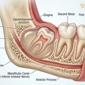
One of the primary anatomical factors contributing to an infection after wisdom tooth removal is the depth and angulation of the impacted tooth. Teeth that are fully bony impacted or positioned horizontally require more extensive surgical intervention, including the reflection of a mucoperiosteal flap and the removal of surrounding cortical bone. This increased surgical trauma naturally elevates the localized inflammatory response and creates a larger dead space where a blood clot must form and stabilize. If this clot is disturbed, breaks down prematurely, or fails to fill the entire void, the exposed bony walls become highly susceptible to bacterial adherence and subsequent infection.
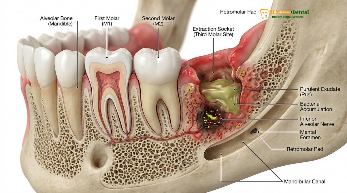
Systemic patient factors also play a profound role in the pathogenesis of post-operative infections. Patients with uncontrolled diabetes mellitus exhibit impaired neutrophil function and reduced peripheral blood flow, which significantly hinders the body’s ability to mount an effective immune response at the surgical site. Similarly, individuals who smoke introduce a dual threat: the physical act of inhaling creates negative intraoral pressure that can dislodge the fragile blood clot, while the chemical toxins in tobacco smoke cause localized vasoconstriction, starving the healing tissues of essential oxygen and immune cells. In severe, neglected cases, a superficial socket infection can progress deeper into the jawbone, eventually presenting with osteomyelitis dental signs, such as deep bone pain, tooth mobility in adjacent teeth, and radiographic evidence of bone destruction[2].
Furthermore, the concept of a “delayed-onset infection” (DOI) is particularly relevant to lower wisdom teeth. Unlike acute infections that appear within the first 48 hours, DOIs typically manifest two to four weeks after the extraction. This phenomenon often occurs when the superficial gum tissue heals and closes over the socket faster than the underlying bone can regenerate. If microscopic food particles or bacterial biofilms are trapped beneath this newly formed mucosal seal, they create an isolated, oxygen-deprived (anaerobic) environment. Over weeks, these trapped bacteria multiply, eventually forming an abscess that ruptures through the healed gum tissue, necessitating secondary surgical intervention.
Worsening Pain vs. Normal Healing Discomfort
Normal healing pain decreases steadily after the second day, whereas infectious pain progressively intensifies, often throbbing and radiating to the ear, temple, or lower jaw.
A critical challenge for patients recovering from oral surgery is distinguishing between the expected physiological discomfort of tissue repair and the pathological pain of an active infection. The body’s natural inflammatory cascade—driven by the release of histamines, prostaglandins, and cytokines—is a necessary component of wound healing. This acute inflammatory phase typically peaks between 48 and 72 hours post-surgery. During this window, patients should expect moderate to severe localized soreness, facial edema (swelling), and mild stiffness in the jaw muscles. However, the defining characteristic of normal healing is a predictable, linear trajectory of improvement: each day following the peak should bring a noticeable reduction in pain and swelling.
Conversely, worsening pain after extraction that begins or intensifies on the third, fourth, or fifth day is a primary clinical red flag. Infectious pain is rarely described as a dull ache; rather, it is frequently characterized as a severe, relentless, throbbing sensation that pulses in time with the patient’s heartbeat. Because the lower wisdom teeth are situated in close proximity to the inferior alveolar nerve, the inflammation associated with an infection can compress this nerve pathway, causing the pain to radiate extensively. Patients often report that the pain shoots up into the ear, across the temporal region, or down along the inferior border of the mandible, making it difficult to pinpoint the exact source of the agony.
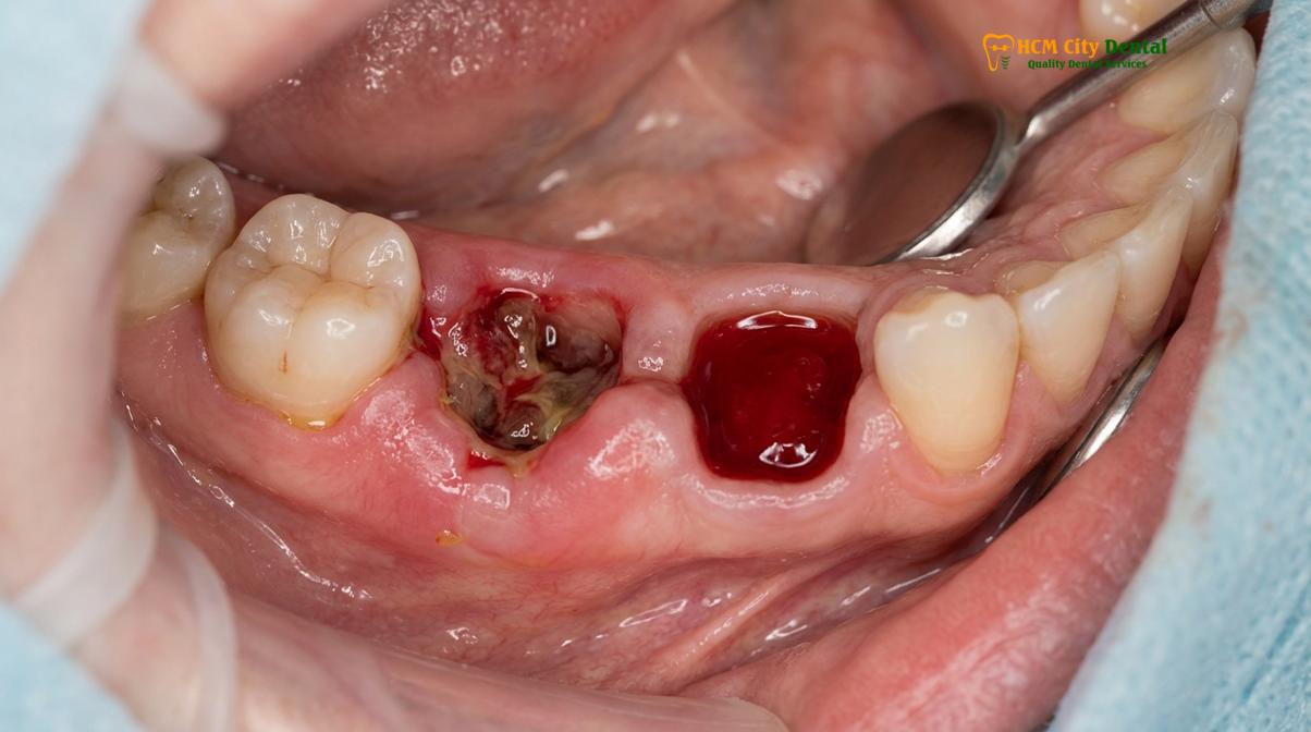
It is also vital to clinically differentiate a true bacterial infection from alveolar osteitis, commonly known as a dry socket. While both conditions cause severe, delayed-onset pain, their underlying mechanisms and treatments differ significantly. A dry socket is a non-infectious complication resulting from the premature fibrinolysis (dissolving) or mechanical dislodgement of the protective blood clot, leaving the underlying bone and nerve endings exposed to the oral environment. While a dry socket is excruciatingly painful, it is not inherently accompanied by pus, systemic fever, or massive facial swelling. An infection, on the other hand, is driven by bacterial proliferation and tissue necrosis[3].
| Clinical Feature | Normal Healing | Dry Socket (Alveolar Osteitis) | Bacterial Socket Infection |
|---|---|---|---|
| Pain Timeline | Peaks at 48 hours, then steadily decreases. | Sudden, severe onset typically on day 3 or 4. | Progressively worsens over several days to weeks. |
| Pain Characteristics | Dull ache, manageable with standard analgesics. | Intense, radiating, exposed bone sensation. | Deep, throbbing, pulsing pressure. |
| Visual Appearance | Dark red/brown stable blood clot present. | Empty socket, visible whitish bone, no clot. | Inflamed, red, puffy gums with yellow/white fluid. |
| Exudate (Discharge) | Slight blood oozing in the first 24 hours. | None. | Purulent discharge (pus) actively seeping. |
| Systemic Symptoms | None. | None. | Low-grade fever, lethargy, swollen lymph nodes. |
If a patient experiences pain that breaks through their prescribed analgesic regimen, or if the pain wakes them from sleep after a period of initial improvement, they must seek immediate evaluation. Relying solely on over-the-counter pain medication to mask progressive symptoms allows the underlying bacterial load to multiply, increasing the risk of more severe anatomical complications.
Early Signs of Socket Infection: Pus, Fever, and Bad Taste
The hallmark indicators of a bacterial socket infection include the visible presence of purulent discharge, a persistent foul odor, and an elevated systemic body temperature.
When bacteria successfully colonize an extraction site, the body’s immune system mounts a vigorous defense. White blood cells, specifically neutrophils and macrophages, rush to the site of the bacterial invasion to engulf and destroy the pathogens. The byproduct of this microscopic battle is purulent exudate, commonly referred to as pus. The presence of pus from wisdom tooth hole is the most definitive, unequivocal sign of an active bacterial infection. This fluid is typically thick, opaque, and ranges in color from pale yellow to yellowish-green. It consists of dead white blood cells, necrotic tissue debris, and both living and dead bacteria.
Patients may notice this discharge spontaneously seeping from the surgical site, or they may experience a sudden rush of fluid if pressure is applied to the surrounding cheek or gum tissue. In some cases, the gum tissue may heal over the top of the infection, trapping the pus beneath the surface. This creates a localized abscess—a fluctuant, highly sensitive swelling on the gum that feels like a fluid-filled blister. As the pressure within the abscess builds, the throbbing pain intensifies until the abscess either ruptures naturally or is surgically drained by a dental professional.
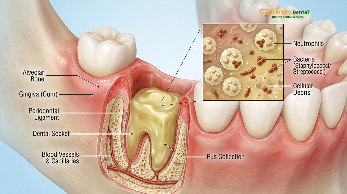
“The appearance of purulent exudate is a clear demarcation line between normal post-operative inflammation and a pathological infection. Once pus is present, mechanical debridement and antimicrobial therapy become absolute clinical necessities to prevent further tissue necrosis.”
Accompanying the physical presence of pus is often a profound alteration in taste and breath. Anaerobic bacteria, which thrive in the deep, oxygen-deprived crevices of an extraction socket, metabolize proteins from trapped food debris and necrotic tissue. This metabolic process releases volatile sulfur compounds (VSCs), such as hydrogen sulfide and methyl mercaptan. Consequently, patients with an infected socket frequently complain of a persistent, metallic, sour, or distinctly foul taste in their mouth that cannot be eliminated by brushing, flossing, or using standard mouthwashes. This halitosis (bad breath) is pervasive and serves as a strong olfactory indicator of bacterial putrefaction.
Furthermore, while localized symptoms dominate the clinical picture, the body may also exhibit a systemic response to the localized infection. The release of pyrogenic cytokines (such as Interleukin-1 and Tumor Necrosis Factor) into the bloodstream signals the hypothalamus to raise the body’s core temperature, resulting in a fever. A low-grade fever (between 99.5°F and 100.9°F / 37.5°C and 38.3°C) occurring several days after the extraction is a strong indicator that the immune system is actively fighting a significant bacterial load. If the fever spikes above 101°F (38.3°C) or is accompanied by severe chills and rigors, it suggests that the infection may be spreading systemically, requiring urgent medical intervention.
Trismus (Lockjaw) and Swollen Lymph Nodes as Warnings
Severe restriction in jaw movement and tender, swollen lymph nodes in the neck indicate that the localized infection is spreading into adjacent muscle tissues and lymphatic channels.
The anatomical positioning of the lower wisdom teeth places them in intimate proximity to several critical structures, including the muscles of mastication and complex fascial planes. When an infection breaches the confines of the alveolar bone, it can easily spread into the adjacent soft tissues. One of the most common and alarming symptoms of this spread is trismus, commonly referred to as lockjaw. Trismus occurs when the inflammatory exudate and bacterial toxins infiltrate the medial pterygoid or masseter muscles, which are responsible for elevating the mandible (closing the mouth). The resulting muscle spasm and severe inflammation physically restrict the patient’s ability to open their mouth[4].
While mild stiffness is a normal part of the post-operative inflammatory phase, infectious trismus is severe and progressive. A patient may find that they can only open their mouth a few millimeters, making it virtually impossible to eat solid food, speak clearly, or maintain adequate oral hygiene. This restriction creates a dangerous cycle: the inability to clean the mouth allows the bacterial load to increase, which in turn worsens the infection and exacerbates the trismus.
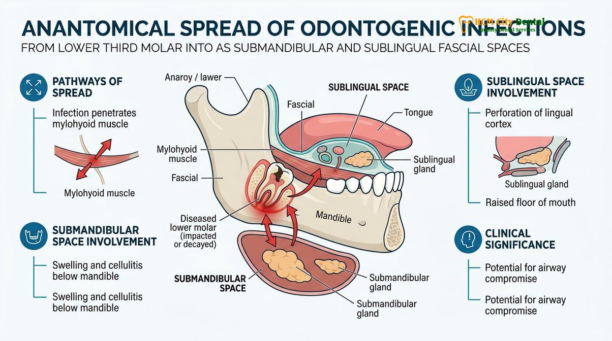
Clinical Warning: If severe trismus is accompanied by rapid swelling in the floor of the mouth (under the tongue) or the submandibular region (under the jawline), it may indicate the onset of Ludwig’s Angina. This is a rare but life-threatening bilateral fascial space infection that can rapidly compromise the airway. Immediate emergency medical evaluation is required.
In addition to muscular involvement, the lymphatic system acts as the body’s drainage and filtration network. The lymphatic vessels draining the lower posterior jaw empty primarily into the submandibular and cervical lymph nodes located along the neck and under the jawline. When a significant infection is present, these nodes become highly active, producing excess lymphocytes to combat the invading bacteria. Clinically, this presents as lymphadenopathy—the lymph nodes become enlarged, palpable, and exquisitely tender to the touch. A patient may feel distinct, painful, marble-sized lumps under their jaw or along the side of their neck.
The combination of progressive trismus, submandibular swelling, and tender lymphadenopathy signifies that the infection is no longer confined to the immediate extraction socket. It has become a regional issue. At this stage, conservative home management is entirely insufficient. The risk of the infection tracking further down the fascial planes of the neck toward the mediastinum (the chest cavity) becomes a genuine clinical concern, necessitating aggressive professional management.
When to See the Surgeon and How Infections are Treated
Recognizing the threshold between manageable post-operative discomfort and a complication requiring professional intervention is vital. Patients should immediately contact their oral surgeon or dentist if they experience any of the definitive signs of infection discussed above: worsening throbbing pain after day three, visible pus, persistent foul taste, fever, severe trismus, or spreading facial swelling. Delaying treatment not only prolongs suffering but significantly increases the complexity and cost of the required clinical interventions.
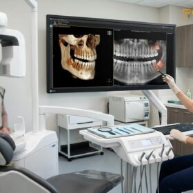
At HCMC Dental Clinic, the clinical workflow for diagnosing and treating an infection after wisdom tooth removal is meticulous and evidence-based. Upon presentation, the clinician will first conduct a thorough visual and tactile examination of the surgical site, assessing for fluctuance, erythema, and purulent discharge. Advanced 3D imaging, such as Cone Beam Computed Tomography (CBCT), may be utilized to evaluate the extent of bone involvement, rule out the presence of retained root fragments, and ensure the infection has not compromised the inferior alveolar nerve canal.
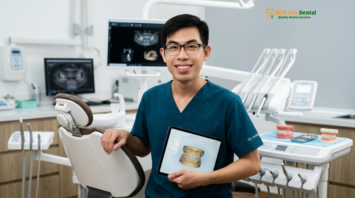
The cornerstone of treating a localized socket infection is establishing proper drainage and removing the source of the bacterial load. Dr. Nguyen Van Cuong notes that relying solely on systemic antibiotics without addressing the localized necrotic debris is a common clinical error that leads to recurrent infections. The treatment protocol typically involves the following steps:
- Profound Local Anesthesia: Because infected tissues are highly acidic, standard local anesthetics can be less effective. The surgeon may utilize specialized regional nerve blocks to ensure the patient is completely comfortable before proceeding.
- Surgical Debridement (Curettage): The surgeon will carefully re-enter the socket using sterile curettes to mechanically scrape away necrotic bone, infected granulation tissue, and any trapped food debris. This process stimulates fresh bleeding, which is necessary to form a new, healthy blood clot.
- Antimicrobial Irrigation: The debrided socket is copiously irrigated using a high-volume syringe filled with a sterile saline solution or a prescription-strength antimicrobial agent, such as 0.12% Chlorhexidine Gluconate. This flushes out microscopic bacteria and lowers the localized bacterial count.
- Pharmacological Intervention: Following mechanical cleaning, the surgeon may place a localized medicated dressing or a Platelet-Rich Fibrin (PRF) membrane into the socket to accelerate tissue regeneration. Systemic antibiotics are then prescribed based on the severity of the infection. A standard first-line regimen often includes Amoxicillin with Clavulanate (Augmentin) to combat beta-lactamase-producing bacteria, or Metronidazole to specifically target aggressive anaerobic strains.
Clinical Case Example: A 28-year-old patient presented to HCMC Dental Clinic in Ho Chi Minh City complaining of severe, radiating jaw pain and a foul taste three weeks after having a lower impacted wisdom tooth removed at a different facility. Clinical examination revealed a delayed-onset infection with a localized abscess beneath the healed gingiva. Dr. Cuong performed a minor incision and drainage under local anesthesia, thoroughly irrigated the socket with chlorhexidine, and prescribed a 7-day course of targeted antibiotics. The patient reported a 90% reduction in pain within 24 hours and achieved complete mucosal healing within two weeks.
It is imperative that patients complete the entire course of prescribed antibiotics, even if their symptoms resolve within a few days. Prematurely discontinuing antibiotic therapy can lead to the survival of the most resilient bacteria, potentially resulting in a rebound infection that is highly resistant to standard pharmacological treatments.
Home Care Rules to Minimize Infection Risk in HCMC
Preventing infections requires meticulous oral hygiene, avoiding negative pressure activities, and adhering to prescribed antimicrobial rinses to maintain a clean surgical environment.
While professional surgical technique is paramount, the patient’s adherence to post-operative home care instructions is equally critical in preventing an infected extraction socket saigon. The oral environment is inherently hostile to wound healing due to constant moisture, mechanical forces from chewing, and a high bacterial load. Therefore, proactive management is required to protect the vulnerable extraction site during the first critical weeks of recovery.
The most important rule of home care is the preservation of the initial blood clot. For the first 24 to 48 hours, patients must strictly avoid any activities that create negative pressure in the oral cavity. This includes smoking, vaping, drinking through a straw, and vigorous spitting. The physical force generated by these actions can easily dislodge the clot, exposing the bone and dramatically increasing the risk of both dry socket and subsequent bacterial infection.
Maintaining a clean surgical site without disrupting the healing tissue requires a delicate balance. Patients should avoid brushing directly over the extraction site for the first few days but must continue to brush their other teeth normally to reduce the overall bacterial load in the mouth. After 24 hours, gentle warm salt water rinses should be initiated. The hypertonic nature of the salt water helps to draw out localized edema and creates a mildly hostile environment for bacterial growth. If the surgeon has prescribed a chlorhexidine mouthwash, it should be used exactly as directed, typically twice daily, to chemically suppress plaque formation[5].
“Effective post-operative care is a partnership between the surgeon and the patient. Meticulous adherence to irrigation protocols and dietary modifications is the most reliable defense against delayed-onset complications.”
Dietary choices also play a significant role in infection prevention, particularly in a vibrant culinary environment like Ho Chi Minh City. Patients should strictly adhere to post-operative dietary guidelines, focusing on soft, nutrient-dense foods that require minimal chewing. It is crucial to avoid hard, crunchy, or granular foods (such as nuts, seeds, or crusty bread) that can easily fracture and become lodged deep within the socket. Furthermore, in the context of local cuisine, patients should temporarily avoid excessively hot, spicy, or acidic broths, which can irritate the raw mucosal tissues and trigger an inflammatory flare-up.
Around the fifth to seventh day post-surgery, the surgeon may provide a curved monoject irrigation syringe. Patients are instructed to gently flush the socket with warm saline or prescribed rinses after every meal. This mechanical flushing is highly effective at removing trapped food debris that would otherwise serve as a substrate for bacterial putrefaction. Understanding proper socket irrigation techniques is essential for patients, especially those who had deep bony impactions, as these sockets can take several weeks to fully fill in with new bone tissue.
By combining expert surgical execution, such as extracting wisdom teeth in older adults with precision, and rigorous patient compliance, the risk of post-operative infections can be minimized, ensuring a smooth and predictable recovery trajectory.
Frequently Asked Questions
How do I know if my wisdom tooth socket is infected?
You can identify an infected socket by monitoring for progressive symptoms that worsen after the third day. Key indicators include severe throbbing pain that does not respond to standard analgesics, visible yellow or white purulent discharge, localized facial swelling that feels warm to the touch, and a persistent foul taste or odor in the mouth. If you experience these symptoms, clinical evaluation is necessary.
What does an infected extraction socket look like?
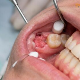
An infected extraction socket typically appears highly inflamed, with deep red or purplish gingival margins surrounding the surgical site. You may observe a pool of yellowish-white fluid (pus) within or seeping from the hole, and the surrounding tissues often look swollen, puffy, and lack the healthy pink appearance of normal healing gums. The normal dark blood clot is usually absent or necrotic.
Can an infection occur 2 weeks after wisdom tooth extraction?
Yes, a delayed-onset infection can absolutely occur two to four weeks after the initial surgery. This typically happens when food debris or bacteria become trapped beneath the healing gum tissue, creating an anaerobic environment that allows opportunistic bacteria to multiply long after the initial surgical trauma has subsided. This requires professional cleaning and irrigation to resolve.
What antibiotics are used to treat a socket infection?
Dentists commonly prescribe broad-spectrum antibiotics such as Amoxicillin combined with Clavulanate (Augmentin) to target the diverse oral microbiome. For patients with penicillin allergies, alternatives like Clindamycin, Azithromycin, or Metronidazole are frequently utilized to effectively eradicate anaerobic bacteria responsible for deep socket infections. The choice depends on the patient’s medical history and the severity of the infection.
How much does it cost to treat a dental infection in Saigon?
The cost to treat a dental infection in Saigon varies depending on the severity of the complication and the required clinical interventions. Minor infections requiring only professional irrigation and an antibiotic prescription are relatively inexpensive, while severe cases necessitating surgical debridement, advanced imaging, or specialized grafting will incur higher fees based on the latest clinic fee schedule. A direct consultation provides an accurate estimate.
References
- Journal of Oral and Maxillofacial Surgery. Delayed-onset infections after lower third molar extraction. (2021).
- International Journal of Oral and Maxillofacial Surgery. Microbiology of odontogenic infections and antibiotic sensitivities. (2019).
- British Dental Journal. Differentiating alveolar osteitis from purulent surgical site infections. (2020).
- Journal of the American Dental Association. Management of fascial space infections of odontogenic origin. (2022).
- Clinical Oral Investigations. Efficacy of chlorhexidine irrigation in preventing post-extraction complications. (2018).