Removing wisdom teeth at 30 or 40 is a safe and highly routine clinical procedure, though it requires more advanced surgical planning than in teenagers. Due to denser bone and fully developed roots, adult extractions demand precise 3D imaging and specialized surgical techniques to ensure a smooth, complication-free recovery.
Clinical Summary:
Wisdom teeth removal in adulthood presents distinct anatomical and physiological challenges compared to adolescent extractions. As patients enter their 30s and 40s, the surrounding alveolar bone becomes highly mineralized (osteosclerosis), and the roots of the third molars reach full development, often curving or approximating critical neurovascular structures like the inferior alveolar nerve. Consequently, the surgical extraction of adult wisdom teeth requires meticulous preoperative assessment, typically involving Cone Beam Computed Tomography (CBCT). While the biological healing process is slower—resulting in a slightly prolonged recovery period and a higher susceptibility to localized osteitis—modern surgical protocols utilizing piezosurgery, autologous blood concentrates, and tailored pharmacological management ensure that adult patients can undergo this necessary procedure safely and comfortably.
Key Takeaways:
- Age is not a contraindication for extraction; pathology and clinical symptoms dictate the necessity of surgery.
- Adult jawbones are significantly denser, requiring advanced surgical techniques like tooth sectioning to minimize trauma.
- Fully formed roots in older patients increase the proximity to major facial nerves, making 3D CBCT imaging essential.
- Recovery in your 30s and 40s takes slightly longer, with a heightened emphasis on preventing dry socket and managing inflammation.
- Leaving asymptomatic impacted teeth unmonitored can lead to silent damage, including cysts and decay on adjacent healthy molars.
Is There an Age Limit for Wisdom Tooth Removal?
There is no strict upper age limit for extracting third molars; however, surgical complexity and healing timelines naturally increase as patients enter their third and fourth decades of life.
A common misconception among patients is that there is a definitive expiration date on the ability to undergo oral surgery. Many individuals wonder, is 40 too old to remove wisdom teeth? Clinically speaking, the answer is no. The decision to extract a third molar is never based solely on a patient’s chronological age, but rather on the presence of active pathology, the risk of impending complications, and the overall systemic health of the individual. While prophylactic removal (extracting teeth before they cause problems) is heavily favored during the late teenage years and early twenties, therapeutic removal (extracting teeth because they are causing active harm) can, and frequently must, be performed at any stage of adulthood.
During adolescence, the roots of the wisdom teeth are only partially formed, typically resembling small, rounded buds. Furthermore, the surrounding alveolar bone is highly vascular, spongy, and pliable. This allows the tooth to be elevated and removed with minimal resistance. However, as a patient transitions into their 30s and 40s, the physiological landscape of the oral cavity undergoes significant changes. The roots complete their apical development, often growing long, divergent, or hooked. Simultaneously, the jawbone undergoes continuous mineralization, becoming rigid and unyielding. These factors combined mean that extracting a tooth at age 40 requires a fundamentally different surgical approach than extracting the same tooth at age 18.
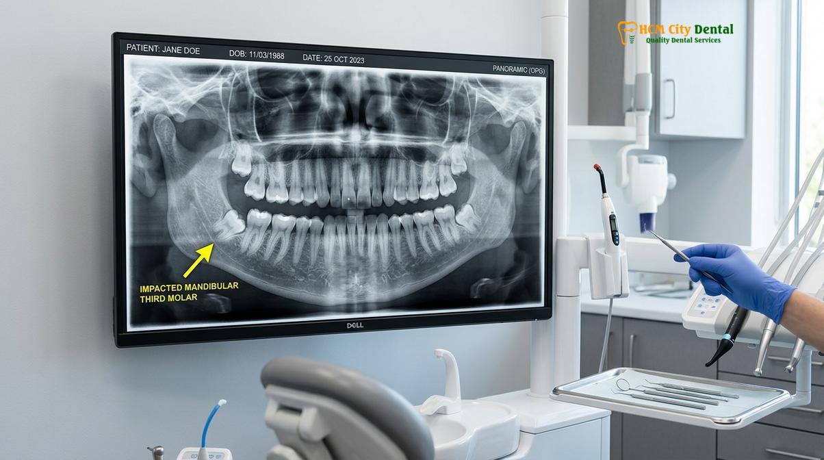
Despite these challenges, modern oral surgery has evolved tremendously. The advent of advanced diagnostic imaging, refined surgical instruments, and superior anesthetic protocols means that older adults can undergo surgical removal of third molars with a high degree of safety and predictability. The clinical focus shifts from simple elevation to strategic surgical planning, ensuring that the surrounding bone and adjacent teeth are preserved while the impacted molar is carefully sectioned and removed. According to established classifications of mandibular third molar impactions, the depth and angulation of the tooth play a far greater role in determining surgical difficulty than the patient’s age alone [4].
Why Adult Wisdom Teeth are Clinically More Challenging
Adult extractions present unique surgical hurdles due to decreased bone elasticity, increased root curvature, and a higher prevalence of adjacent periodontal defects that complicate the procedure.
When evaluating the necessity of surgery in older patients, clinicians must carefully weigh the adult wisdom teeth extraction risks against the dangers of leaving a pathological tooth in place. The challenges of adult extractions are multifaceted, encompassing anatomical, biological, and systemic factors. One of the primary difficulties is the phenomenon of ankylosis. Over decades of microscopic function and inflammation, the periodontal ligament—the fibrous tissue that acts as a shock absorber between the tooth root and the bone—can atrophy. This leads to a direct fusion of the tooth’s cementum to the surrounding alveolar bone. An ankylosed wisdom tooth cannot simply be “pulled”; it must be meticulously freed from the bone using specialized surgical drills or ultrasonic instruments.
Furthermore, the periodontal health of the adjacent second molar is a critical consideration in adult patients. In many cases, a partially impacted wisdom tooth creates a deep, inaccessible pocket between itself and the second molar. Over the course of 10 to 20 years, chronic bacterial accumulation in this pocket leads to localized periodontitis, destroying the bone that supports the healthy second molar. When the wisdom tooth is finally removed in the patient’s 30s or 40s, the surgeon must not only extract the offending tooth but also manage the resulting bony defect to ensure the long-term survival of the adjacent molar.
Clinical Warning: Ignoring asymptomatic impacted wisdom teeth can be hazardous. A lack of pain does not equate to an absence of disease. Silent pathologies, such as dentigerous cysts or external root resorption of the adjacent second molar, can progress for years without causing discomfort until severe, irreversible damage has occurred.
Systemic health also plays a more prominent role in adult extractions. Patients in their 30s, 40s, and beyond are more likely to present with comorbidities such as hypertension, type 2 diabetes, or cardiovascular disease. They may also be taking medications, such as anticoagulants or bisphosphonates, which directly impact surgical hemostasis and bone healing. A comprehensive preoperative medical evaluation is therefore mandatory to tailor the anesthetic approach and post-operative pharmacological regimen, ensuring patient safety throughout the perioperative period.
Anatomical Considerations: Denser Bone and Fully Formed Roots
The transition from adolescent to adult bone involves significant mineralization, resulting in a highly dense mandible that resists expansion and necessitates advanced surgical sectioning techniques.
To truly understand the complexities of adult extractions, one must examine the microanatomy of the aging mandible. The concept of bone density wisdom teeth adults is a central theme in oral surgery. The human jawbone is composed of an outer layer of dense cortical bone and an inner core of trabecular (spongy) bone. In youth, the trabecular spaces are large and filled with vascular marrow, allowing the bone to flex slightly under pressure. By the time a patient reaches their late 30s, the cortical plates have thickened, and the trabecular bone has undergone osteosclerosis—a physiological hardening and densification of the bone matrix.
This increased density means that the bone will not yield or expand when an elevator instrument is applied to the tooth. Applying excessive force to dense adult bone risks fracturing the mandible or shearing off the crown of the tooth, leaving the roots embedded. Therefore, the surgical paradigm shifts from “forceful elevation” to “strategic space creation.” Surgeons must perform an ostectomy (the careful removal of surrounding bone) to create a pathway for the tooth’s exit.
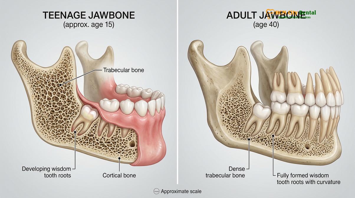
Coupled with dense bone are the fully developed roots of adult wisdom teeth. Unlike the blunt, incomplete roots of a teenager, adult roots are fully elongated. They frequently exhibit dilaceration (severe curvature) or hypercementosis (a bulbous thickening of the root tip). To safely extract such a tooth without damaging the surrounding structures, the surgeon will perform an odontotomy, or tooth sectioning. By dividing the tooth into multiple smaller fragments—separating the crown from the roots, and dividing divergent roots from one another—each piece can be removed individually along its specific path of withdrawal. Modern techniques increasingly utilize piezoelectric surgery (Piezotome), which uses ultrasonic micro-vibrations to cut bone and tooth structure with extreme precision while preserving adjacent soft tissues, significantly reducing post-operative trauma compared to traditional rotary burs [2].
“The primary difference in treating a 40-year-old versus an 18-year-old lies in respecting the rigidity of the bone. We do not force the tooth out; rather, we meticulously dismantle the tooth within its socket to ensure the surrounding jawbone and nerves remain entirely undisturbed.”
— Dr. Nguyen Van Cuong, Lead Surgeon
Nerve Proximity and CBCT Imaging for Older Patients
Fully elongated roots in older adults frequently approximate or intertwine with the inferior alveolar canal, making three-dimensional radiographic assessment an absolute clinical necessity.
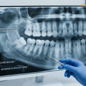
One of the most critical risk factors in adult wisdom tooth extraction is the anatomical relationship between the tooth roots and the major sensory nerves of the face. The Inferior Alveolar Nerve (IAN) travels through a bony canal within the lower jaw, providing sensation to the lower lip, chin, and teeth. The Lingual Nerve, running along the inner aspect of the jaw, provides sensation and taste to the anterior two-thirds of the tongue. Because the roots of adult wisdom teeth are fully grown, they often extend deep into the mandible, sometimes resting directly upon, or even wrapping around, the IAN canal.
In younger patients with incomplete root development, the distance between the tooth bud and the nerve is usually substantial, providing a wide margin of safety. In adults, this margin is frequently non-existent. Surgical trauma to these nerves—whether from direct instrumentation, compression during elevation, or post-operative swelling—can result in paresthesia (altered sensation), dysesthesia (painful sensation), or anesthesia (complete numbness) of the lip or tongue. While often temporary, these neurological complications can occasionally be permanent, profoundly affecting a patient’s quality of life [5].
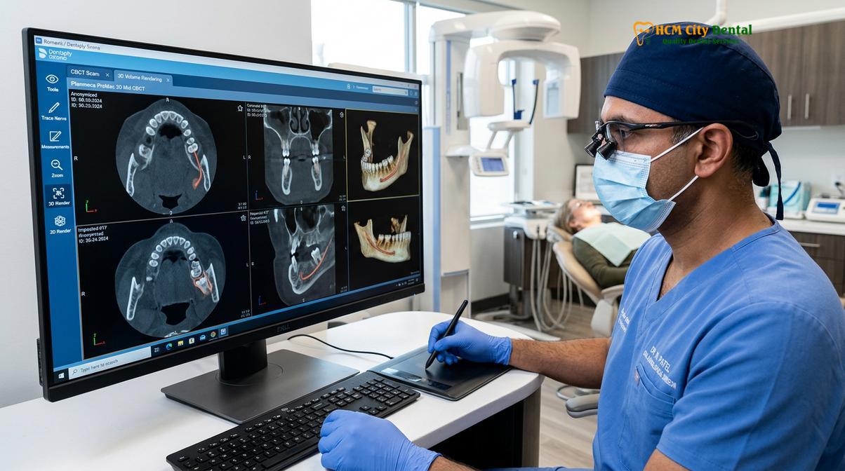
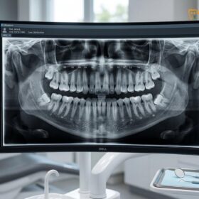
To mitigate these risks, standard two-dimensional panoramic X-rays are often insufficient for adult patients. A 2D image can show that the nerve and the root overlap, but it cannot determine whether the nerve lies to the cheek side (buccal), the tongue side (lingual), or directly between the roots. This is where Cone Beam Computed Tomography (CBCT) becomes indispensable. A 3D CBCT scan allows the surgeon to visualize the exact spatial relationship between the tooth and the nerve in sub-millimeter slices. If the scan reveals an exceptionally high risk of nerve injury, the surgeon may alter the treatment plan, perhaps opting for a coronectomy (removing only the crown of the tooth and intentionally leaving the roots buried) rather than a complete extraction. For a deeper understanding of these neurological considerations, patients can review our comprehensive inferior alveolar nerve complications guide.
Recovery Comparison: Teenagers vs. Adults in Their 30s and 40s
Post-operative healing in mature adults is characterized by a prolonged inflammatory phase and slower osteogenesis, requiring meticulous aftercare and advanced healing protocols.
Setting realistic expectations for the post-operative period is crucial for patient satisfaction. When discussing wisdom teeth age 35 recovery, it is important to acknowledge that the biological healing capacity of the human body naturally decelerates with age. Cellular turnover is slower, local blood supply to the dense cortical bone is reduced, and the immune system’s inflammatory response tends to be more pronounced and prolonged.
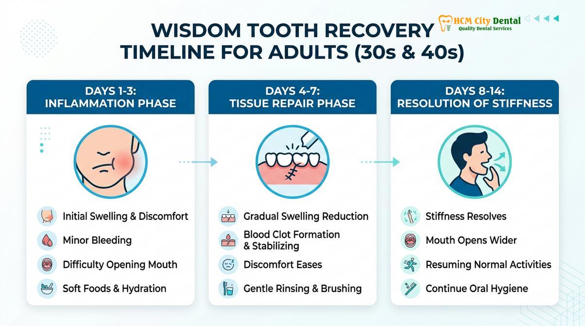
For a teenager, the acute inflammatory phase (characterized by swelling and discomfort) typically peaks at 48 hours and resolves rapidly by day 4 or 5. They can often return to normal activities, including school and sports, within a week. Conversely, an adult in their late 30s or 40s should anticipate a more protracted recovery. Swelling may persist for 5 to 7 days, and trismus (jaw stiffness) can linger for up to two weeks. The surgical site itself takes longer to granulate and fill with new bone, requiring strict adherence to dietary restrictions and oral hygiene protocols for an extended period [1].
| Recovery Metric | Teenagers (16-21) | Adults in their 30s | Adults 40 and Over |
|---|---|---|---|
| Peak Swelling Duration | 2 – 3 days | 3 – 5 days | 4 – 7 days |
| Resolution of Jaw Stiffness | 3 – 5 days | 7 – 10 days | 10 – 14 days |
| Risk of Alveolar Osteitis | Low (approx. 3-5%) | Moderate (approx. 8-12%) | Higher (requires strict protocols) |
| Return to Normal Diet | 5 – 7 days | 10 – 14 days | 14+ days |
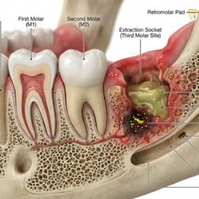
One of the most significant concerns during adult recovery is the increased incidence of alveolar osteitis, commonly known as dry socket. This painful condition occurs when the protective blood clot within the extraction socket fails to form properly or is prematurely dislodged, exposing the underlying bone and nerve endings. Because adult bone is less vascular, the initial clot formation is more fragile. To combat this, modern surgical protocols highly recommend the use of autologous blood concentrates, such as Platelet-Rich Fibrin (PRF). By drawing a small amount of the patient’s blood and centrifuging it, the surgeon can create a dense fibrin membrane rich in growth factors. Placing this PRF membrane into the socket immediately after extraction significantly accelerates soft tissue healing, reduces post-operative pain, and drastically lowers the risk of dry socket [3]. Patients seeking more information on preventing this complication should consult our detailed alveolar osteitis prevention guide.
When Extraction is Absolutely Necessary for Adults
Surgical intervention becomes mandatory when impacted third molars trigger recurrent pericoronitis, cause irreversible damage to adjacent teeth, or develop cystic lesions.
While prophylactic removal is debated in older adults, therapeutic extraction is non-negotiable when active pathology is present. The most common indication for wisdom tooth removal in adults is recurrent pericoronitis. This is an acute, painful infection of the operculum—the flap of gum tissue that partially covers an erupting or impacted wisdom tooth. Because this area is impossible to keep clean with a toothbrush, food debris and bacteria become trapped beneath the tissue, leading to severe inflammation, purulent discharge (pus), foul breath, and an inability to fully open the mouth. While antibiotics can temporarily suppress the infection, the only definitive cure is the surgical removal of the offending tooth.
Another critical indication is the presence of distal cervical caries (decay) on the adjacent second molar. An impacted wisdom tooth leaning against the second molar creates a plaque trap. Over time, this leads to deep cavities on the back surface of the healthy second molar—an area that is notoriously difficult for a dentist to restore. If left untreated, the patient risks losing both the wisdom tooth and the essential second molar. Furthermore, the follicular sac that surrounds an impacted wisdom tooth can occasionally undergo cystic degeneration, forming a dentigerous cyst. These fluid-filled lesions silently expand within the jawbone, hollowing out the mandible and potentially causing pathological fractures or displacing adjacent teeth.
Clinical Case Study: Managing Adult Impaction
A 42-year-old male presented to HCMC Dental Clinic in Ho Chi Minh City complaining of a dull, throbbing ache in his lower left jaw. He had retained his wisdom teeth, believing they were harmless as they had never caused pain in his youth. A 3D CBCT scan revealed a horizontally impacted third molar that had caused severe external root resorption on the adjacent second molar, alongside a developing 1.5cm dentigerous cyst. The surgical team, led by Dr. Cuong, performed a complex extraction utilizing piezosurgery to carefully section the tooth and enucleate the cyst without damaging the inferior alveolar nerve. The socket was grafted with PRF to promote optimal healing. This case underscores the critical importance of radiographic monitoring for retained adult wisdom teeth.
When to See a Doctor for Adult Wisdom Teeth
Recognizing the signs of wisdom tooth pathology early can prevent localized issues from escalating into severe systemic infections. Adults who have retained their third molars should be vigilant for specific clinical symptoms. You should seek immediate evaluation from a qualified oral surgeon if you experience sudden, severe pain radiating from the back of the jaw to the ear, noticeable swelling of the cheek or submandibular lymph nodes, difficulty swallowing, or an unexplained bad taste in the mouth accompanied by localized bleeding gums.
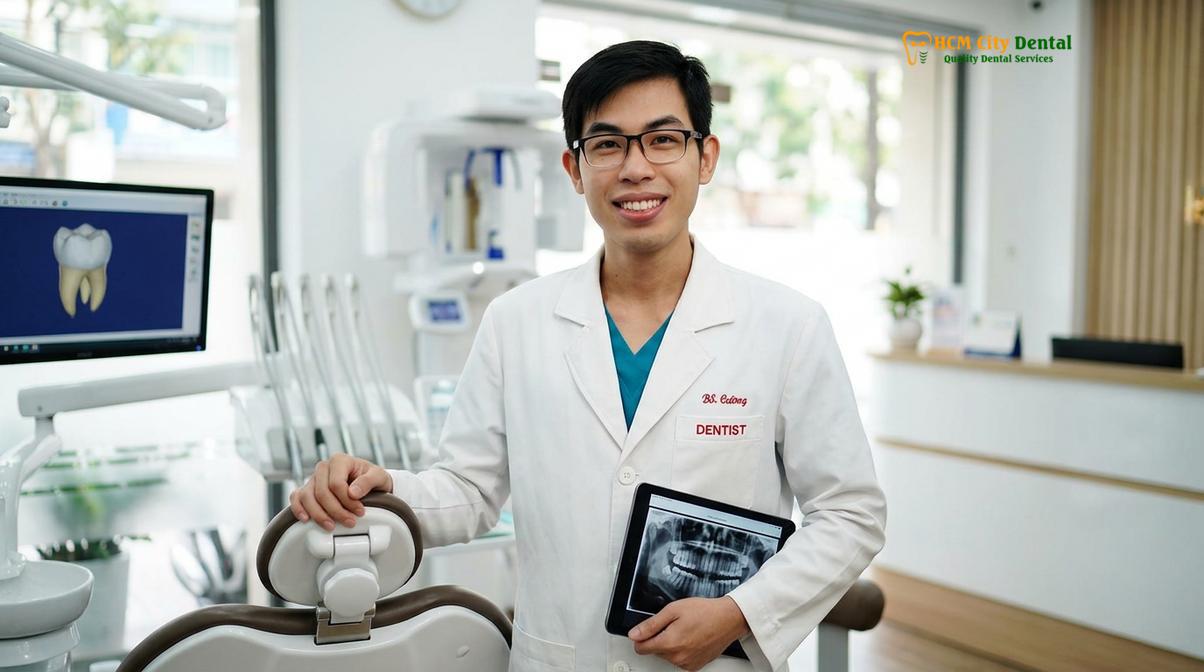
For patients experiencing dental anxiety regarding surgery later in life, modern clinics offer various pharmacological management options to ensure comfort. Depending on the complexity of the extraction and the patient’s medical history, procedures can be performed under local anesthesia supplemented with inhalation analgesia (laughing gas) or deeper intravenous conscious sedation. A thorough consultation is essential to determine the safest and most effective approach.
“Delaying intervention when active symptoms arise only allows the infection to compromise more surrounding bone. Timely, precise surgical management is the key to preserving the overall structural integrity of the adult dental arch.”
— Dr. Nguyen Van Cuong, HCMC Dental Clinic
If you are experiencing discomfort or simply need a comprehensive evaluation of your retained third molars, seeking care at specialized oral surgery centers ensures you have access to the necessary 3D imaging and surgical expertise required for complex adult cases.
Frequently Asked Questions
Am I too old to have my wisdom teeth removed?
No, you are never strictly too old to have your wisdom teeth removed if they are causing pathology. While extractions in your 30s, 40s, or beyond are surgically more complex due to denser bone, experienced oral surgeons routinely perform these procedures safely. The decision is based on clinical necessity, such as infection, decay, or cyst formation, rather than your chronological age. A thorough medical evaluation ensures the procedure is tailored to your specific health profile.
Is recovery harder for wisdom teeth in your 30s?
Yes, recovery typically takes slightly longer in your 30s compared to your teenage years. Because adult bone is denser and cellular turnover is slower, the inflammatory phase is prolonged. Patients in their 30s may experience swelling and stiffness for 7 to 10 days, whereas teenagers often recover in 3 to 5 days. Strict adherence to post-operative care, including proper diet and oral hygiene, minimizes these challenges and promotes steady healing.
Why do adults have more complications with wisdom teeth?
Adults face a higher risk of complications primarily due to anatomical changes over time. The jawbone becomes highly mineralized and less pliable, while the tooth roots fully form and often curve or anchor deeply into the bone. Additionally, these elongated roots are more likely to sit in close proximity to the inferior alveolar nerve, increasing the risk of surgical trauma. Systemic health factors also play a larger role in adult healing.
Is it safe to leave impacted wisdom teeth if they don’t hurt?
Leaving asymptomatic impacted wisdom teeth requires careful, ongoing radiographic monitoring, as a lack of pain does not guarantee an absence of disease. Silent pathologies, such as dentigerous cysts, root resorption of adjacent molars, or hidden periodontal pockets, can develop without symptoms. If imaging reveals potential risks, prophylactic extraction may still be recommended to prevent severe future damage to the surrounding jawbone and healthy teeth.
How long does jaw stiffness last for older patients?
Post-operative jaw stiffness, known as trismus, can last anywhere from 7 to 14 days for patients in their 30s and 40s. This occurs due to inflammation of the masticatory muscles and the surgical trauma required to navigate dense adult bone. Applying warm compresses after the first 48 hours and performing gentle jaw stretching exercises, as directed by your surgeon, can significantly accelerate the resolution of stiffness and restore normal function.
References
- Journal of Oral and Maxillofacial Surgery. Recovery after third molar extraction. (2021).
- International Journal of Oral and Maxillofacial Surgery. Piezotome vs rotary drill in wisdom teeth removal. (2019).
- British Dental Journal. Dry socket incidence and prevention with PRF. (2020).
- Journal of the American Dental Association. Mandibular third molar impaction classifications. (2022).
- Clinical Oral Investigations. Lingual nerve protection during surgical extraction. (2018).