Undergoing a wisdom tooth extraction while managing diabetes and taking blood thinners requires precise clinical coordination. Dentists prioritize local bleeding control over stopping anticoagulants, while ensuring blood glucose levels are stable to promote healing and prevent post-operative infections.
Clinical Summary:
Managing patients with concurrent diabetes and anticoagulant therapy during oral surgery involves a delicate balance between achieving hemostasis and preventing infection. Current clinical protocols strongly favor maintaining blood thinner regimens to protect cardiovascular health, utilizing advanced local hemostatic agents like tranexamic acid and collagen sponges to control bleeding. Simultaneously, strict glycemic control must be verified prior to surgery, as elevated blood sugar impairs immune function and delays socket closure. Multidisciplinary coordination between the dental surgeon and the primary care physician is essential to ensure safe, predictable outcomes for medically complex patients.
Key Takeaways:
- Most dental extractions can be safely performed without interrupting prescribed blood thinner medications.
- Local hemostatic measures, such as advanced suturing and oxidized cellulose, effectively manage post-operative bleeding risks.
- Fasting blood glucose levels should ideally be below 180 mg/dL for elective oral surgical procedures.
- Uncontrolled diabetes significantly increases the risk of post-extraction infections and delayed tissue regeneration.
- Seamless communication between your dental surgeon and medical doctor is critical for verifying INR levels and HbA1c stability.
Why Diabetes and Blood Thinners Require Special Dental Protocols
Patients with both diabetes and cardiovascular conditions face a dual surgical challenge: managing prolonged bleeding risks while ensuring proper tissue regeneration in an environment susceptible to infection.
The intersection of metabolic and cardiovascular diseases presents a unique clinical scenario in oral and maxillofacial surgery. When a patient requires a Tooth Extraction, the surgical team must account for two distinct, yet overlapping, physiological challenges. On one hand, anticoagulant or antiplatelet medications intentionally inhibit the body’s ability to form blood clots, which is a necessary step in the initial phase of wound healing. On the other hand, diabetes mellitus—particularly when poorly controlled—alters the inflammatory response, impairs microcirculation, and compromises the cellular mechanisms required to rebuild tissue [1].
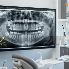
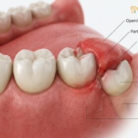
Dr. Nguyen Van Cuong emphasizes that treating these patients requires a shift from standard extraction protocols to a highly customized, medically integrated approach. The primary objective is to perform the surgery atraumatically, minimizing tissue disruption, while simultaneously deploying proactive measures to stabilize the surgical site. The presence of wisdom teeth diabetes complications, such as chronic pericoronitis (inflammation of the gum tissue surrounding a partially erupted tooth), further complicates the scenario, as pre-existing infection can exacerbate both bleeding and glycemic instability.
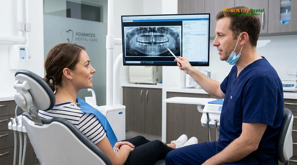
Furthermore, the physiological stress of anticipating and undergoing dental surgery can trigger the release of endogenous catecholamines (like adrenaline and cortisol). These stress hormones naturally elevate blood glucose levels and increase blood pressure, which can indirectly increase the bleeding risk tooth extraction presents. Therefore, modern dental protocols for these high-risk demographics focus heavily on profound local anesthesia, anxiety management, and meticulous surgical planning to ensure the procedure is as swift and atraumatic as possible.
Managing Blood Thinners Before and After Oral Surgery
Current clinical guidelines strongly recommend continuing most anticoagulant and antiplatelet therapies during minor oral surgeries to prevent severe thromboembolic events, relying instead on local bleeding control.
Historically, patients on blood thinners were routinely instructed to cease their medication for several days prior to a dental extraction. However, extensive medical research has fundamentally shifted this paradigm. The risk of experiencing a life-threatening thromboembolic event—such as a stroke, deep vein thrombosis, or myocardial infarction—due to the temporary cessation of anticoagulants is significantly higher than the risk of prolonged, yet manageable, oral bleeding [2].
“The risk of a life-threatening thromboembolic event from stopping anticoagulants far outweighs the risk of manageable local bleeding following a dental extraction. Systemic protection must always take precedence over localized surgical convenience.”
The management strategy depends heavily on the specific classification of the blood-thinning medication:
- Vitamin K Antagonists (e.g., Warfarin): For patients on Warfarin, the standard protocol involves checking the International Normalized Ratio (INR) within 24 to 72 hours before the surgery. An INR of 4.0 or below is generally considered safe for routine and surgical extractions without altering the dosage.
- Direct Oral Anticoagulants (DOACs): Medications such as Rivaroxaban (Xarelto), Apixaban (Eliquis), and Dabigatran (Pradaxa) have shorter half-lives and do not require routine INR monitoring. Dentists typically schedule the extraction at the medication’s “trough” level—usually 12 to 24 hours after the last dose—without requiring the patient to skip a dose.
- Antiplatelet Agents (e.g., Aspirin, Clopidogrel): Patients on single or dual antiplatelet therapy for stents or prior cardiac events should almost never stop their medication for dental surgery. The bleeding associated with these drugs is typically capillary in nature and easily controlled with local measures.
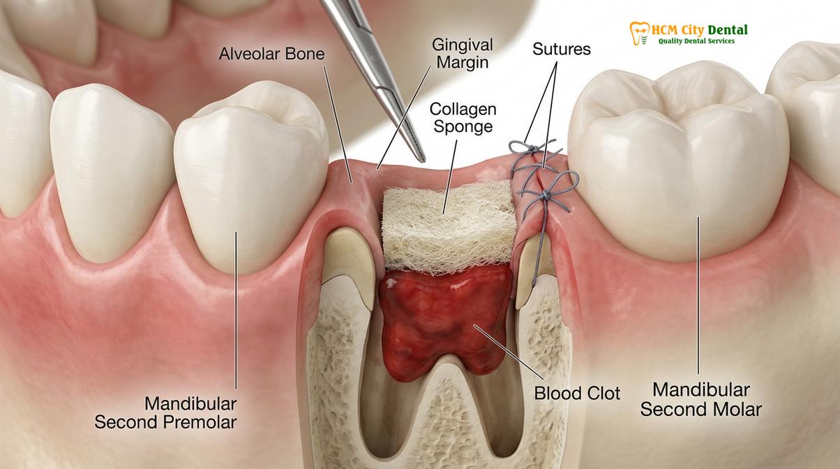
When planning dental surgery blood thinners saigon clinics follow strict international protocols to ensure patient safety. The focus remains entirely on local hemostasis, ensuring that the patient’s cardiovascular protection remains uninterrupted throughout the perioperative period.
Controlling Bleeding: Hemostatic Agents and PRF Technology
Advanced local hemostatic techniques, including tranexamic acid and Platelet-Rich Fibrin, provide predictable bleeding control directly at the extraction site without altering systemic medications.
Because systemic blood thinners are maintained, the dental surgeon must employ a robust arsenal of local hemostatic agents to encourage clot formation and stabilize the extraction socket. The foundation of local hemostasis begins with an atraumatic surgical technique, preserving as much surrounding bone and soft tissue as possible. Following the removal of the tooth, mechanical compression using sterile gauze is the first line of defense.
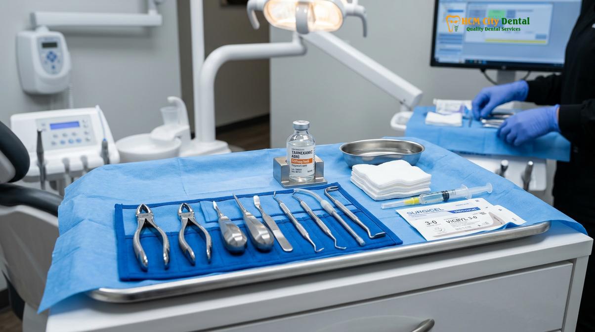
When mechanical pressure is insufficient, clinicians utilize specialized biocompatible materials placed directly into the socket. Oxidized regenerated cellulose (such as Surgicel) and absorbable gelatin sponges (such as Gelfoam) act as physical scaffolds. They provide a matrix that traps platelets and red blood cells, artificially initiating the coagulation cascade even in the presence of systemic anticoagulants [3].
| Hemostatic Agent | Mechanism of Action | Clinical Application |
|---|---|---|
| Oxidized Regenerated Cellulose | Lowers local pH, causing artificial clot formation and acting as a physical scaffold. | Placed directly into the bleeding socket before suturing. |
| Tranexamic Acid (TXA) 5% | Antifibrinolytic; prevents the premature breakdown of the formed blood clot. | Used as a post-operative mouth rinse or soaked onto compression gauze. |
| Absorbable Gelatin Sponge | Absorbs blood and expands, providing mechanical pressure and a matrix for clotting. | Packed into the socket, often combined with topical thrombin. |
| Platelet-Rich Fibrin (PRF) | Autologous blood concentrate that releases growth factors and forms a dense fibrin mesh. | Grafted into the socket to accelerate healing and seal the wound. |
One of the most significant advancements in managing high-risk extractions is the use of Platelet-Rich Fibrin (PRF). By drawing a small amount of the patient’s own blood and centrifuging it prior to surgery, the dentist creates a dense, fibrin-rich membrane loaded with leukocytes and growth factors. When packed into the extraction site, PRF not only acts as a powerful biological hemostatic plug but also accelerates tissue regeneration, which is particularly beneficial for diabetic patients with compromised healing capabilities.
Additionally, the surgical site is often secured using tension-free suturing techniques. Sutures physically approximate the gingival tissues, reducing the exposed surface area of the wound and holding the hemostatic agents securely in place.
Diabetic Wound Healing and Preventing Post-Extraction Infections
Elevated blood glucose impairs immune function and collagen synthesis, making strict glycemic control essential for preventing infections and ensuring proper socket closure.
Diabetes mellitus profoundly impacts the body’s ability to heal following oral surgery. The primary culprit is chronic hyperglycemia, which triggers a cascade of detrimental cellular events. High blood sugar impairs the chemotaxis and phagocytosis of neutrophils—the white blood cells responsible for migrating to the surgical site and destroying invading bacteria. Consequently, diabetic patients are at a significantly higher risk of developing post-operative infections [4].
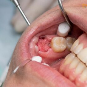
Furthermore, elevated glucose levels lead to the formation of Advanced Glycation End-products (AGEs). These molecules cross-link with collagen, the primary structural protein required to rebuild the gum tissue and bone inside the extraction socket. This abnormal cross-linking results in friable, weak tissue that is prone to breakdown. This delayed wound healing diabetic dental patients experience increases the window of vulnerability for complications, making them more susceptible to conditions like Dry Socket, where the blood clot fails to form or is prematurely lost.
Clinical Warning: Blood Glucose Thresholds for Dental Surgery
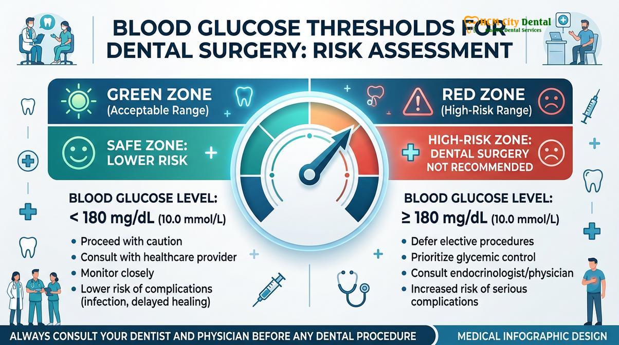
Elective dental extractions should generally be postponed if a patient’s fasting blood glucose exceeds 180 mg/dL or if their HbA1c is above 8.0%. For emergency extractions involving acute infection, a random blood glucose level up to 234 mg/dL may be acceptable, provided prophylactic antibiotics and strict post-operative monitoring are implemented.
To mitigate these risks, dental surgeons emphasize the importance of pre-operative glycemic control. Patients are advised to maintain their regular diet and insulin or oral hypoglycemic medication schedule on the day of surgery to prevent hypoglycemic episodes in the dental chair. The use of prophylactic antibiotics may be considered on a case-by-case basis, particularly if the diabetes is poorly controlled or if the extraction is highly complex.
Coordinating Care: Dental Surgeon and Primary Physician Integration
Safe surgical outcomes rely on seamless communication between the dental team and the patient’s medical providers to verify INR stability and HbA1c levels prior to treatment.
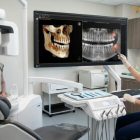
The successful management of a patient with concurrent diabetes and anticoagulant therapy cannot be achieved in isolation. It requires a multidisciplinary approach, integrating the expertise of the dental surgeon, the primary care physician, and potentially a cardiologist or endocrinologist. Before any surgical intervention, a comprehensive medical consultation is mandatory.
The dental team will typically send a formal medical clearance request to the patient’s physician. This communication outlines the planned surgical procedure, the anticipated level of bleeding, and the proposed local hemostatic measures. In return, the physician provides the most recent HbA1c results, current INR readings (if applicable), and confirmation that the patient’s systemic conditions are stable enough to withstand minor oral surgery [5].
Clinical Case Review: Multidisciplinary Management
A 62-year-old patient presented to HCMC Dental Clinic in Ho Chi Minh City requiring the surgical extraction of an impacted lower wisdom tooth. The patient had Type 2 Diabetes (HbA1c 7.4%) and was taking Rivaroxaban for atrial fibrillation. Dr. Nguyen Van Cuong coordinated with the patient’s cardiologist, confirming that the DOAC should not be stopped. The surgery was scheduled 18 hours after the last medication dose. Utilizing an atraumatic piezosurgery technique, PRF grafting, and precise suturing, the tooth was removed with zero intraoperative complications. The patient experienced normal clot formation and healed uneventfully, demonstrating the efficacy of integrated medical-dental protocols.
This collaborative approach ensures that all potential risk factors are identified and mitigated before the patient ever sits in the surgical chair. It also provides a clear roadmap for managing any unexpected perioperative emergencies, ensuring that the patient’s overall health is never compromised for the sake of dental treatment.
Safe Recovery Guidelines for High-Risk Patients in HCMC
Post-operative care for medically compromised patients focuses on protecting the fragile blood clot, monitoring for prolonged oozing, and maintaining strict oral hygiene to prevent infection.
The recovery phase is arguably the most critical period for patients managing diabetes and taking blood thinners. Because the systemic medications are still active, the local hemostatic measures placed during surgery must be carefully protected by the patient at home. Strict adherence to post-operative instructions is non-negotiable.
For the first 24 hours, patients must avoid any actions that create negative pressure in the mouth, such as using a straw, spitting forcefully, or vigorous rinsing. These actions can easily dislodge the fragile blood clot, leading to secondary bleeding. If a Wisdom Teeth Removal IV Sedation protocol was used, patients must rest with their head elevated to reduce blood pressure in the maxillofacial region.
“Patient compliance during the first 72 hours dictates the success of the healing process. For those on anticoagulants, protecting the surgical site from mechanical disruption is the single most effective way to prevent secondary hemorrhage.”
Diabetic patients must pay special attention to their blood sugar levels during the recovery week. The stress of surgery and changes in dietary intake (moving to a soft food diet) can cause unexpected fluctuations in glucose levels. Maintaining tight glycemic control during this window is vital to support immune function and collagen synthesis. Patients should consume nutrient-dense, soft foods that do not cause rapid spikes in blood sugar, and they must continue their prescribed diabetic medications unless otherwise directed by their physician.
When to Seek Immediate Medical Attention
While minor oozing (saliva tinged pink with blood) is entirely normal for the first 24 to 48 hours, patients on blood thinners must be able to distinguish between normal healing and active hemorrhage. It is crucial to monitor the surgical site closely and recognize the warning signs that require immediate clinical intervention.

You should contact your dental surgeon or seek emergency medical care if you experience any of the following:
- Active, Uncontrolled Bleeding: Bleeding that rapidly fills the mouth, forms large, dark clots, or does not subside after 45 minutes of firm, continuous pressure with a sterile gauze pad.
- Signs of Systemic Infection: A fever exceeding 101°F (38.3°C), chills, or a general feeling of severe malaise, which may indicate that the diabetic immune response is overwhelmed.
- Progressive Swelling: While some swelling is normal, swelling that continues to worsen after 72 hours, spreads down the neck, or causes difficulty swallowing or breathing is a medical emergency.
- Severe, Unrelenting Pain: Pain that is not managed by prescribed analgesics and worsens after the third day, which is a primary indicator of dry socket or deep space infection.
At HCMC Dental Clinic, our surgical team provides comprehensive 24/7 post-operative support for medically complex patients, ensuring that any deviations from normal healing are addressed immediately based on sound clinical diagnostics.
Frequently Asked Questions
Can I have a wisdom tooth extracted if I am diabetic?
Yes, diabetic patients can safely undergo wisdom tooth extraction provided their blood glucose levels are well-controlled prior to the procedure. Dentists typically require a fasting blood sugar reading below 180 mg/dL for elective surgeries to minimize the risk of infection and ensure proper wound healing. Your dentist will likely request a recent HbA1c test and may consult with your endocrinologist to ensure your condition is stable enough for oral surgery.
Do I need to stop taking my blood thinners before dental surgery?
In most cases, you do not need to stop taking your blood thinners before a routine or surgical tooth extraction. Clinical guidelines strongly recommend continuing anticoagulant therapy, as the risk of a severe cardiovascular event far outweighs the manageable risk of local bleeding. Your dentist will use specialized local techniques to control bleeding at the site without compromising your systemic health.
How does high blood sugar affect extraction healing?
High blood sugar impairs the body’s immune response and disrupts collagen synthesis, which significantly delays the healing of the extraction socket. Elevated glucose levels also reduce blood flow to the surgical site, increasing the likelihood of post-operative infections and complications like dry socket. Maintaining strict glycemic control in the weeks surrounding your surgery is critical for predictable tissue regeneration.
What precautions are taken to prevent bleeding during surgery?
Dentists utilize advanced local hemostatic measures rather than altering your systemic medication. These precautions include placing oxidized cellulose or collagen sponges into the socket, applying precise suturing techniques, and using tranexamic acid mouth rinses to stabilize the blood clot. In many cases, Platelet-Rich Fibrin (PRF) is also grafted into the site to accelerate clotting and tissue repair.
Are diabetics more prone to dry socket?
Yes, patients with uncontrolled diabetes have a higher susceptibility to developing dry socket following an extraction. Poor glycemic control compromises microcirculation and delays the formation of a stable blood clot, leaving the underlying bone exposed and vulnerable to inflammation. Strict adherence to post-operative care and maintaining stable blood sugar levels are the best defenses against this painful complication.
References
- Journal of Oral and Maxillofacial Surgery. Management of dental extractions in patients on anticoagulants. (2021).
- American Dental Association. Clinical practice guidelines for oral surgery in diabetic patients. (2020).
- International Journal of Oral and Maxillofacial Surgery. Local hemostatic agents in oral surgery. (2019).
- British Dental Journal. The impact of glycemic control on post-extraction healing. (2022).
- Clinical Oral Investigations. Platelet-rich fibrin applications in medically compromised patients. (2023).