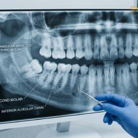
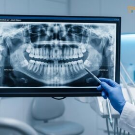
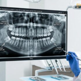
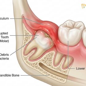
Dry socket after wisdom teeth removal is a painful dental condition where the protective blood clot at the extraction site fails to form or dislodges prematurely. This exposes underlying bone and nerves to air, food, and bacteria, causing intense, radiating pain that requires professional clinical intervention.
Clinical Summary:
Alveolar osteitis, commonly known as dry socket, is the most frequent complication following third molar extractions. It occurs when the initial blood clot is lost, exposing the alveolar bone and sensitive nerve endings. Clinical management focuses on immediate pain relief through medicated intra-socket dressings and antimicrobial irrigation. Prevention relies heavily on strict adherence to post-operative care, avoiding negative intraoral pressure (such as using straws or smoking), and utilizing advanced surgical techniques like Platelet-Rich Fibrin (PRF) to stabilize the healing environment and promote rapid cellular regeneration.
Key Takeaways:
- Dry socket occurs in approximately 2% to 5% of all tooth extractions, but the risk increases significantly with impacted lower wisdom teeth.
- The condition typically manifests 3 to 5 days post-surgery, characterized by a sudden escalation in throbbing, radiating pain.
- Visual inspection often reveals an empty socket with exposed whitish bone rather than a dark, stabilizing blood clot.
- Smoking, vaping, and drinking through straws are the primary mechanical and chemical triggers for premature clot dislodgement.
- Professional intervention using eugenol-based medicated dressings provides rapid symptomatic relief while the body initiates secondary healing.
What is Dry Socket (Alveolar Osteitis)?
Dry socket is a localized inflammation of the jawbone occurring when the essential blood clot in an extraction site dissolves or falls out, leaving nerves and bone completely unprotected.
In clinical dentistry, dry socket is formally diagnosed as alveolar osteitis. To understand this condition, one must first understand the normal physiological healing process following the surgical removal of third molars. When a tooth is extracted, the body immediately initiates a coagulation cascade. Platelets aggregate at the surgical site, and a complex web of fibrin forms to trap red blood cells, creating a stable blood clot. This clot serves as a biological bandage; it stops the bleeding, protects the underlying alveolar bone, shields the exposed nerve endings of the periodontal ligament, and provides a foundational matrix for new bone and soft tissue to grow[1].
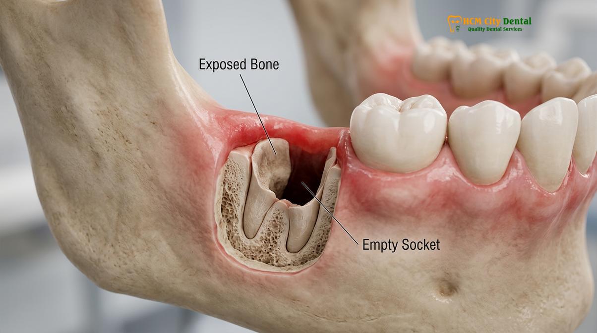
Alveolar osteitis occurs when this critical blood clot either fails to form entirely, is mechanically dislodged, or undergoes premature lysis (dissolving) before the socket has had time to fill with protective granulation tissue. Once the clot is lost, the bony walls of the socket are directly exposed to the harsh oral environment. Every breath of air, sip of liquid, and microscopic food particle that enters the empty socket directly stimulates the unprotected nerve endings, resulting in severe hyperalgesia (extreme sensitivity to pain).
“The initial blood clot is the cornerstone of post-extraction healing. It acts as a biological matrix that guides fibroblasts and osteoblasts into the surgical site. When this matrix is lost prematurely, the body must heal via secondary intention, which is a significantly slower and more painful process.” — Dr. Nguyen Van Cuong
The pathophysiology of clot breakdown is often linked to localized fibrinolysis. Trauma during a difficult extraction can trigger the release of tissue activators that convert plasminogen into plasmin, an enzyme that aggressively breaks down fibrin networks. Furthermore, certain anaerobic bacteria present in the oral cavity can release their own fibrinolytic enzymes, accelerating the destruction of the clot. This is why maintaining pristine oral hygiene and managing pre-existing gum tissue inflammation prior to surgery is paramount.
When Does Dry Socket Usually Occur? (The 3-5 Day Window)
The critical window for dry socket development is typically three to five days post-extraction, as this is when the blood clot is most vulnerable to premature breakdown before granulation tissue forms.
Understanding the timeline of post-operative healing is crucial for distinguishing between normal surgical recovery and the onset of alveolar osteitis. Immediately following the extraction (Days 1 to 2), patients naturally experience localized swelling, mild oozing, and surgical pain. During this initial phase, the blood clot is actively forming and cross-linking to the bony walls of the socket. It is highly unusual for a dry socket to present on the first day, as the clot is usually still intact and the surgical site remains under the influence of local anesthetics and initial inflammatory mediators.
The danger zone emerges between Day 3 and Day 5. By this time, normal surgical pain should be steadily decreasing. However, if the clot undergoes fibrinolysis or is mechanically dislodged, the patient will experience a sudden, sharp reversal in their recovery trajectory. The pain will escalate dramatically rather than subside. This specific timeline is a hallmark diagnostic indicator for clinicians.
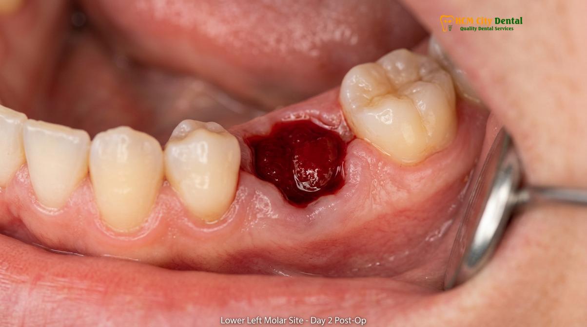
During this 3-to-5-day window, the clot is in a transitional state. It has not yet been replaced by durable granulation tissue (which typically begins forming around Day 7), making it highly susceptible to external forces. If a patient creates negative intraoral pressure or introduces chemical irritants during this fragile period, the clot can easily detach. Once a patient passes the one-week mark without symptoms, the risk of developing a dry socket drops to near zero, as the socket walls are sufficiently covered by new epithelial tissue[2].
Classic Symptoms of Dry Socket: Throbbing Pain & Bad Odor
Hallmark symptoms include severe, radiating pain that worsens after a few days, an empty-looking socket with visible bone, foul breath, and an unpleasant taste in the mouth.
The clinical presentation of alveolar osteitis is distinct and highly symptomatic. The most prominent feature is the nature of the pain. Unlike the dull, localized ache typical of normal post-operative healing, dry socket pain signs manifest as a deep, severe, throbbing agony. Because the trigeminal nerve branches are directly exposed, this pain frequently radiates along the nerve pathways. Patients commonly report that the pain shoots from the extraction site in the lower jaw up into their ear, eye, temple, or down into their neck on the affected side of the face[5].
Visual inspection of the surgical site provides further confirmation. In a healthy healing process, the socket is filled with a dark red or brownish clot. In a dry socket, the site appears as a cavernous, empty hole. Upon closer examination with clinical lighting, the clinician can often see the whitish-gray surface of the bare alveolar bone at the base and walls of the socket.
Clinical Warning: Radiating pain that suddenly intensifies 72 hours after surgery and does not respond to over-the-counter analgesics (like ibuprofen or acetaminophen) is a primary indicator of alveolar osteitis. Do not attempt to probe the empty socket with toothpicks or cotton swabs, as this will exacerbate nerve irritation and risk introducing secondary bacterial infections.
Beyond pain, patients frequently experience secondary symptoms related to the stagnation of debris within the empty socket. Without the clot to fill the void, food particles and saliva accumulate in the bony defect. Anaerobic bacteria rapidly colonize this trapped debris, breaking it down and releasing volatile sulfur compounds. This biochemical process results in severe halitosis (bad breath) and a persistent, foul, or metallic taste in the mouth that cannot be resolved by normal brushing. If you experience these symptoms, seeking Emergency Dental Care is highly recommended for prompt palliative intervention.
How to Prevent Dry Socket: Critical Home Rules
Prevention requires strict avoidance of negative pressure in the mouth, maintaining gentle oral hygiene, adhering to a soft diet, and resting to ensure the blood clot remains securely in place.
While some factors predisposing a patient to dry socket (such as the anatomical difficulty of an impacted mandibular molar) are beyond their control, the vast majority of preventative measures rely entirely on patient compliance with post-operative instructions. The overarching goal of home care is to protect the mechanical integrity of the blood clot while minimizing bacterial load in the oral cavity.
The physics of intraoral pressure play a massive role in clot retention. Any action that creates a vacuum or negative suction inside the mouth can forcefully pull the clot away from the bone. Therefore, patients must absolutely avoid using straws for at least one week post-surgery. Similarly, vigorous spitting, aggressive swishing of mouthwash, or playing wind instruments can generate enough force to dislodge the clot. When rinsing is required (usually starting 24 hours after surgery), patients should use a gentle, passive head-tilting motion rather than active cheek-muscle swishing.
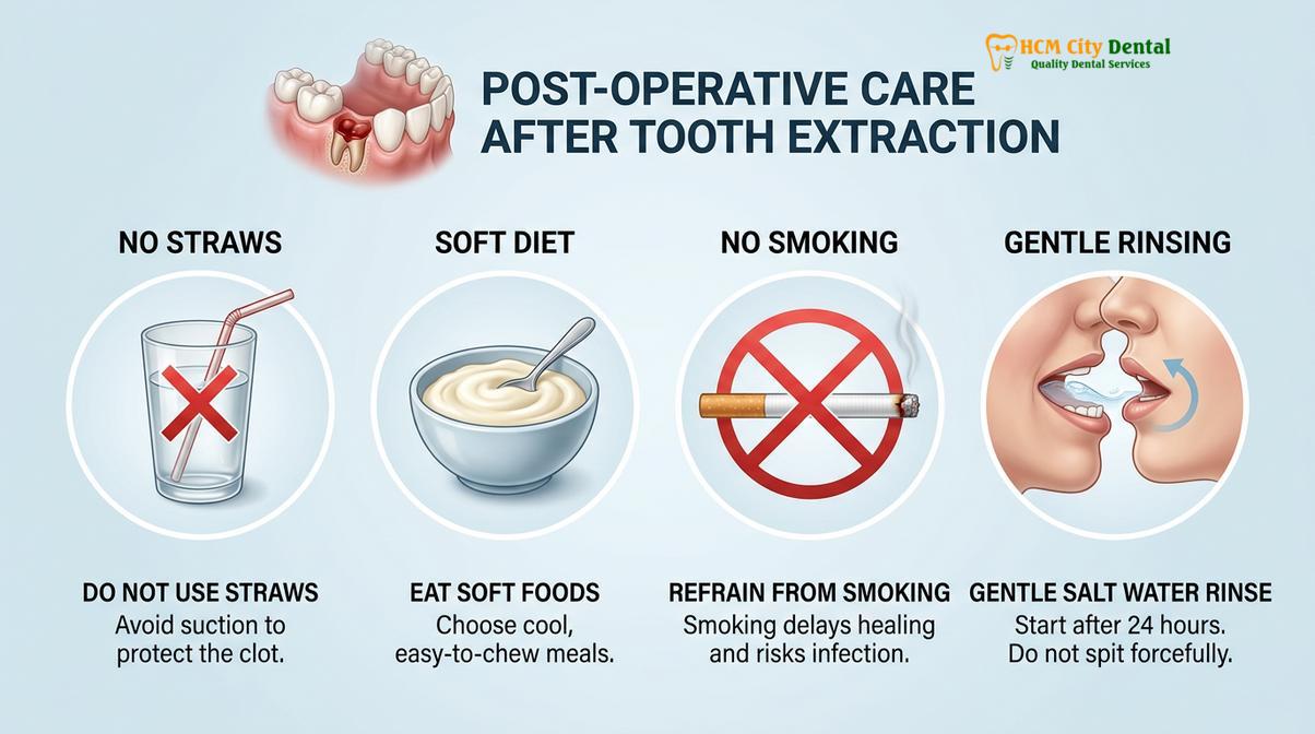
Dietary modifications are equally critical to prevent dry socket extraction complications. Hard, crunchy, or sharp foods (like chips, nuts, or crusty bread) can physically traumatize the surgical site or leave sharp fragments lodged in the healing socket. Patients must adhere to a strict soft-food diet (yogurt, applesauce, mashed potatoes, lukewarm broths) for the first few days, gradually reintroducing solid foods as tolerated, and always chewing on the side opposite the extraction.
| Post-Op Action | Clinical Recommendation | Physiological Rationale |
|---|---|---|
| Using Straws | Strictly Avoid (7-10 days) | Creates negative intraoral pressure that physically dislodges the fragile fibrin clot. |
| Oral Rinsing | Gentle head-tilting only | Vigorous swishing disrupts the clot; gentle saline soaks reduce bacterial load safely. |
| Physical Activity | Rest for 48-72 hours | Elevated heart rate and blood pressure can cause secondary bleeding and clot displacement. |
| Dietary Choices | Soft, lukewarm foods | Prevents mechanical trauma to the socket and avoids thermal breakdown of the clot. |
Furthermore, utilizing advanced ultrasonic extraction techniques can minimize surrounding bone trauma, thereby reducing the localized inflammatory response and lowering the baseline risk of clot failure.
The Impact of Smoking and Vaping on Socket Clot Formation
Smoking and vaping introduce harmful chemicals that restrict blood flow and create negative suction pressure, drastically increasing the risk of dislodging the fragile blood clot.
The correlation between tobacco use and alveolar osteitis is one of the most well-documented phenomena in oral surgery. Patients who smoke are statistically three to four times more likely to develop a dry socket compared to non-smokers. This elevated risk is driven by a combination of mechanical forces and severe chemical interference with the body’s natural healing mechanisms.
Mechanically, the act of inhaling smoke or vapor requires the patient to purse their lips and draw inward, creating a strong vacuum effect inside the oral cavity. This suction is often more than enough to rip the newly formed blood clot away from the socket walls. Many patients ask, “can i vape after wisdom tooth removal?” assuming that because vaping doesn’t involve combustion, it is safe. Clinically, the mechanical suction required for vaping is identical to smoking, carrying the exact same risk of physical clot dislodgement.
Chemically, the effects are even more detrimental. Nicotine is a potent vasoconstrictor. When introduced into the bloodstream, it causes the peripheral blood vessels—including the microvasculature supplying the alveolar bone and gingival tissues—to narrow significantly. This localized ischemia (lack of blood flow) deprives the surgical site of the oxygen, nutrients, and immune cells required to sustain the blood clot and initiate tissue regeneration[3].
“Nicotine-induced vasoconstriction severely impairs angiogenesis at the extraction site. Furthermore, carbon monoxide from combustible tobacco binds to hemoglobin with a higher affinity than oxygen, creating a hypoxic environment that actively promotes the breakdown of the fibrin matrix and delays osteoblast activity.” — Clinical Oral Surgery Guidelines
For patients wondering about smoking after wisdom teeth extraction, clinical protocols mandate complete cessation for a minimum of 72 hours post-surgery, though abstaining for a full 7 to 10 days is strongly advised to ensure the socket has transitioned from a fragile clot to stable granulation tissue.
How PRF (Platelet-Rich Fibrin) Minimizes Dry Socket Risk
Applying Platelet-Rich Fibrin (PRF) during surgery utilizes the patient’s own concentrated growth factors to accelerate tissue regeneration, significantly reducing the likelihood of clot failure.
In modern oral and maxillofacial surgery, the prevention of alveolar osteitis has evolved beyond simple post-operative instructions to include proactive, biologically driven surgical techniques. One of the most effective advancements is the use of Platelet-Rich Fibrin (PRF). This autologous biomaterial is created from the patient’s own blood, drawn immediately prior to the extraction procedure.
The blood is placed into a specialized centrifuge and spun at specific protocols to separate the red blood cells from the plasma and platelets. The result is a dense, yellowish, gel-like membrane composed of a highly cross-linked fibrin matrix, heavily concentrated with platelets, leukocytes, and vital growth factors (such as PDGF, TGF-beta, and VEGF). Unlike a natural blood clot, which is fragile and easily dissolved by oral enzymes, the PRF membrane is structurally robust and highly resistant to premature breakdown.
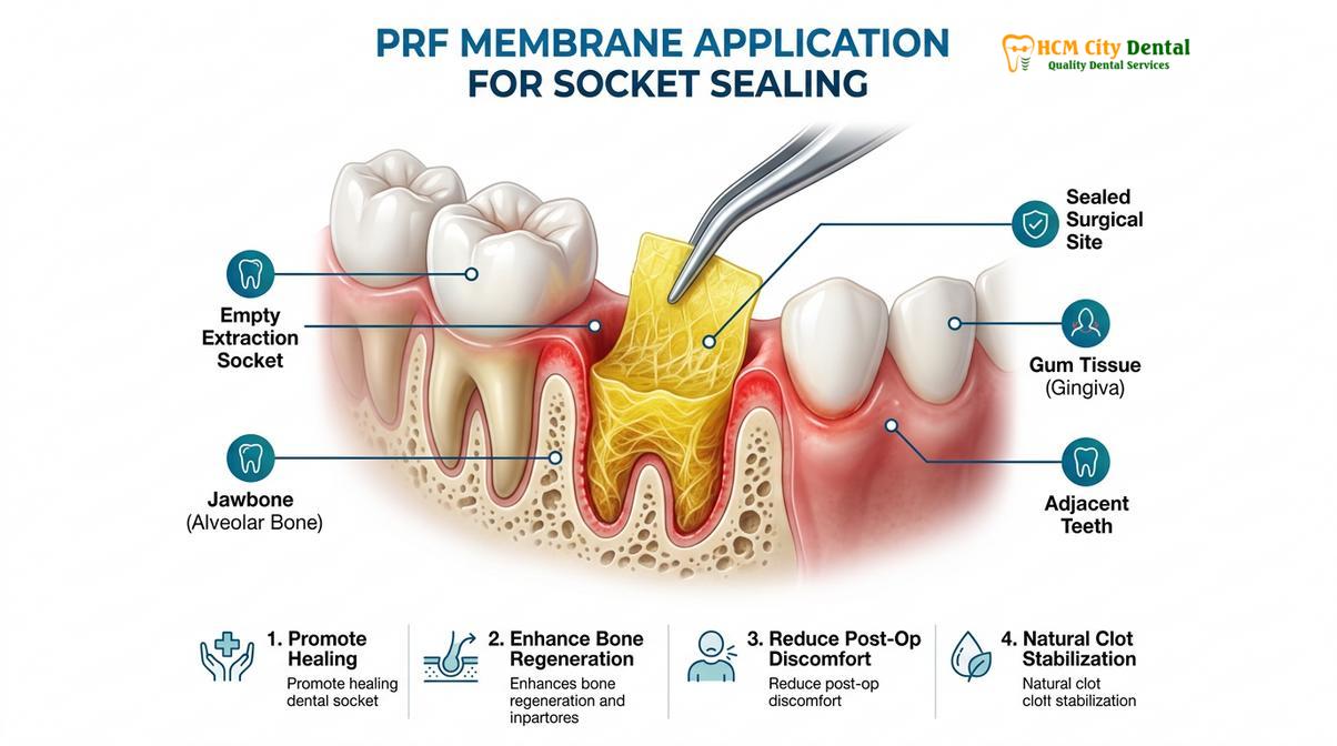
At HCMC Dental Clinic, Dr. Nguyen Van Cuong emphasizes that utilizing PRF protocols significantly alters the healing trajectory. Once the wisdom tooth is removed, the surgeon carefully packs the empty socket with the PRF membrane and sutures the gingival tissue over it. This membrane acts as an immediate, secure biological seal over the exposed bone. Because it is packed with concentrated growth factors, it actively stimulates rapid angiogenesis (new blood vessel formation) and accelerates the migration of fibroblasts to the site.
Clinical studies demonstrate that the application of PRF drastically reduces the incidence of dry socket, minimizes post-operative pain and swelling, and provides a superior scaffold for long-term bone regeneration in the extraction defect. It is particularly recommended for patients with a history of difficult extractions, smokers, or those undergoing the removal of deeply impacted mandibular third molars.
Professional Treatments at HCMC Dental for Dry Socket
Clinical treatment involves gently irrigating the socket to remove debris and packing it with a medicated dressing to provide immediate, soothing pain relief while secondary healing occurs.
Despite the best preventative efforts, alveolar osteitis can still occur. When it does, patients often search for how to treat dry socket at home. It is critical to understand that true dry socket cannot be cured with home remedies; it requires professional palliative care to manage the severe pain and prevent secondary infections while the body heals via secondary intention.
Patients seeking alveolar osteitis treatment saigon often visit our facility for immediate relief. The clinical workflow for treating a dry socket is straightforward, highly effective, and focuses entirely on symptom management. First, the clinician will gently irrigate the empty socket using a sterile saline or chlorhexidine solution. This step is crucial to flush out any impacted food debris, necrotic tissue, and bacterial colonies that are contributing to the pain and halitosis.
Clinical Case Review: A 24-year-old patient presented to HCMC Dental Clinic in Ho Chi Minh City complaining of severe, radiating jaw pain four days after a lower wisdom tooth extraction. Examination revealed a classic dry socket with exposed alveolar bone. The site was gently irrigated with antimicrobial saline, and an Alvogyl dressing was placed. The patient reported a 90% reduction in pain within 15 minutes of application, allowing them to resume normal sleep and hydration.
Once the socket is clean, the dentist will place a medicated dressing directly into the bony defect. The most common dressing used globally is Alvogyl, a fibrous paste containing eugenol (a potent analgesic and obtundent derived from clove oil), butamben (a topical anesthetic), and iodoform (a broad-spectrum antimicrobial agent)[4]. The eugenol acts rapidly to soothe the exposed nerve endings, providing profound and almost immediate pain relief.

This medicated dressing acts as a physical barrier, replacing the lost blood clot and shielding the bone from oral stimuli. Depending on the severity of the condition, the dressing may need to be replaced every 48 to 72 hours until the patient’s pain naturally subsides and new granulation tissue begins to cover the exposed bone. In cases where the extraction site is adjacent to an adjacent molar decay, the clinician will also ensure that no secondary pulpal inflammation is contributing to the pain profile.
When to See a Doctor
While post-operative discomfort is expected, certain clinical signs warrant immediate professional evaluation. You should contact your oral surgeon or dentist if you experience pain that suddenly worsens 3 to 5 days after the extraction and is unresponsive to prescribed analgesics. Additionally, if the pain is accompanied by a high fever, significant facial swelling that worsens after the third day, difficulty swallowing or breathing, or visible pus discharging from the extraction site, these are red flags for a spreading bacterial infection rather than a simple dry socket. Prompt clinical diagnostics are essential to differentiate between alveolar osteitis and more severe complications like osteomyelitis or deep fascial space infections.
Frequently Asked Questions
What does dry socket look and feel like?
Dry socket looks like an empty hole where the tooth was extracted, often revealing whitish underlying bone instead of a dark blood clot. It feels like a severe, throbbing ache that radiates from the extraction site to your ear, eye, or temple, typically beginning a few days after surgery. The pain is usually accompanied by a foul taste and bad breath due to trapped debris.
How can I avoid getting dry socket?
You can avoid dry socket by strictly following post-operative instructions: do not smoke, avoid drinking through straws, chew on the opposite side of your mouth, and refrain from vigorous rinsing or spitting. Maintaining a soft diet and keeping your head elevated also helps stabilize the forming blood clot. Following these guidelines minimizes the mechanical and chemical disruptions that lead to clot failure.
How long after wisdom teeth extraction can I smoke or vape?
Clinicians strongly advise waiting at least 72 hours before smoking or vaping, though abstaining for a full week is optimal for healing. The suction creates negative pressure that can dislodge the clot, while nicotine constricts blood vessels, severely delaying the cellular regeneration necessary for socket closure and increasing the risk of complications.
Does drinking through a straw cause dry socket?
Yes, drinking through a straw is a primary mechanical cause of dry socket. The suction required to pull liquid through a straw creates significant negative intraoral pressure, which can easily dislodge the fragile blood clot from the extraction site before it has organized into stable granulation tissue. Patients should drink directly from a cup during the initial healing phase.
What will a dentist do to fix dry socket?
A dentist will gently irrigate the empty socket with an antimicrobial solution to remove trapped debris, then pack the site with a medicated dressing containing analgesic and antiseptic agents like eugenol. This provides almost immediate pain relief and protects the exposed bone while secondary healing occurs. The dressing may be replaced every few days until symptoms resolve.
References
- Journal of Oral and Maxillofacial Surgery. Pathogenesis and management of alveolar osteitis. (2021).
- International Journal of Oral and Maxillofacial Surgery. The efficacy of PRF in preventing dry socket. (2020).
- British Dental Journal. Smoking and its effects on post-extraction healing. (2019).
- Journal of the American Dental Association. Clinical protocols for intra-socket medicated dressings. (2022).
- Clinical Oral Investigations. Trigeminal nerve pain pathways in alveolar osteitis. (2018).