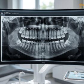
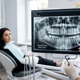
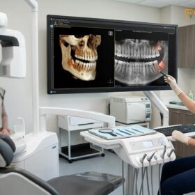
CBCT wisdom teeth mapping is an advanced three-dimensional imaging technique used to visualize the exact position of impacted third molars, their complex root structures, and their precise proximity to vital anatomical landmarks like the inferior alveolar nerve. This high-resolution mapping allows oral surgeons to plan highly predictable, minimally invasive extractions while mitigating the risk of neurological injury.
Clinical Summary:
Cone Beam Computed Tomography (CBCT) has fundamentally transformed the diagnostic and surgical planning phases for impacted third molars. Unlike traditional 2D panoramic radiographs, which suffer from inherent magnification and superimposition, CBCT provides a volumetric, 1:1 undistorted view of the maxillofacial region. This technology enables the precise mapping of the inferior alveolar nerve (IAN) and the maxillary sinus floor in relation to complex, multi-rooted tooth anatomies. Dr. Nguyen Van Cuong emphasizes that utilizing a 3d cbct dental scan saigon is a critical standard of care for high-risk impactions. By revealing the exact buccal-lingual position of the nerve, CBCT dictates the safest surgical approach—whether that involves a complete extraction, strategic tooth sectioning, or a nerve-sparing coronectomy—thereby significantly reducing the incidence of postoperative paresthesia and ensuring optimal clinical outcomes.
Key Takeaways:
- CBCT provides a 1:1 undistorted, three-dimensional view of wisdom teeth and surrounding jawbone anatomy.
- Precise nerve mapping significantly reduces the risk of inferior alveolar nerve (IAN) injury during complex extractions.
- 3D imaging reveals hidden root curvatures, dilacerations, and variations in localized bone density.
- Radiation exposure from modern dental CBCT is minimal, strictly localized, and follows the ALARA safety principle.
- High-risk impactions often require CBCT diagnostics to determine if a nerve-sparing coronectomy is safer than full extraction.
The Limitation of 2D Panoramic X-Rays for Wisdom Teeth
Traditional 2D panoramic X-rays compress complex three-dimensional jaw anatomy into a flat image, creating superimposition and distortion that can obscure the true spatial relationship between wisdom tooth roots and vital nerves.
For decades, the orthopantomogram (OPG), commonly known as a panoramic X-ray, has been the standard initial screening tool for evaluating third molars. While it provides an excellent broad overview of the entire dentition, the maxilla, and the mandible, it possesses inherent physical limitations due to its two-dimensional nature. When an X-ray beam passes through the curved structure of the human jaw and projects it onto a flat sensor, the resulting image suffers from magnification errors—often ranging between 15% and 30%—and the superimposition of anatomical structures[1].
In the context of lower wisdom teeth, this superimposition becomes a critical clinical challenge. On a 2D radiograph, the roots of an impacted third molar may appear to intersect or overlap directly with the mandibular canal, which houses the inferior alveolar nerve (IAN). Radiographic markers such as the darkening of the tooth root, the interruption of the radiopaque borders of the canal, or the sudden diversion of the canal’s path are classic warning signs of nerve proximity. However, because a panoramic image lacks depth (the buccal-lingual dimension), it is impossible to determine whether the nerve is positioned in front of the root, behind the root, or directly entangled between the root apices.
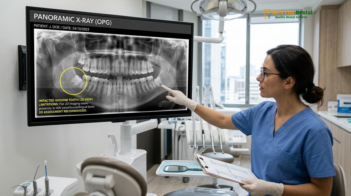
Relying solely on 2D imaging for high-risk impactions forces the surgeon to operate with a degree of anatomical uncertainty. If a root is severely curved (dilacerated) in a direction not visible on the flat film, applying standard elevation forces can lead to root fracture or inadvertent pressure on the underlying nerve. Furthermore, in the upper jaw, 2D X-rays often fail to accurately depict the thickness of the bone separating the upper wisdom teeth from the floor of the maxillary sinus, increasing the risk of an oroantral communication (a hole between the mouth and the sinus cavity) during surgical tooth extraction.
What is a 3D CBCT (Cone Beam Computed Tomography) Scan?
A CBCT scan utilizes a cone-shaped X-ray beam that rotates around the patient, capturing hundreds of high-resolution images that are reconstructed into a highly accurate, undistorted 3D model of the maxillofacial region.
Cone Beam Computed Tomography (CBCT) represents a paradigm shift in dentomaxillofacial radiology. Unlike conventional medical CT scanners that use a high-output fan-shaped beam and capture images in multiple continuous slices, a dental CBCT machine uses a divergent cone-shaped beam. As the scanner rotates a single time around the patient’s head—usually taking less than 30 seconds—it captures a series of hundreds of 2D projection images from various angles. Advanced software algorithms then reconstruct these projections into a complete volumetric dataset[2].
The resulting 3D model is composed of isotropic voxels (three-dimensional pixels that are perfectly cubical). This isotropic nature ensures that the images are geometrically accurate and completely free of the magnification and distortion inherent in panoramic X-rays. Clinicians can slice through this digital model in multiple planes: axial (top-down), coronal (front-back), and sagittal (side-to-side). This multi-planar reconstruction allows the surgeon to view the anatomy from literally any angle, providing a 1:1 representation of the patient’s actual bone structure and dentition.
“The introduction of Cone Beam Computed Tomography into oral surgery has transitioned our approach from reactive problem-solving during surgery to proactive, precision-guided surgical planning, fundamentally elevating the standard of patient safety.”
For wisdom tooth evaluation, a cone beam ct dentist will typically utilize a “small” or “medium” Field of View (FOV). A small FOV restricts the X-ray beam strictly to the area of the impacted tooth, providing the highest possible resolution (often down to 0.076 millimeters) while minimizing radiation exposure to surrounding tissues. This level of microscopic detail is what makes CBCT an indispensable tool for complex oral and maxillofacial surgeries.
Mapping the Inferior Alveolar Nerve to Avoid Injury
CBCT allows oral surgeons to trace the exact three-dimensional pathway of the inferior alveolar nerve, determining its precise buccal-lingual position relative to the roots of an impacted wisdom tooth.
The most significant risk associated with the removal of lower mandibular third molars is injury to the inferior alveolar nerve (IAN) or the lingual nerve. The IAN supplies sensory innervation to the lower lip, chin, and lower teeth on that side of the face. If this nerve is bruised, stretched, or severed during extraction, the patient may experience paresthesia (altered sensation), dysesthesia (painful sensation), or permanent anesthesia (complete numbness) in the affected areas[3].
Proper wisdom teeth root nerve mapping using CBCT is the most effective method to mitigate this risk. When a 2D X-ray indicates a high risk of nerve overlap, the 3D scan is employed to reveal the true anatomical relationship. The surgeon can digitally trace the mandibular canal through the bone, observing exactly where it travels. In many cases, the CBCT reveals that while the nerve appeared to intersect the tooth on the 2D film, it is actually situated safely to the buccal (cheek) or lingual (tongue) side of the roots, separated by a distinct layer of cortical bone.
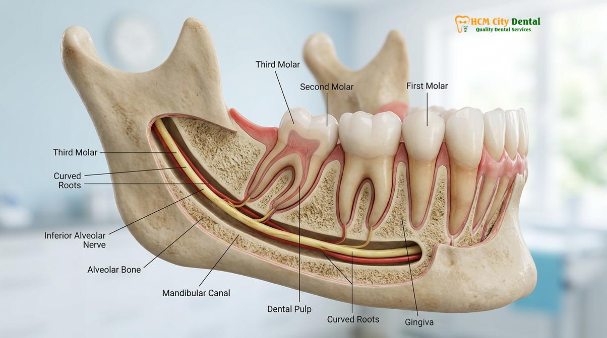
However, in cases where the CBCT confirms that the nerve is directly in contact with the root, or worse, that the root has grown completely around the nerve (a true “dumbbell” or grooved root morphology), the surgical plan must be drastically altered. Dr. Nguyen Van Cuong notes that in these high-risk scenarios, attempting a standard extraction could be catastrophic. Instead, the precise mapping provided by the CBCT allows the surgeon to confidently recommend an alternative procedure, such as a coronectomy, where the crown of the tooth is removed but the roots are intentionally left behind to preserve the nerve.
Clinical Case Review: Nerve Preservation
A 26-year-old patient presented to HCMC Dental Clinic in Ho Chi Minh City with a deeply impacted lower right wisdom tooth. The initial panoramic X-ray showed the tooth roots completely overlapping the mandibular canal, suggesting extreme risk. A localized CBCT scan was performed, revealing that the nerve was not just adjacent to the tooth, but actually passing through a physical hole (fenestration) between the two fused roots. Armed with this 3D mapping, the surgical team opted for a precise coronectomy rather than a full extraction. The patient recovered smoothly with zero neurological deficits, a successful outcome directly attributed to advanced 3D diagnostics.
How 3D Scans Help Detect Root Curvatures and Bone Densities
Beyond nerve mapping, 3D imaging exposes hidden root dilacerations, accessory roots, and the exact density of the surrounding jawbone, enabling the surgeon to calculate the precise angle of elevation required.
Wisdom teeth are notorious for their unpredictable root morphology. Unlike anterior teeth which typically have straight, single roots, third molars can have multiple roots that curve sharply (dilaceration), hook at the ends, or splay outward into the surrounding bone. On a 2D X-ray, a root that curves directly toward or away from the camera lens will appear deceptively short and straight. If a surgeon attempts to elevate such a tooth without knowing about the hidden curvature, the root tip is highly likely to fracture and remain embedded deep in the socket.
CBCT imaging eliminates this blind spot. By rotating the 3D model, the surgeon can visualize every curve, hook, and accessory root. This dictates the strategy for tooth sectioning. Instead of applying excessive force to remove the tooth in one piece, the surgeon uses a surgical handpiece or a piezoelectric device to divide the tooth into smaller, manageable sections, allowing each curved root to be removed along its natural path of withdrawal[4].
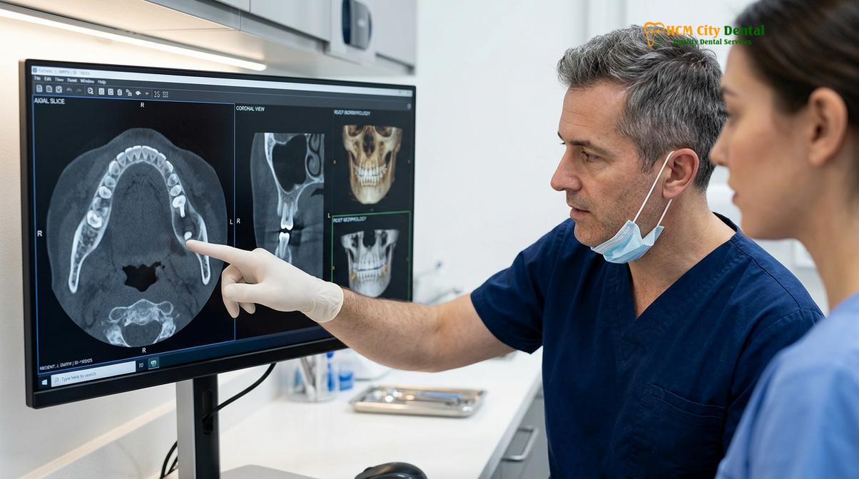
Furthermore, CBCT provides critical data regarding localized bone density. The software can measure the density of the cortical (outer) and cancellous (inner) bone surrounding the tooth. If the bone is exceptionally dense—common in older adult patients—the surgeon knows that the tooth will not yield easily to elevation, and more extensive bone guttering (removal of bone around the crown) will be necessary. Conversely, in the upper jaw, identifying thin or porous bone helps the surgeon avoid applying forces that could fracture the maxillary tuberosity.
| Diagnostic Feature | 2D Panoramic X-Ray | 3D CBCT Scan |
|---|---|---|
| Image Dimension | Flat, compressed 2D image | Volumetric, 360-degree 3D model |
| Magnification/Distortion | 15% – 30% distortion rate | 1:1 ratio, zero distortion (isotropic) |
| Nerve Proximity | Shows overlap, cannot confirm depth | Exact buccal-lingual nerve mapping |
| Root Morphology | Hides buccal/lingual curvatures | Reveals all dilacerations and extra roots |
| Bone Density Assessment | Poor to moderate estimation | Highly accurate density visualization |
Safety, Radiation Dosage, and Procedure of CBCT Scans
Dental CBCT scans are highly safe, exposing patients to a fraction of the radiation of medical CTs, and the scanning procedure is entirely painless, non-invasive, and completed in under a minute.
A common concern among patients is the level of radiation exposure associated with 3D imaging. It is crucial to distinguish between a traditional medical CT scan (used in hospitals for full-body or head trauma imaging) and a dental Cone Beam CT. Because the dental CBCT uses a focused, cone-shaped beam and captures the entire volume in a single rotation, the radiation dose is exponentially lower.
Radiation is measured in microsieverts (µSv). A standard dental CBCT scan of the jaw typically exposes the patient to between 30 and 100 µSv, depending on the machine settings and the chosen Field of View (FOV). To put this into perspective, the average person absorbs about 3,000 µSv of natural background radiation from the environment every year. The dose from a localized dental CBCT is roughly equivalent to the cosmic radiation received during a cross-country commercial flight. Dental professionals strictly adhere to the ALARA principle (As Low As Reasonably Achievable), ensuring that 3D imaging is only prescribed when the diagnostic benefits significantly outweigh the minimal exposure risks[5].
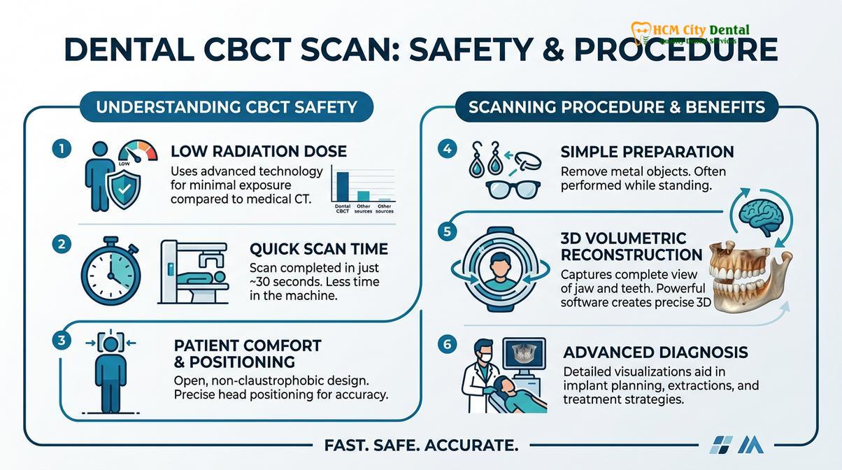
The procedure itself is remarkably straightforward and comfortable. The patient is asked to stand or sit in the machine and bite down gently on a sterile plastic guide to ensure the jaws are properly aligned and stationary. The clinician will position laser guides to target the specific area of interest. Once activated, the C-arm of the scanner rotates 360 degrees around the patient’s head. The actual scanning time is usually between 10 to 20 seconds. There is no enclosed tube, making it completely comfortable for patients who suffer from claustrophobia.
Important Clinical Safety Notice
While dental CBCT radiation is extremely low, it is standard medical protocol to avoid elective radiographic imaging during pregnancy unless absolutely necessary for emergency infection control. Always inform your dental provider if you are pregnant, suspect you might be pregnant, or are undergoing specific radiation therapies, so that appropriate lead shielding and clinical modifications can be implemented.
CBCT Scan Costs at Top Dental Clinics in Saigon
The cost of a CBCT scan in Ho Chi Minh City is highly competitive, offering patients access to world-class 3D diagnostic imaging at a fraction of the price found in Western healthcare systems.
For international patients and expatriates, the economics of dental care in Vietnam present a significant advantage. In countries like the United States, Australia, or the UK, a dedicated dental CBCT scan can cost anywhere from $250 to $600 USD, often billed separately from the consultation and the surgical extraction itself. This high cost sometimes deters patients from receiving the optimal diagnostic workup they need for safe surgery.
Conversely, the cbct price dental vietnam is remarkably accessible. At top-tier facilities like HCMC Dental Clinic, a high-resolution 3D scan typically ranges from $20 to $50 USD. This affordability does not come at the expense of quality; leading clinics in Saigon utilize the exact same state-of-the-art European and American imaging equipment (such as Sirona, Planmeca, or Morita) found in premium Western practices.
“The accessibility of advanced 3D imaging in Vietnam ensures that clinical safety is never compromised by financial constraints. Every patient with a high-risk impaction can receive the precise nerve mapping required for a predictable surgical outcome.”
When planning your treatment, it is important to view the CBCT scan not as an optional add-on, but as a critical investment in your surgical safety. The detailed anatomical map it provides allows the surgeon to operate faster, more efficiently, and with less trauma to the surrounding tissues, which directly translates to a smoother and less painful recovery after wisdom tooth extraction. For a comprehensive breakdown of surgical fees, patients can review the comparative wisdom tooth cost across Southeast Asia.
When to See a Doctor for Wisdom Tooth Evaluation
Wisdom teeth do not always cause immediate pain, but asymptomatic impactions can silently damage adjacent teeth or form destructive cysts over time. It is highly recommended to seek a professional clinical evaluation if you experience any of the following symptoms:
- Persistent Jaw Aching: A dull, throbbing pain at the back of the jaw that worsens when chewing or opening the mouth wide.
- Gum Swelling and Redness: Inflammation of the gum tissue overlying a partially erupted tooth (pericoronitis), which can trap food and bacteria.
- Referred Pain: Unexplained headaches, earaches, or tension in the temporomandibular joint (TMJ) caused by the pressure of erupting third molars.
- Bad Taste or Odor: Chronic halitosis or a foul taste originating from the back of the mouth, indicating a localized infection.
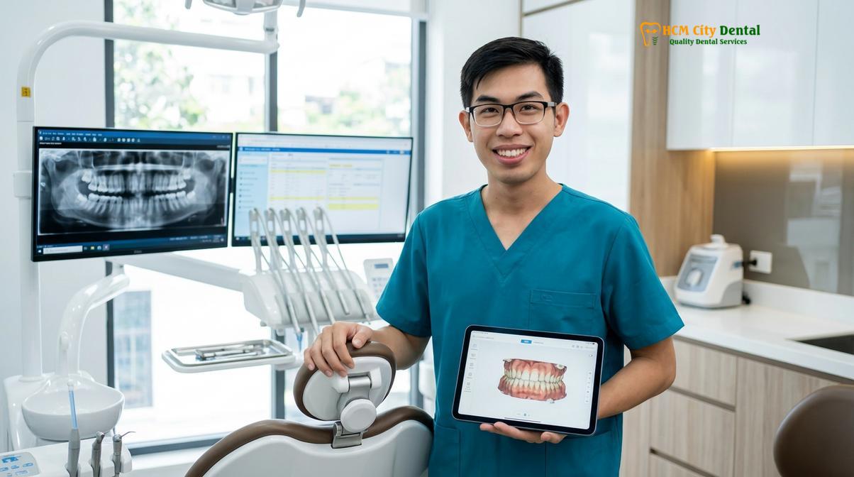
Early diagnostic imaging is the key to preventing severe complications. If you have been told your wisdom teeth are close to the nerve, or if you are experiencing recurrent flare-ups, scheduling a consultation with an oral surgeon is the safest course of action. At HCMC Dental Clinic in Ho Chi Minh City, our specialists utilize advanced 3D CBCT mapping to provide a clear, evidence-based assessment of your unique anatomy, ensuring that any proposed surgical intervention is planned with the highest degree of precision and care.
Frequently Asked Questions
Why do I need a 3D CBCT scan for my wisdom teeth?
A 3D CBCT scan is required to precisely map the inferior alveolar nerve and complex root structures that are hidden on flat 2D X-rays. This three-dimensional visualization allows your oral surgeon to plan a safe extraction path, significantly reducing the risk of permanent nerve damage or sinus complications. By seeing the exact buccal-lingual depth, the surgeon can decide whether to extract the tooth whole, section it, or perform a coronectomy.
How much radiation is in a dental CBCT scan?
A dental CBCT scan emits a very low dose of radiation, typically ranging from 30 to 100 microsieverts depending on the field of view. This is substantially less than a conventional medical CT scan and is equivalent to the background radiation you would experience during a short commercial flight. Modern machines utilize pulsed X-ray technology to further minimize exposure while maintaining high-resolution imagery.
How much does a CBCT scan cost in Ho Chi Minh City?
The cost of a CBCT scan in Ho Chi Minh City generally ranges from $20 to $50 USD, depending on the clinic and the required field of view. This highly competitive pricing makes advanced diagnostic imaging accessible while maintaining strict international standards for radiological safety and accuracy. Many premium clinics include the scan as part of a comprehensive surgical consultation package.
How long does a cone beam CT scan take?
The actual scanning process for a cone beam CT takes less than 30 seconds to complete. During this brief time, the machine rotates around your head to capture hundreds of images, though the entire appointment, including positioning and image reconstruction, usually takes about 10 to 15 minutes. The procedure is entirely painless and non-claustrophobic.
Can a panoramic X-ray show nerve overlap?
A panoramic X-ray can show an apparent overlap between the wisdom tooth roots and the nerve canal, but it cannot determine true three-dimensional proximity. Because it is a flat 2D image, structures that appear to intersect may actually be safely separated by millimeters of bone in the buccal-lingual dimension. Only a 3D scan can confirm if the root and nerve are truly in physical contact.
References
- Journal of Oral and Maxillofacial Surgery. Diagnostic accuracy of CBCT compared to panoramic radiography in predicting IAN exposure. (2021).
- International Journal of Oral and Maxillofacial Surgery. Does additional cone beam computed tomography decrease the risk of inferior alveolar nerve injury? (2017).
- Clinical Oral Investigations. Necessity of 3D visualization for the removal of lower wisdom teeth. (2012).
- Dentomaxillofacial Radiology. Assessment of impacted and partially impacted lower third molars with panoramic radiography compared to MRI and CBCT. (2018).
- Journal of the American Dental Association. Guidelines for the use of cone-beam computed tomography in oral and maxillofacial surgery. (2022).