Wisdom teeth extraction carries a small but notable nerve risk, primarily involving the inferior alveolar and lingual nerves. While temporary numbness occurs in a minority of cases, permanent damage is exceptionally rare. Advanced 3D imaging and specialized surgical techniques significantly minimize these risks during lower third molar removal.
Clinical Summary:
The proximity of mandibular third molars to major facial nerves necessitates precise surgical planning to mitigate the wisdom teeth extraction nerve risk. Nerve injury during extraction typically manifests as altered sensation in the lower lip, chin, or tongue. Most sensory deficits resolve spontaneously within weeks as the nerve tissue regenerates. However, high-risk impactions require advanced diagnostic protocols, such as Cone Beam Computed Tomography (CBCT), to map nerve pathways accurately in three dimensions. In cases where the root intimately wraps the nerve canal, alternative procedures like coronectomy are indicated to eliminate the risk of mechanical trauma. Comprehensive preoperative assessment, combined with meticulous surgical execution, ensures optimal safety and predictable recovery outcomes for patients undergoing complex extractions.
Key Takeaways:
- The inferior alveolar nerve (IAN) and lingual nerve are the primary neural structures at risk during lower wisdom tooth surgery.
- Temporary sensory changes occur in roughly 1% to 5% of cases, while permanent deficits affect less than 1% of patients.
- Symptoms of nerve involvement include persistent numbness, tingling, or burning sensations in the lip, chin, or tongue.
- 3D CBCT imaging serves as the clinical gold standard for evaluating the anatomical relationship between tooth roots and nerve canals.
- Coronectomy provides a highly effective, conservative alternative to full extraction for teeth deemed high-risk for nerve trauma.
Understanding the Risk of Nerve Injury in Wisdom Tooth Surgery
The risk of nerve injury during wisdom tooth removal stems from the anatomical proximity of lower molar roots to the mandibular nerve canal. Careful surgical execution and preoperative imaging are essential to prevent mechanical trauma to these delicate neural structures.
The surgical removal of impacted mandibular third molars is one of the most frequently performed procedures in oral and maxillofacial surgery. While the vast majority of these extractions proceed without significant complications, the anatomical positioning of the lower wisdom teeth introduces a specific set of clinical challenges. The primary concern among both practitioners and patients is the potential for a wisdom tooth nerve injury. This risk is inherently tied to the developmental trajectory of the third molars, which often erupt late and find themselves wedged against dense cortical bone, adjacent teeth, and vital neurovascular bundles[1].
To fully grasp the nature of this risk, it is necessary to understand the biomechanics of tooth extraction. The process involves the expansion of the alveolar bone and the severing of the periodontal ligament to elevate the tooth from its socket. When a tooth is deeply impacted—whether mesioangular, distoangular, horizontal, or vertical—the forces required for elevation must be carefully controlled. If the roots of the tooth are in direct contact with or wrapped around the nerve canal, the mechanical pressure exerted during elevation can be transferred directly to the nerve sheath. This compression, even if momentary, can disrupt the delicate axonal pathways responsible for transmitting sensory information from the lower face to the brain.
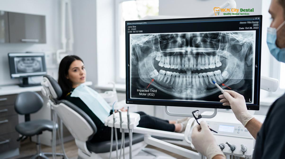
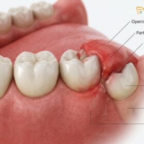
Dr. Nguyen Van Cuong, a leading specialist at HCMC Dental Clinic, emphasizes that the incidence of nerve injury is directly correlated with the complexity of the impaction and the age of the patient. As individuals age, the surrounding jawbone becomes denser and less pliable, while the roots of the wisdom teeth complete their formation, often growing deeper into the mandible. Consequently, prophylactic removal of wisdom teeth in young adults is frequently recommended to avoid the heightened risks associated with fully formed roots and calcified bone in older patients. However, every case requires a highly individualized diagnostic approach to weigh the benefits of extraction against the potential neurological risks.
“Informed consent is a cornerstone of ethical oral surgery. Patients must be fully educated about the anatomical realities of their specific impaction. While the statistical probability of permanent nerve damage is exceedingly low, acknowledging and planning for this risk through advanced imaging is what separates standard care from exceptional clinical practice.”
It is also important to differentiate between the risks associated with upper (maxillary) and lower (mandibular) wisdom teeth. Upper wisdom teeth rarely pose a threat to major sensory nerves; instead, their primary anatomical risk involves the maxillary sinus cavity. Lower wisdom teeth, conversely, are the exclusive source of the nerve risks discussed in this context. The surgical approach to a lower third molar must account for the density of the external oblique ridge, the trajectory of the roots, and the precise location of the neurovascular structures. By employing minimally invasive techniques, such as ultrasonic extraction techniques, surgeons can selectively remove bone while preserving soft tissue and nerve integrity.
The Inferior Alveolar Nerve & Lingual Nerve Pathways
Two primary nerves are vulnerable during lower third molar extraction: the inferior alveolar nerve, which supplies sensation to the lower lip and chin, and the lingual nerve, which provides feeling and taste to the anterior two-thirds of the tongue.
A comprehensive understanding of the regional anatomy is paramount for any clinician performing mandibular surgery. The trigeminal nerve, the fifth cranial nerve, is the largest and most complex of the cranial nerves, responsible for providing sensory innervation to the face and motor innervation to the muscles of mastication. The mandibular division of the trigeminal nerve (V3) gives rise to several branches, two of which are of critical importance during wisdom tooth extraction: the inferior alveolar nerve (IAN) and the lingual nerve.
The inferior alveolar nerve enters the mandible through the mandibular foramen, located on the medial surface of the mandibular ramus. It then travels anteriorly through the mandibular canal, a bony tunnel that runs beneath the roots of the lower molars. As it courses through this canal, it provides sensory branches to the mandibular teeth and the surrounding alveolar bone. Upon reaching the mental foramen, typically located near the premolars, the nerve bifurcates. One branch continues anteriorly within the bone as the incisive nerve, while the other exits the bone as the mental nerve, supplying sensation to the lower lip, chin, and the labial gingiva of the anterior teeth. When the roots of a third molar extend deeply into the jaw, they can impinge upon the mandibular canal, placing the IAN at direct risk of compression or laceration during surgery[2].
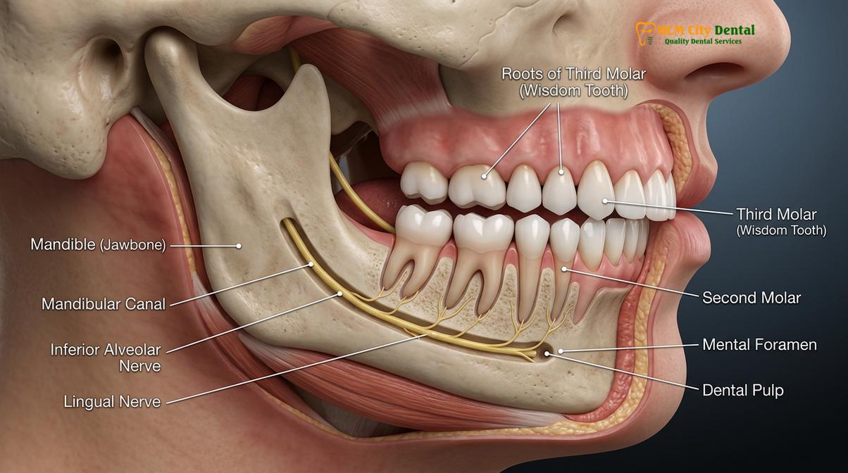
The lingual nerve, another major branch of the mandibular nerve, follows a different trajectory. It descends through the pterygomandibular space and runs along the medial (lingual) aspect of the mandible, closely hugging the periosteum near the roots of the second and third molars. Unlike the IAN, which is encased in a protective bony canal, the lingual nerve travels through soft tissue, making it highly vulnerable to surgical trauma. Its anatomical position is notoriously variable; in a significant percentage of the population, the nerve rests at or even above the crest of the alveolar bone in the third molar region. The lingual nerve is responsible for general sensation to the anterior two-thirds of the tongue, the floor of the mouth, and the lingual gingiva. Furthermore, it carries special sensory fibers (chorda tympani) that facilitate taste perception.
| Nerve Structure | Anatomical Location | Sensory Function | Primary Surgical Risk Factors |
|---|---|---|---|
| Inferior Alveolar Nerve (IAN) | Encased within the bony mandibular canal beneath the molar roots. | Sensation to the lower teeth, lower lip, and chin. | Deep root impaction, hooked roots, excessive elevation force. |
| Lingual Nerve | Soft tissue along the inner (lingual) aspect of the lower jaw. | Sensation and taste to the anterior two-thirds of the tongue. | Lingual flap reflection, aggressive bone removal, anatomical variations. |
Injury to either of these nerves can profoundly impact a patient’s quality of life. While IAN injuries are more commonly associated with the direct manipulation of the tooth roots, lingual nerve injuries often result from the reflection of the surgical flap or the use of rotary instruments near the lingual cortical plate. To mitigate these risks, modern surgical protocols emphasize the avoidance of lingual flap elevation whenever possible and the use of specialized retractors to protect the soft tissues during bone removal. Understanding these distinct pathways allows the surgeon to tailor the extraction technique, ensuring that the mechanical forces applied are directed safely away from these critical neural corridors.
Symptoms of Temporary vs Permanent Nerve Damage
Nerve damage symptoms range from mild tingling to complete numbness in the affected areas. While temporary neuropraxia resolves within weeks to months, permanent neurotmesis involves lasting sensory deficits requiring specialized management.
When a patient experiences a nerve injury following a Tooth Extraction, the clinical presentation can vary widely depending on the severity and mechanism of the trauma. In the immediate postoperative period, it is entirely normal for the patient to experience profound numbness due to the administration of local anesthesia. However, if this numbness persists beyond 24 to 48 hours, it is indicative of potential nerve involvement. The symptoms of nerve damage are broadly categorized into three types of altered sensation: anesthesia (complete loss of feeling), paresthesia (an abnormal sensation such as tingling or “pins and needles” without a clear stimulus), and dysesthesia (an unpleasant, painful, or burning sensation triggered by normal stimuli).
The medical classification of nerve injuries, originally described by Seddon, provides a framework for understanding the prognosis of these sensory deficits. The mildest form of injury is neuropraxia, which involves a temporary conduction block without any structural damage to the nerve axon or its surrounding myelin sheath. This typically occurs due to mild compression or stretching of the nerve during the elevation of the tooth. Patients with neuropraxia often report a numb lip after extraction, but the prognosis is excellent. Sensation usually begins to return within a few days to a few weeks, as the localized inflammation subsides and normal nerve conduction is restored[3].
A more severe injury is axonotmesis, where the internal nerve fibers (axons) are damaged, but the outer connective tissue sheath (epineurium) remains intact. This type of injury triggers a process known as Wallerian degeneration, where the damaged portion of the nerve fiber dies off and must slowly regenerate from the site of the injury. Because the outer sheath is intact, it acts as a guide tube for the regenerating nerve fibers. Nerve regeneration is a slow process, occurring at a rate of approximately 1 to 2 millimeters per day. Consequently, recovery from axonotmesis can take several months, and patients may experience fluctuating symptoms, including periods of intense tingling or hypersensitivity, as the nerve pathways re-establish themselves.
The most severe, albeit rarest, form of nerve injury is neurotmesis. This involves the complete physical severing of the nerve, including both the axons and the surrounding connective tissue sheaths. Neurotmesis can occur if a surgical drill inadvertently lacerates the nerve or if a severely hooked root tears the nerve during extraction. In these cases, spontaneous recovery is highly unlikely because the regenerating nerve fibers have no anatomical guide to follow, often resulting in the formation of a painful neuroma. Permanent nerve damage is generally diagnosed if there is no clinical improvement after six to twelve months of careful monitoring. While permanent anesthesia of the lip or tongue is a significant complication, the brain exhibits a remarkable degree of neuroplasticity, and many patients eventually adapt to the altered sensation over time.
Clinical Warning: If you experience severe, electric-shock-like pain, a complete loss of taste, or difficulty articulating words due to tongue numbness lasting more than 48 hours after surgery, contact your oral surgeon immediately. Early clinical evaluation is critical for documenting the baseline deficit and determining if pharmacological intervention, such as a short course of systemic corticosteroids, is warranted to reduce acute nerve inflammation.
How 3D CBCT Scans Prevent Nerve Injury
Cone Beam Computed Tomography (CBCT) provides a three-dimensional view of the jawbone, allowing oral surgeons to precisely map the distance between wisdom tooth roots and the nerve canal prior to surgery.
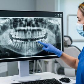
The evolution of dental imaging has fundamentally transformed the safety protocols surrounding third molar surgery. Historically, oral surgeons relied exclusively on two-dimensional panoramic radiographs (OPG) to assess the position of wisdom teeth. While a panoramic X-ray provides an excellent general overview of the dentition and the jaws, it suffers from inherent limitations, primarily the superimposition of three-dimensional anatomical structures onto a flat plane. This lack of depth perception can make it exceedingly difficult to determine the exact spatial relationship between the roots of a wisdom tooth and the inferior alveolar nerve canal.
Certain radiographic markers on a 2D panoramic image act as red flags, indicating a high probability of nerve proximity. These signs include the darkening of the tooth root where it crosses the canal, the interruption or loss of the white cortical lines defining the canal, the diversion or narrowing of the canal itself, and the sudden narrowing or deflection of the tooth root. When any of these signs are present, the standard of care dictates the utilization of advanced three-dimensional imaging to achieve accurate nerve damage dental prevention. This is where Cone Beam Computed Tomography (CBCT) becomes an indispensable diagnostic tool.
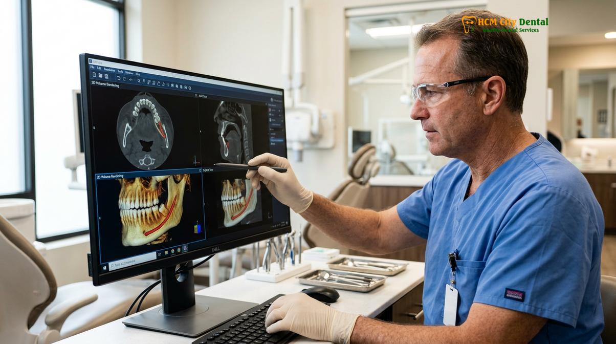
CBCT technology utilizes a cone-shaped X-ray beam that rotates around the patient’s head, capturing hundreds of high-resolution images in a matter of seconds. Advanced software then reconstructs these images into a highly detailed, manipulable 3D model of the maxillofacial region. Unlike conventional medical CT scans, CBCT delivers a significantly lower dose of radiation while providing superior spatial resolution for hard tissue structures. At facilities like HCMC Dental Clinic in Ho Chi Minh City, the integration of CBCT imaging into the preoperative workflow allows surgeons to visualize the exact trajectory of the mandibular canal in the sagittal, coronal, and axial planes[4].
The clinical value of a CBCT scan lies in its ability to eliminate anatomical guesswork. The surgeon can measure the precise distance in millimeters between the root apex and the superior border of the nerve canal. Furthermore, the scan reveals whether the nerve is positioned buccally (towards the cheek), lingually (towards the tongue), or directly inferior to the roots. This three-dimensional mapping dictates the surgical approach. For instance, if the CBCT reveals that the nerve is positioned on the buccal aspect of the roots, the surgeon will modify the bone removal and elevation techniques to direct forces away from the buccal plate, thereby safeguarding the nerve. By providing a clear, unambiguous roadmap of the surgical site, CBCT imaging drastically reduces the incidence of unexpected intraoperative complications and ensures a higher standard of patient safety.
Coronectomy: Partial Root Removal for High-Risk Teeth
When a wisdom tooth root is intimately entwined with the mandibular nerve canal, a coronectomy is performed to remove the crown while leaving the root intact, thereby eliminating the risk of nerve trauma.
Despite the precision afforded by 3D imaging, there are clinical scenarios where the anatomical relationship between the wisdom tooth and the inferior alveolar nerve is so intimate that a complete extraction poses an unacceptably high risk of permanent neurological damage. In cases where the roots are hooked around the nerve, or where the nerve canal actually passes through the roots of the tooth, the mechanical force required to deliver the roots would inevitably crush or sever the nerve fibers. In these specific, high-risk situations, an alternative surgical procedure known as a coronectomy is indicated.
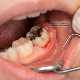
A coronectomy, also referred to as an intentional partial odontectomy, involves the surgical amputation and removal of the crown of the tooth, while deliberately leaving the vital root complex undisturbed within the alveolar bone. The rationale behind this procedure is straightforward: by eliminating the crown, the surgeon removes the source of the clinical problem—such as pericoronitis management, food impaction, or damage to adjacent molars—without disturbing the delicate neural structures located at the root apices. The coronectomy wisdom teeth protocol has gained widespread acceptance in the oral surgery community as a highly effective, conservative approach to managing high-risk impactions.
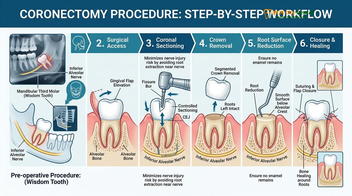
The surgical technique for a coronectomy requires meticulous precision. After administering profound local anesthesia, the surgeon raises a mucoperiosteal flap to expose the crown of the impacted tooth. Using a specialized surgical fissure bur, the tooth is sectioned horizontally, typically at a 45-degree angle, just below the cementoenamel junction (CEJ). The crown is then carefully elevated and removed. The critical next step involves reducing the remaining root structure so that it sits at least 3 to 4 millimeters below the crest of the surrounding alveolar bone. This deep reduction is essential to allow the bone to heal over the roots and prevent future exposure or infection. Finally, the surgical site is thoroughly irrigated, and the soft tissue flap is sutured closed to achieve primary healing.
Clinical Case Review: A 28-year-old patient presented to HCMC Dental Clinic with recurrent pain from a horizontally impacted lower right wisdom tooth. A 2D panoramic X-ray showed the roots overlapping the mandibular canal. A subsequent CBCT scan confirmed that the roots were grooved by the nerve, indicating an extreme risk of IAN injury upon extraction. Dr. Nguyen Van Cuong recommended a coronectomy. The crown was successfully sectioned and removed, leaving the roots intact. The patient experienced normal postoperative healing with zero numbness or altered sensation in the lower lip, validating the efficacy of this conservative surgical approach.
It is important to note that a coronectomy is not suitable for every patient. Contraindications include teeth with active root infections, mobile teeth, or teeth that are horizontally impacted in a way that makes sectioning the crown impossible without mobilizing the roots. Following a successful coronectomy, it is common for the retained roots to migrate slightly upward within the bone over the first few years. In the vast majority of cases, this migration is asymptomatic and ceases once the roots are fully encased in new bone. In the rare event that the roots migrate to the surface and become exposed, a secondary surgery may be required to remove them; however, by this time, the roots have typically moved far enough away from the nerve canal that the extraction can be performed safely without neurological risk.
Special Medical Considerations (Pregnancy, Diabetes & Blood Thinners)
Systemic health conditions require tailored extraction protocols to ensure patient safety, manage bleeding risks, and promote optimal wound healing without compromising nerve integrity.
The management of wisdom teeth extraction nerve risk cannot be viewed in isolation from the patient’s overall systemic health. Various medical conditions and pharmacological treatments can significantly influence the surgical approach, the choice of anesthesia, and the postoperative healing trajectory. A thorough preoperative medical history is a mandatory component of the clinical assessment, allowing the dental team to modify their protocols to accommodate specific physiological vulnerabilities.
Pregnancy introduces a unique set of clinical considerations. Hormonal fluctuations during pregnancy can exacerbate gingival inflammation, making conditions like pericoronitis more frequent and severe. When evaluating wisdom teeth removal during pregnancy, the timing of the intervention is critical. Elective surgical procedures are generally deferred until after delivery. However, if a patient experiences acute infection or severe pain that cannot be managed conservatively, extraction may be necessary. The second trimester is universally considered the safest period for dental surgery. During this window, the risk of fetal developmental issues is minimized, and the mother is generally more comfortable than in the third trimester, where supine hypotensive syndrome (a drop in blood pressure caused by the weight of the uterus compressing the inferior vena cava) becomes a concern. Obstetricians and dental surgeons collaborate closely to select local anesthetics, such as lidocaine, which have a proven safety profile, and to avoid systemic medications that could cross the placental barrier[5].
Patients with diabetes mellitus require meticulous perioperative management. Chronic hyperglycemia impairs leukocyte function, reducing the body’s ability to fight off bacterial pathogens, and compromises microvascular circulation, leading to delayed wound healing. A wisdom tooth extraction diabetes protocol begins with verifying the patient’s HbA1c levels to ensure their metabolic state is adequately controlled. Morning appointments are preferred, as endogenous cortisol levels are higher, providing better tolerance to surgical stress, and the risk of hypoglycemia is reduced following a normal breakfast and standard medication dosing. Prophylactic antibiotics may be prescribed to mitigate the heightened risk of postoperative socket infections, which can indirectly cause nerve irritation due to localized swelling and purulent exudate.
The management of patients on anticoagulant or antiplatelet therapy (blood thinners) focuses on balancing the risk of postoperative hemorrhage against the risk of a thromboembolic event if the medication is altered. Historically, patients were routinely advised to stop their blood thinners prior to dental surgery. However, current clinical guidelines strongly advocate for maintaining therapeutic anticoagulation for minor oral surgeries, including extractions. For a blood thinners tooth extraction, the surgeon employs robust local hemostatic measures rather than altering systemic medications. These measures include the use of absorbable gelatin sponges, oxidized regenerated cellulose, or advanced biological adjuncts like Platelet-Rich Fibrin (PRF) placed directly into the extraction socket. Meticulous suturing techniques are then utilized to achieve primary closure and stabilize the blood clot, ensuring a safe recovery without exposing the patient to cardiovascular risks.
When to See a Doctor
Prompt clinical evaluation is necessary if postoperative numbness persists beyond 48 hours, or if the patient experiences severe, unmanageable pain and signs of systemic infection.
Navigating the postoperative recovery phase requires vigilance and clear communication between the patient and the clinical team. While a certain degree of swelling, discomfort, and limited jaw opening (trismus) is expected following the surgical removal of impacted wisdom teeth, specific symptoms warrant immediate professional evaluation. The most critical indicator of a potential complication is the duration and nature of altered sensation in the maxillofacial region.
As previously established, profound numbness in the lower lip, chin, or tongue is a normal consequence of the local anesthetic administered during the procedure. This pharmacological block typically wears off within 4 to 8 hours, depending on the specific agent used and the patient’s individual metabolism. If the patient wakes up the morning after surgery and still experiences a complete lack of sensation, or a persistent “pins and needles” feeling in these areas, they should contact their oral surgeon. While this does not necessarily indicate permanent damage, early documentation and assessment are vital for establishing a baseline and determining if anti-inflammatory interventions are appropriate.
Beyond neurological concerns, patients must be alert to signs of acute infection or alveolar osteitis (dry socket). If the initial surgical pain begins to subside but is suddenly replaced by a severe, throbbing ache radiating up to the ear or down the neck on the third or fourth postoperative day, this is a classic presentation of a dry socket. Furthermore, the onset of a high fever, difficulty swallowing, or swelling that continues to increase aggressively after 72 hours are red flags for a spreading bacterial infection. In any of these scenarios, seeking prompt evaluation at a specialized facility like HCMC Dental Clinic ensures that complications are managed swiftly, preventing further tissue damage and supporting a safe, predictable return to oral health.
“Patient education does not end when the surgery is completed. Empowering patients to recognize the difference between normal healing physiology and the early warning signs of nerve irritation or infection is a critical component of comprehensive postoperative care.”
Frequently Asked Questions
Frequently Asked Questions
What are the chances of nerve damage during wisdom tooth extraction?
The overall risk of temporary nerve damage during lower wisdom tooth extraction ranges from 1% to 5%, while permanent injury occurs in less than 1% of cases. Advanced imaging and careful surgical planning significantly reduce these probabilities for high-risk impactions. The specific risk profile for an individual depends heavily on the anatomical position of the tooth roots relative to the mandibular canal, the patient’s age, and the density of the surrounding jawbone.
What does wisdom tooth nerve damage feel like?
Nerve damage typically feels like persistent numbness, tingling, or a burning sensation in the lower lip, chin, or tongue. It resembles the feeling of local anesthesia that has not worn off, sometimes accompanied by altered taste perception. Patients may also experience dysesthesia, where normal touch stimuli, such as brushing teeth or drinking cold water, are perceived as uncomfortable or mildly painful electrical shocks.
Is it safe to get a wisdom tooth extracted during pregnancy?
Yes, wisdom tooth extraction can be safely performed during pregnancy, ideally during the second trimester. Obstetricians and dental surgeons collaborate to select safe local anesthetics and avoid medications that could cross the placental barrier. While elective surgeries are generally postponed until after childbirth, acute infections or severe pain caused by an impacted wisdom tooth must be addressed promptly to protect the systemic health of both the mother and the developing fetus.
What precautions should diabetic patients take for dental extraction?
Diabetic patients must ensure their blood glucose levels are well-controlled prior to surgery to minimize infection risks and delayed healing. Prophylactic antibiotics and morning appointments are often recommended to maintain stable metabolic function during the procedure. Furthermore, patients should adhere to their normal dietary and medication routines on the day of surgery to prevent intraoperative hypoglycemic episodes, and closely monitor their surgical sites for any signs of delayed tissue closure.
Is a coronectomy safer than a full extraction?
For teeth where the roots are intimately entangled with the mandibular nerve canal, a coronectomy is considered safer than full extraction. By leaving the root tips undisturbed in the bone, the surgeon eliminates the risk of direct mechanical nerve trauma. This conservative approach effectively resolves the clinical symptoms associated with the impacted crown, such as infection or decay, while providing a highly predictable method for preserving the integrity of the inferior alveolar nerve.
References
- Journal of Oral and Maxillofacial Surgery. Inferior alveolar nerve injury in third molar surgery. (2021).
- International Journal of Oral and Maxillofacial Surgery. Coronectomy vs total extraction for high-risk teeth. (2019).
- British Dental Journal. Lingual nerve protection techniques during mandibular extractions. (2020).
- Journal of the American Dental Association. CBCT imaging in preoperative third molar assessment. (2022).
- Clinical Oral Investigations. Management of systemic conditions in oral surgery. (2018).