A mesioangular wisdom tooth is a third molar that erupts at a forward angle, tilting directly toward the adjacent second molar. This common anatomical anomaly creates a severe trap for food and bacteria, frequently leading to hidden decay, chronic gum inflammation, and irreversible damage to neighboring teeth if left untreated.
Clinical Summary:
Mesioangular impaction is the most prevalent type of wisdom tooth misalignment, characterized by a forward tilt that wedges the crown beneath the contour of the second molar. This positioning disrupts the natural eruption pathway and creates an un-cleanable periodontal pocket. Clinically, this leads to pericoronitis, distal cervical caries on the adjacent tooth, and localized alveolar bone loss. Treatment requires a precise surgical extraction protocol, often involving mucoperiosteal flap elevation and tooth sectioning, to safely remove the impacted molar while preserving the structural integrity of the jawbone and surrounding nerves. Proactive diagnostic imaging, particularly CBCT, is essential for mapping the root anatomy and planning a safe surgical approach.
Key Takeaways:
- A mesioangular impaction features a forward tilt, typically between 30 and 60 degrees, pressing against the second molar.
- This angulation creates a high-risk plaque trap, making the adjacent tooth highly susceptible to hidden, deep-root cavities.
- Chronic inflammation around the partially erupted crown frequently leads to painful pericoronitis and periodontal bone loss.
- Surgical removal involves sectioning the tooth to relieve mechanical pressure and protect the inferior alveolar nerve.
- Early extraction, guided by 3D imaging, prevents irreversible damage to the neighboring dentition and ensures smoother recovery.
What is Mesioangular Wisdom Tooth Impaction?
Mesioangular impaction is a condition where the third molar is angled forward, preventing full eruption and causing the tooth to collide with the back of the second molar.
The human jaw has undergone significant evolutionary changes, often resulting in inadequate arch space to accommodate the final set of molars. When a third molar attempts to erupt into a space that is too small, it frequently deviates from a vertical path. A mesioangular impaction occurs when the crown of the wisdom tooth tilts forward, toward the anterior (front) of the mouth. This specific angulation is statistically the most common type of mandibular (lower jaw) impaction encountered in clinical practice.
The severity of a mesioangular impaction is classified based on the depth of the tooth within the jawbone and its relationship to the ascending ramus of the mandible. In a soft tissue impaction, the crown has penetrated the bone but remains covered by a flap of dense gingival tissue known as an operculum. In a hard tissue (bony) impaction, the tooth remains partially or completely encased within the alveolar bone. The forward tilt, which can range from a mild 15-degree inclination to a severe 75-degree angle, dictates the complexity of the clinical presentation [1].
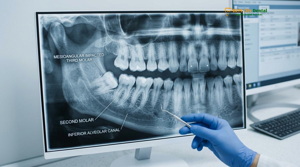
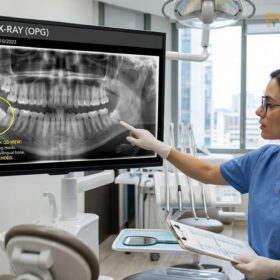
Because the crown is wedged beneath the protective contour of the adjacent tooth, normal eruption is mechanically impossible. The tooth remains in a state of arrested development, exerting continuous, low-grade pressure on the surrounding structures. Advanced diagnostic imaging, specifically Cone Beam Computed Tomography (CBCT), is utilized to assess the exact degree of the tilt, the morphology of the roots, and the proximity of the tooth to critical anatomical landmarks such as the inferior alveolar nerve canal.
The Clinical Mechanics of Forward-Tilted Wisdom Teeth
The forward-tilted wisdom tooth alters normal dental biomechanics by directing eruptive forces horizontally into the adjacent tooth rather than vertically into the oral cavity.
To understand the destructive potential of a forward tilted wisdom tooth, one must examine the biomechanics of tooth eruption. Normally, a tooth erupts vertically, guided by the periodontal ligament and the resorption of overlying bone. However, when the tooth is angled mesially (forward), the eruptive force vector is misdirected. Instead of pushing through the gum tissue, the crown of the third molar drives directly into the distal (back) surface of the second molar.
This mechanical collision creates a “wedge effect.” The hardest substance in the human body—dental enamel—is forced against the root surface of the adjacent tooth, which is covered by softer cementum. Over time, this constant pressure can lead to external root resorption, a pathological process where the body’s osteoclasts begin to break down the root structure of the healthy second molar [2]. Furthermore, the abnormal positioning disrupts the integrity of the dental arch, potentially contributing to late mandibular incisor crowding, although the exact correlation remains a subject of ongoing orthodontic research.
“The biomechanical stress exerted by a mesioangular impaction is not merely a static problem; it is an active, continuous force that compromises the periodontal attachment apparatus of the adjacent molar, necessitating proactive clinical intervention.”
The density of the surrounding mandibular bone also plays a crucial role. The external oblique ridge and the dense cortical bone of the lower jaw offer significant resistance. As the tooth attempts to move forward, it becomes locked between this dense bone and the unyielding second molar. This mechanical locking makes spontaneous correction impossible and sets the stage for a cascade of secondary dental pathologies.
Why Mesioangular Teeth Lead to Food Trapping & Cavities
The abnormal angle creates an inaccessible V-shaped crevice that traps food and bacteria, leading to aggressive decay on both the wisdom tooth and the adjacent molar.
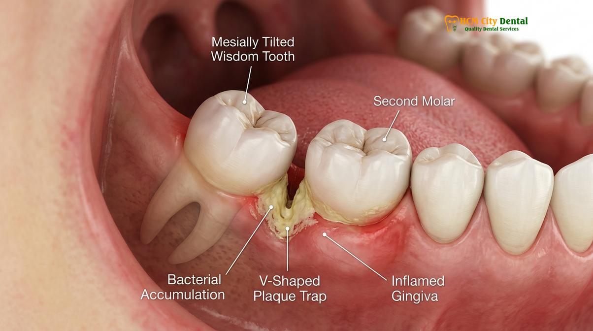
One of the most critical clinical concerns associated with a mesioangular impaction is its propensity to act as a massive plaque trap. Because the tooth is tilted forward, it creates a deep, V-shaped anatomical defect between its crown and the back of the second molar. This area is entirely inaccessible to standard oral hygiene tools. Toothbrush bristles cannot penetrate the tight contact point, and traditional dental floss cannot navigate the complex subgingival contours.
As a result, food debris and cariogenic (cavity-causing) bacteria, primarily Streptococcus mutans, accumulate rapidly in this stagnant environment. The bacteria metabolize fermentable carbohydrates from trapped food, producing lactic acid. Because this acid is shielded from the neutralizing effects of saliva, it rapidly demineralizes the enamel and dentin. This process leads to Distal Cervical Caries (DCC)—a highly destructive form of tooth decay that attacks the neck and root of the second molar [3].
Clinical Warning: Silent Decay
Decay caused by a mesioangular impaction often progresses without any pain until it reaches the dental pulp of the second molar. By the time a patient experiences a toothache, the adjacent tooth may require a complex root canal or, in severe cases, extraction alongside the wisdom tooth.
The decay associated with a mesioangular wisdom tooth is notoriously difficult to restore. Because the cavity forms deep below the gum line and often extends onto the root surface, achieving a dry, isolated field for a dental filling is highly challenging. In many cases, the only way to save the second molar is to first perform a mesioangular wisdom tooth extraction to gain access to the decayed area. This highlights the importance of prophylactic evaluation before irreversible damage occurs.
Risk of Periodontal Pockets on the Second Molar
Chronic bacterial accumulation around the tilted tooth destroys the supporting alveolar bone, creating deep periodontal pockets that threaten the stability of the second molar.
Beyond dental caries, the soft tissues and bone surrounding a mesioangular impaction second molar are at severe risk. The presence of a partially erupted crown creates a persistent communication between the oral cavity and the deeper periodontal structures. The gingival tissue overlying the tooth (the operculum) frequently becomes inflamed and infected, a condition known as pericoronitis. This acute inflammation is characterized by severe pain, swelling, purulent discharge (pus), and trismus (difficulty opening the mouth).
However, the chronic, low-grade inflammation is often more destructive in the long term. The persistent bacterial load triggers a localized immune response. Inflammatory cytokines, such as interleukins and tumor necrosis factor, stimulate osteoclast activity, leading to the resorption of the alveolar bone situated between the second and third molars [4]. As the bone recedes, a deep periodontal pocket forms.
| Clinical Parameter | Healthy Second Molar | Adjacent to Mesioangular Impaction |
|---|---|---|
| Probing Depth | 1.0 mm – 3.0 mm | 5.0 mm – 9.0+ mm |
| Bleeding on Probing | Absent | Severe (Profuse bleeding) |
| Bone Level | Intact alveolar crest | Vertical bone defect (crater) |
| Tissue Tone | Firm, stippled, pink | Edematous, erythematous, flaccid |
A deep periodontal pocket compromises the clinical attachment level of the second molar. Even if the wisdom tooth is eventually removed, the bone defect left behind may not fully regenerate, leaving the second molar permanently vulnerable to root sensitivity, mobility, and future periodontal disease. Therefore, timely extraction is not just about removing a problematic tooth; it is a critical periodontal preservation strategy for the adjacent dentition.
Surgical Steps for Extracting Mesioangular Teeth
Extraction requires a meticulous surgical approach, utilizing flap elevation, precise bone removal, and tooth sectioning to safely deliver the impacted molar without damaging surrounding structures.
The surgical management of a mesioangular wisdom tooth is a highly technical procedure that demands a thorough understanding of maxillofacial anatomy. Unlike a simple extraction where a tooth is elevated and pulled, an impacted third molar must be strategically dismantled and removed in sections to navigate the mechanical obstructions and protect the adjacent second molar and the inferior alveolar nerve.
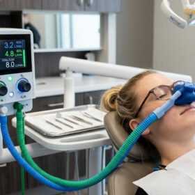
The procedure begins with the administration of profound local anesthesia, often supplemented with conscious sedation to ensure patient comfort. The surgeon then designs a mucoperiosteal flap—typically a triangular or envelope flap—to expose the underlying jawbone and the impacted crown. Once the surgical site is visualized, the surgeon must address the bone overlying the tooth. Using a specialized surgical handpiece or an ultrasonic Piezotome device, a precise trough (gutter) is created in the buccal (cheek side) and distal bone to relieve the hard tissue impaction [5].
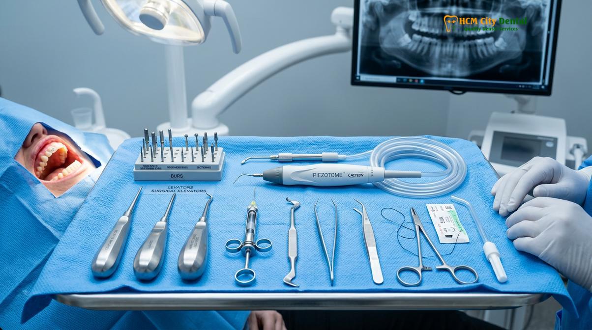
The most critical step in a mesioangular wisdom tooth extraction is odontectomy, or tooth sectioning. Because the crown is wedged under the second molar, attempting to elevate the tooth in one piece would exert catastrophic force on the adjacent tooth or risk fracturing the mandible. The surgeon uses a surgical bur to section the tooth, typically separating the crown from the roots, or splitting the tooth longitudinally. The distal portion of the crown is removed first, creating space to elevate the remaining roots upward and backward, following the natural curvature of the root anatomy.
At HCMC Dental Clinic in Ho Chi Minh City, our surgical teams utilize advanced 3D CBCT imaging to map the exact trajectory of the roots prior to surgery. This allows the surgeon to plan the precise angle of sectioning, minimizing trauma to the surrounding bone and significantly reducing the risk of nerve paresthesia. Once all fragments are meticulously delivered, the socket is thoroughly debrided, irrigated with sterile saline, and the mucosal flap is repositioned and secured with sutures to initiate primary healing.
Post-Surgical Healing and Gum Recovery
Optimal recovery depends on the stabilization of the blood clot, meticulous oral hygiene, and the biological regeneration of bone within the extraction socket.
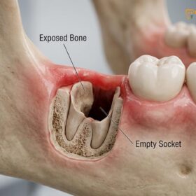
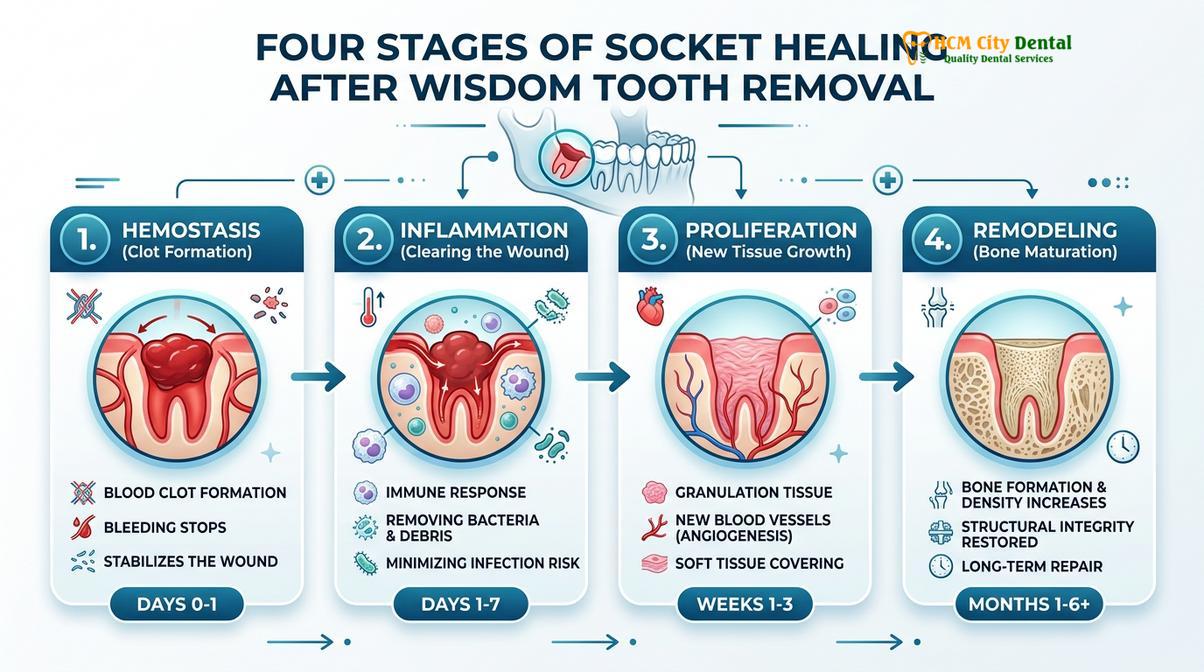
The healing process following the removal of a mesioangular impaction is a complex biological cascade. Immediately after the extraction, hemostasis occurs, and a stable blood clot forms within the socket. This clot is the foundational matrix for all subsequent healing; it protects the exposed bone and serves as a scaffold for the migration of fibroblasts and osteoblasts. Protecting this clot during the first 72 hours is paramount to preventing alveolar osteitis, commonly known as dry socket.
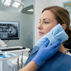
During the inflammatory phase, patients will experience localized swelling and mild to moderate discomfort, which peaks around the second or third day post-operation. This is a normal physiological response as the body increases blood flow to the area to deliver immune cells and growth factors. To enhance this healing phase, modern surgical protocols often incorporate the use of Platelet-Rich Fibrin (PRF). By drawing a small amount of the patient’s blood and centrifuging it, a concentrated membrane of growth factors is created and placed directly into the extraction socket, accelerating soft tissue closure and reducing post-operative pain.
Clinical Case Review: Preserving the Second Molar
A 24-year-old patient presented to HCMC Dental Clinic with severe localized pain and a deep periodontal pocket behind the lower right second molar. CBCT imaging revealed a deeply impacted mesioangular wisdom tooth causing a 7mm bone defect. Dr. Nguyen Van Cuong performed a minimally invasive surgical extraction utilizing tooth sectioning and placed a PRF membrane in the distal defect. At the 6-month follow-up, periodontal probing depths had reduced to a healthy 3mm, demonstrating successful bone regeneration and the preservation of the adjacent second molar.
As the weeks progress, the proliferation and remodeling phases take over. The blood clot is gradually replaced by granulation tissue, which eventually mineralizes into woven bone. The complete regeneration of the alveolar bone, particularly the distal defect behind the second molar, can take several months. Patients are instructed on specific oral hygiene practices, including the use of a curved irrigation syringe to gently flush the socket after meals, ensuring that food debris does not disrupt the maturing tissue.
When to See a Doctor
While some impacted wisdom teeth remain asymptomatic for years, a mesioangular impaction carries a high statistical probability of developing clinical complications. It is crucial to seek professional dental evaluation if you experience specific red flags. Persistent, throbbing pain in the back of the jaw, localized swelling of the gums, or a recurring bad taste and odor in the mouth are strong indicators of active infection or decay.
Additionally, if you experience difficulty opening your mouth fully (trismus) or notice swelling extending to the cheek or lymph nodes in the neck, immediate clinical intervention is required to manage the acute infection. Do not wait for severe pain to develop. Proactive evaluation using panoramic or 3D imaging can identify a problematic forward-tilted tooth long before it causes irreversible damage to the adjacent dentition.
“Early diagnostic imaging is the cornerstone of preventive oral surgery. Identifying a mesioangular impaction before it compromises the second molar allows us to perform a safer, more predictable extraction with a significantly smoother recovery profile.” — Dr. Nguyen Van Cuong
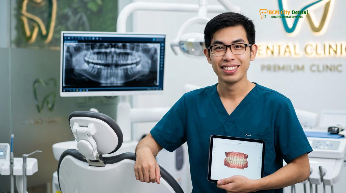
For a comprehensive evaluation of your wisdom teeth and to discuss personalized surgical options, we recommend scheduling a consultation. Advanced diagnostic tools and expert surgical care are essential for managing complex impactions safely. Learn more about our specialized protocols for Tooth Extraction to ensure your long-term oral health is protected.
Frequently Asked Questions
What is mesioangular wisdom tooth impaction?
A mesioangular wisdom tooth impaction occurs when the third molar is tilted forward toward the front of the mouth. This abnormal angulation causes the tooth to wedge against the adjacent second molar, preventing it from erupting normally and creating a high-risk zone for bacterial accumulation, decay, and localized gum infections. Because the tooth cannot erupt vertically, it remains trapped in the jawbone or soft tissue, requiring surgical intervention to resolve the mechanical obstruction.
Is mesioangular extraction easier than horizontal extraction?
Generally, a mesioangular extraction is considered slightly less complex than a fully horizontal impaction, but it still requires careful surgical intervention. The difficulty depends heavily on the depth of the impaction, the root anatomy, and the proximity to the inferior alveolar nerve, which must be evaluated via a 3D CBCT scan. While the angle may be slightly more favorable for elevation than a tooth lying completely flat, the need for bone removal and tooth sectioning remains a critical part of the surgical workflow.
Can a mesioangular wisdom tooth cause cavities on the next tooth?
Yes, a forward-tilted wisdom tooth is a primary cause of distal cervical caries on the adjacent second molar. The V-shaped gap between the two teeth traps food and plaque that cannot be reached by a toothbrush or floss, leading to rapid and often silent decay on the neighboring tooth. Because this decay occurs below the gum line and near the root, it is highly destructive and can lead to the loss of the second molar if the wisdom tooth is not removed promptly.
How do you clean a forward-tilted wisdom tooth?
Cleaning a forward-tilted wisdom tooth requires specialized tools such as an end-tuft brush, a water flosser, and antimicrobial mouthwashes. While these tools can help flush out superficial debris from the gum pocket, they cannot completely remove plaque trapped deep beneath the contact point, which is why extraction is usually recommended. Maintaining rigorous hygiene can temporarily manage inflammation, but it does not solve the underlying anatomical defect caused by the impaction.
What is the cost of mesioangular wisdom tooth extraction in HCMC?
The cost of mesioangular wisdom tooth extraction in HCMC varies based on the surgical complexity, the type of anesthesia used, and the specific clinical protocols required. Currently, patients can expect competitive pricing that reflects advanced surgical techniques, 3D imaging, and comprehensive post-operative care at specialized dental centers. During your initial consultation at HCMC Dental Clinic, a detailed treatment plan and transparent fee schedule will be provided based on your specific diagnostic imaging.
References
- Journal of Oral and Maxillofacial Surgery. Biomechanics of third molar impaction and eruption pathways. (2021).
- International Journal of Oral and Maxillofacial Surgery. Distal cervical caries in second molars associated with mesioangular impactions. (2020).
- Journal of Periodontology. Periodontal attachment loss adjacent to impacted third molars. (2019).
- British Dental Journal. Surgical sectioning techniques for complex mandibular impactions. (2022).
- Clinical Oral Investigations. Alveolar bone regeneration following third molar extraction. (2023).