A horizontal wisdom tooth is a third molar that grows sideways at a 90-degree angle, remaining fully or partially trapped beneath the gums. This severe impaction pushes directly into the adjacent second molar, causing intense pain, root damage, and high infection risk, requiring prompt surgical extraction.
Clinical Summary:
Horizontal impaction is the most complex form of wisdom tooth development, characterized by a 90-degree angulation parallel to the jawbone. Because the tooth cannot erupt vertically, it exerts continuous mechanical pressure on the adjacent second molar, leading to root resorption, pericoronitis, and potential cyst formation. Surgical removal by an experienced oral surgeon is the definitive treatment. This procedure often involves precise bone removal and tooth sectioning to safely extract the molar without damaging the inferior alveolar nerve or surrounding bone architecture.
Key Takeaways:
- Horizontal impaction occurs when the third molar develops entirely sideways, parallel to the jawbone.
- It is widely considered the most painful and structurally damaging type of wisdom tooth impaction.
- Continuous eruptive pressure can destroy the roots of the healthy adjacent second molar.
- Surgical extraction requires sectioning the tooth into smaller pieces to protect surrounding nerves.
- Early diagnostic imaging and proactive removal prevent irreversible damage and complex orthodontic shifting.
What is Horizontal Wisdom Tooth Impaction?
Horizontal wisdom tooth impaction happens when the third molar develops entirely sideways, lying parallel to the jawbone rather than emerging vertically. This structural anomaly prevents normal eruption and traps the tooth deep within the alveolar bone.
The human jaw has undergone significant evolutionary changes, gradually becoming smaller over thousands of years. As a result, the third molars—commonly known as wisdom teeth—often lack the necessary arch space to erupt properly into the oral cavity. When a tooth is completely blocked from entering the mouth, it is classified as impacted. Among the various classifications of impaction, the horizontal alignment is the most extreme and clinically challenging.
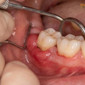
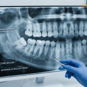
In a horizontal impaction, the tooth’s long axis is perpendicular to the adjacent second molar. Instead of pointing upward toward the chewing surface, the crown of the sideways wisdom tooth points directly forward, burying itself into the roots of the neighboring tooth. This condition is most frequently observed in the mandibular (lower) jaw, where the bone is exceptionally dense and the anatomical space is highly restricted [1].
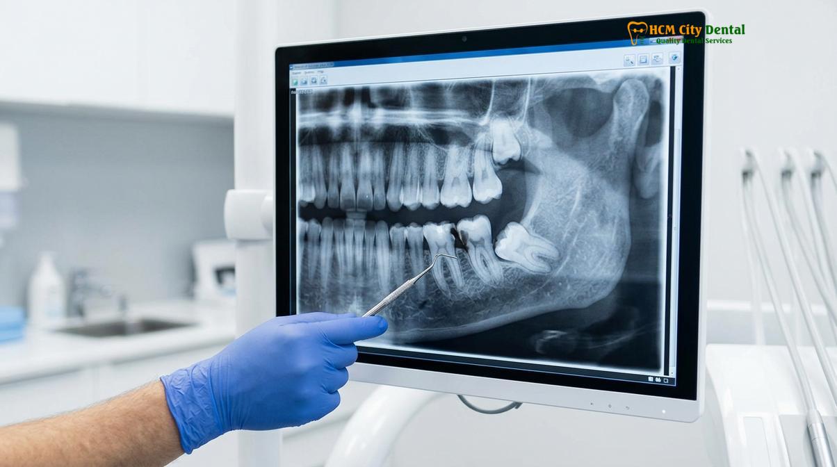
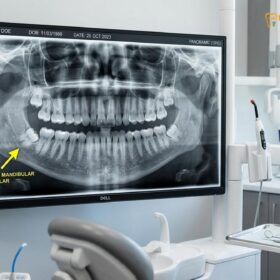
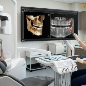
According to clinical diagnostics, these teeth can be either partially bony impacted (where a small portion of the crown breaches the bone but remains covered by gum tissue) or completely bony impacted (where the entire tooth is encased in the jawbone). Because they cannot be seen during a standard visual examination, advanced radiographic imaging, such as a panoramic X-ray or a 3D Cone Beam Computed Tomography (CBCT) scan, is mandatory to accurately diagnose the exact angulation and depth of the tooth.
Dr. Nguyen Van Cuong, a leading specialist in oral surgery, frequently notes that patients are often unaware they possess a horizontal impaction until secondary symptoms arise. “Because the tooth is buried deep within the bone, it can silently cause structural damage for years before the patient feels the first sign of pain,” Dr. Cuong explains. This underscores the critical importance of routine dental imaging during late adolescence to monitor third molar development before complications occur.
For a comprehensive overview of how different types of impactions are managed, you can explore our detailed guide on Tooth Extraction, which outlines the clinical protocols for safely addressing these complex dental anomalies.
Why Horizontal Wisdom Teeth are a High-Risk Condition
A sideways wisdom tooth is considered high-risk because it remains buried under the gumline, creating a breeding ground for severe bacterial infections and structural jaw damage. It rarely resolves without surgical intervention.
The presence of a horizontally impacted tooth introduces a multitude of severe risks to the oral environment. Unlike vertical impactions that might eventually find their way through the gums, a horizontal tooth is physically incapable of erupting. As the tooth attempts to push forward, it creates a chronic inflammatory response within the surrounding soft tissues and bone.
One of the most common and painful complications is pericoronitis. This occurs when the tooth partially breaches the bone but remains covered by a flap of gum tissue known as an operculum. This flap creates a deep, inaccessible pocket where food debris, plaque, and aggressive anaerobic bacteria accumulate. Because the patient cannot clean beneath this tissue, the area becomes highly susceptible to acute infection, leading to severe swelling, purulent discharge (pus), and intense pain that can radiate to the ear and neck [2].
Clinical Warning: Ignoring an asymptomatic horizontal wisdom tooth is highly discouraged. Even if you do not currently feel pain, the tooth can silently develop dentigerous cysts or cause irreversible root resorption to the adjacent molar, leading to the loss of multiple teeth.
Furthermore, the developmental sac that surrounds the wisdom tooth during its formation can fill with fluid, forming a dentigerous cyst. If left untreated, this cyst can slowly expand, hollowing out the jawbone and compromising its structural integrity. In rare but documented cases, the cellular lining of these cysts can mutate into benign but aggressive tumors, such as ameloblastomas, which require extensive maxillofacial surgery to remove.
The proximity of the lower horizontal wisdom teeth to the inferior alveolar nerve is another major risk factor. This nerve supplies sensation to the lower lip, chin, and teeth. As the roots of the wisdom tooth grow, they can wrap around or press against this nerve canal. Any inflammation or infection in this area can cause nerve compression, resulting in paresthesia (numbness or tingling) in the lower face. Therefore, proactive horizontal wisdom teeth removal is often the most prudent clinical recommendation to prevent these cascading complications.
How Horizontal Teeth Damage Adjacent Second Molars
The horizontal growth trajectory forces the wisdom tooth crown directly into the root of the second molar. This continuous mechanical pressure causes irreversible root resorption, severe decay, and eventual loss of the healthy adjacent tooth.
The second molar is a crucial tooth for mastication (chewing) and maintaining the stability of the dental arch. Unfortunately, it is the primary victim of a wisdom tooth pushing second molar. The biomechanics of tooth eruption generate significant force. When a wisdom tooth is aligned horizontally, all of this eruptive force is directed laterally into the distal (back) surface of the second molar.
This continuous pressure triggers a biological process known as osteoclastic activity. Osteoclasts are cells that break down bone and tooth structure. As the wisdom tooth presses against the second molar, it causes the roots of the second molar to dissolve—a condition called external root resorption. This damage is permanent; once the root structure is lost, it cannot regenerate, often leading to the necessary extraction of the otherwise healthy second molar [3].
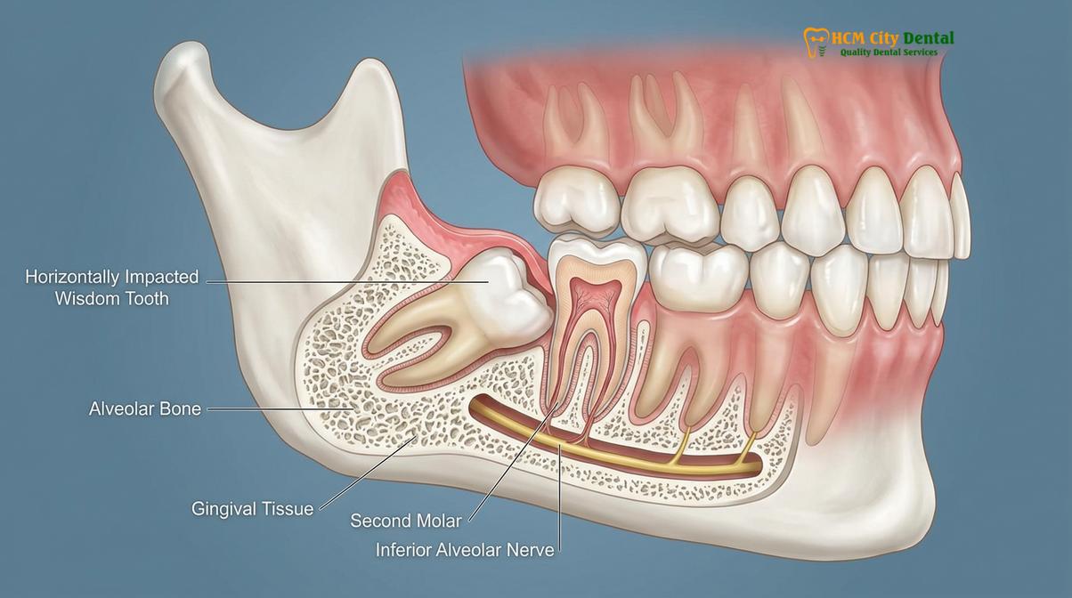
“The horizontal impaction acts as a slow-moving battering ram against the second molar. By the time the patient experiences pain from root resorption, the damage to the adjacent tooth is often irreversible, highlighting the need for early radiographic screening.”
In addition to root resorption, the physical contact point between the two teeth creates a highly vulnerable area for dental caries (tooth decay). Because the wisdom tooth is lodged against the back of the second molar below the gumline, it creates a “plaque trap.” It is anatomically impossible for a toothbrush or dental floss to reach this contact point. Consequently, aggressive decay can rapidly develop on the root surface of the second molar. Root caries are notoriously difficult to treat because they occur below the bone level, often rendering the tooth unrestorable.
Furthermore, the pressure exerted by the impacted tooth can contribute to late anterior crowding. While the exact degree to which wisdom teeth cause front teeth to shift is debated in orthodontics, the localized pressure undoubtedly disrupts the alignment of the posterior teeth, potentially undoing years of expensive orthodontic treatment.
Surgical Extraction Process for Sideways Teeth
Removing a horizontally impacted tooth requires a precise surgical approach involving gum incision, bone removal, and tooth sectioning. This minimally invasive technique ensures the tooth is extracted safely without traumatizing the jawbone or nearby nerves.
The extraction of a horizontal wisdom tooth is not a simple “pulling” procedure; it is a meticulous oral surgery. Because the tooth is locked beneath the bone and behind the second molar, the surgeon must create a pathway for its removal. The primary goal of horizontal impaction surgery is to extract the tooth while preserving the maximum amount of surrounding bone and protecting the inferior alveolar nerve and lingual nerve.
The procedure begins with profound anesthesia. Depending on the patient’s anxiety level and the complexity of the impaction, this may involve local anesthesia with nerve blocks, intravenous (IV) sedation, or general anesthesia. Once the surgical site is completely numb, the surgeon raises a mucoperiosteal flap—gently peeling back the gum tissue to expose the underlying jawbone.
Next, the surgeon must perform an ostectomy (bone removal). Using a specialized surgical handpiece or an advanced ultrasonic Piezotome device, the surgeon carefully removes the dense bone covering the crown of the wisdom tooth. The Piezotome is particularly advantageous because it cuts hard tissue (bone) using ultrasonic micro-vibrations while leaving soft tissue (nerves and blood vessels) completely unharmed [4].
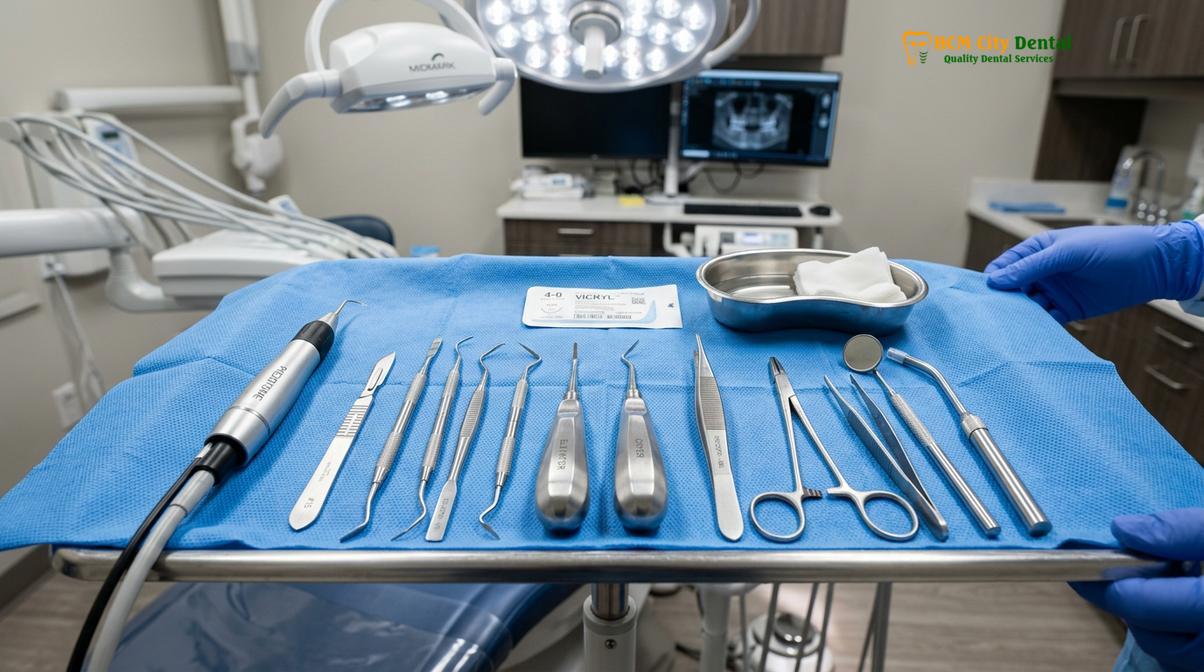
Once the crown is exposed, the most critical step occurs: odontotomy, or tooth sectioning. Attempting to elevate a horizontal tooth in one piece would require massive bone removal and risk fracturing the jaw. Instead, the surgeon uses a high-speed surgical bur to divide the tooth into smaller, manageable segments. Typically, the crown is severed from the roots and removed first. Then, the roots are carefully elevated and extracted individually.
| Surgical Phase | Clinical Action | Primary Objective |
|---|---|---|
| 1. Diagnostic Imaging | 3D CBCT Scan analysis | Map nerve proximity and exact tooth angulation. |
| 2. Anesthesia | Inferior Alveolar Nerve Block / Sedation | Ensure profound pain control and patient comfort. |
| 3. Flap Elevation | Incision and reflection of gingival tissue | Expose the surgical site and underlying alveolar bone. |
| 4. Ostectomy | Removal of overlying bone (Piezotome/Bur) | Create an access pathway to the impacted crown. |
| 5. Odontotomy | Sectioning the tooth into multiple pieces | Allow removal without excessive force or jaw trauma. |
| 6. Socket Debridement | Curettage and irrigation of the extraction site | Remove follicle remnants and prevent cyst formation. |
In cases where the roots of the horizontal tooth are intimately entwined with the inferior alveolar nerve, a specialized technique called a coronectomy may be considered. This involves removing only the crown of the tooth and intentionally leaving the roots in place to avoid nerve injury. For more details on this nerve-sparing approach, read our clinical guide on Tooth Extraction techniques for high-risk cases.
Recovery and Bone Healing After Horizontal Extraction
Recovery from horizontal extraction typically takes one to two weeks, with the underlying jawbone requiring several months to fully regenerate. Proper post-operative care is crucial to prevent dry socket and ensure smooth tissue healing.
The recovery phase following the removal of a horizontal wisdom tooth is more intensive than a standard extraction due to the surgical trauma inflicted on the bone and soft tissues. Understanding the biological healing timeline is essential for managing expectations and preventing post-operative complications.
Immediately after surgery, the primary objective is hemostasis (stopping the bleeding). A blood clot must form within the empty socket. This clot serves as a biological bandage, protecting the exposed bone and nerve endings. Patients are instructed to bite down firmly on sterile gauze for the first hour. During the first 48 to 72 hours, swelling and localized discomfort will peak. This is a normal inflammatory response as the body rushes white blood cells and healing factors to the surgical site.
Clinical Case Review: A 24-year-old patient visited HCMC Dental Clinic in Ho Chi Minh City presenting with severe jaw pain. Dr. Nguyen Van Cuong diagnosed a deeply impacted horizontal wisdom tooth causing acute pericoronitis. Following a precise surgical extraction utilizing 3D CBCT mapping and PRF (Platelet-Rich Fibrin) therapy, the patient experienced minimal post-operative swelling and returned to normal dietary habits within 6 days, demonstrating the efficacy of advanced surgical protocols.
To optimize healing, many modern clinics utilize PRF (Platelet-Rich Fibrin). A small sample of the patient’s blood is spun in a centrifuge to isolate growth factors, which are then placed directly into the extraction socket. PRF significantly accelerates soft tissue healing and drastically reduces the risk of alveolar osteitis, commonly known as dry socket [5].
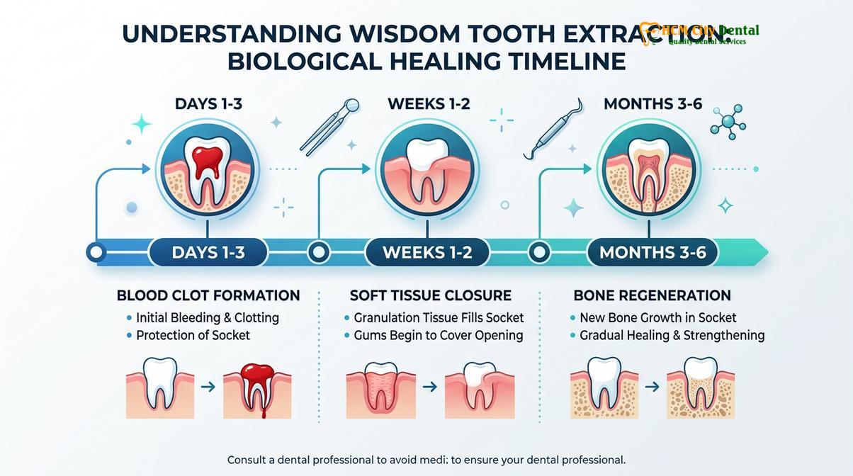
By the end of the first week, the soft gum tissue will begin to close over the extraction site, and the sutures will either dissolve or be removed by the dentist. However, the underlying bone healing is a much longer process. It takes approximately three to six months for osteoblasts (bone-forming cells) to completely fill the surgical void with dense, new alveolar bone. During this period, patients must maintain excellent oral hygiene to prevent secondary infections. If you experience worsening pain or foul-tasting discharge after the first few days, it is critical to recognize the signs of complications. Learn more about identifying these issues in our guide on Tooth Extraction infections.
“The success of a complex wisdom tooth surgery relies 50% on the surgeon’s skill and 50% on the patient’s strict adherence to post-operative care instructions. Protecting the initial blood clot is the absolute foundation of a smooth recovery.”
For a detailed day-by-day breakdown of what to expect during the healing process, please refer to our comprehensive timeline on how to Recover After Wisdom Tooth Extraction.
Cost of Horizontal Wisdom Tooth Removal in Vietnam
The cost of removing a horizontal wisdom tooth in Vietnam is highly competitive compared to Western countries, offering premium surgical care, advanced 3D imaging, and expert oral surgeons at a fraction of the price.
When facing the prospect of complex oral surgery, financial considerations are a significant factor for many patients. The cost of extracting a horizontally impacted wisdom tooth is inherently higher than a simple extraction due to the surgical expertise required, the use of advanced imaging (CBCT), and the necessary anesthesia protocols.
However, Vietnam has emerged as a premier destination for high-quality dental tourism. Clinics in major urban centers like Ho Chi Minh City offer world-class surgical environments equipped with the latest European and American dental technologies. The cost of horizontal impaction surgery in Vietnam is often 60% to 70% less than the equivalent procedure in the United States, Australia, or the United Kingdom, without any compromise in clinical safety or sterilization standards.
The total fee for the procedure generally encompasses the pre-operative 3D CBCT scan, the surgical extraction itself, the type of anesthesia administered (local vs. IV sedation), and post-operative medications. At HCMC Dental Clinic, we pride ourselves on transparent pricing and comprehensive care plans tailored to each patient’s specific anatomical needs. For a detailed breakdown of pricing structures and how they compare regionally, review our analysis on Tooth Extraction costs across Southeast Asia.
When to See a Doctor
Because horizontal wisdom teeth are entirely hidden beneath the gums, relying solely on visual inspection at home is impossible. It is highly recommended that all young adults undergo a comprehensive radiographic evaluation between the ages of 17 and 21 to assess the trajectory of their third molars. Early detection allows for proactive surgical planning before the roots fully develop and anchor themselves deep into the jawbone.
You should seek immediate clinical evaluation if you experience any of the following acute symptoms: severe, throbbing pain in the back of the jaw; sudden swelling of the cheek or submandibular lymph nodes; difficulty opening your mouth fully (trismus); a persistent bad taste or foul odor originating from the back of the mouth; or unexplained numbness in your lower lip or chin. These are classic signs of active infection, cyst expansion, or nerve compression requiring urgent surgical intervention.
Furthermore, if you are planning extended travel or relocating to an area with limited access to specialized dental care, addressing impacted wisdom teeth beforehand is a crucial preventive measure. Changes in cabin pressure during flights can exacerbate the pain of an infected impacted tooth. For more information on travel considerations post-surgery, consult our guidelines on Tooth Extraction and flight safety.
Frequently Asked Questions
Is horizontal wisdom tooth removal difficult?
Yes, it is considered one of the most complex dental extractions. Because the tooth lies flat and is often encased in jawbone, it requires a specialized surgical approach, including precise bone removal and tooth sectioning, to safely extract without damaging surrounding structures. The procedure demands the expertise of a skilled oral surgeon utilizing advanced instruments like a Piezotome to ensure a minimally invasive and safe outcome.
Does a horizontal wisdom tooth always need to be removed?
In almost all clinical cases, yes. Even if it is currently asymptomatic, a horizontal wisdom tooth will eventually cause root damage to the adjacent molar, cyst formation, or severe bacterial infection, making proactive surgical removal highly recommended by oral surgeons. Waiting until pain develops often means that irreversible damage has already occurred to the neighboring healthy teeth or jawbone.
Can a horizontal wisdom tooth damage my other teeth?
Absolutely. The sideways growth exerts continuous mechanical pressure on the adjacent second molar, leading to irreversible root resorption, severe decay in hard-to-clean areas, and potential shifting or crowding of your entire dental arch. The physical contact point below the gumline creates a trap for bacteria, making the second molar highly susceptible to deep root cavities that are difficult or impossible to restore.
How long does recovery take after horizontal wisdom tooth surgery?
Initial soft tissue recovery takes about 7 to 14 days, during which swelling and discomfort gradually subside. However, the underlying jawbone will take three to six months to completely fill in and remodel into dense, healthy bone tissue. Adhering strictly to post-operative guidelines, such as maintaining a soft diet and avoiding smoking, is critical to ensuring the blood clot remains stable during the first crucial week.
What is the cost of removing a horizontal wisdom tooth in Saigon?
The cost in Saigon typically ranges based on the surgical complexity, anesthesia type, and the clinic’s technology. It remains significantly more affordable than in Western countries while maintaining world-class surgical standards and utilizing advanced 3D imaging. Patients traveling to Vietnam for dental care can expect premium surgical environments and expert care at a fraction of the international cost.
References
- Journal of Oral and Maxillofacial Surgery. Recovery after third molar extraction. (2021).
- International Journal of Oral and Maxillofacial Surgery. Piezotome vs rotary drill in wisdom teeth removal. (2019).
- British Dental Journal. Dry socket incidence and prevention with PRF. (2020).
- Journal of the American Dental Association. Mandibular third molar impaction classifications. (2022).
- Clinical Oral Investigations. Lingual nerve protection during surgical extraction. (2018).