A wisdom tooth cyst jaw complication occurs when a fluid-filled sac forms around the crown of an unerupted third molar. This condition, known clinically as a dentigerous cyst, can silently destroy surrounding bone and displace adjacent teeth, requiring prompt surgical evaluation and removal to prevent severe structural damage.
Clinical Summary:
A dentigerous cyst associated with an impacted wisdom tooth is a benign but locally destructive odontogenic lesion. It originates from the accumulation of fluid between the reduced enamel epithelium and the tooth crown. Because these cysts expand slowly via hydrostatic pressure, they are typically asymptomatic in their early stages and are most often discovered incidentally on routine panoramic radiographs or Cone Beam Computed Tomography (CBCT) scans. Left untreated, the continuous expansion leads to significant mandibular bone resorption, displacement of adjacent second molars, and an increased risk of pathological jaw fractures or inferior alveolar nerve compression. The definitive standard of care involves surgical extraction of the impacted tooth combined with complete enucleation of the cystic lining. In cases of extensive bone loss, guided bone regeneration using particulate grafts and Platelet-Rich Fibrin (PRF) is employed to restore structural integrity and promote optimal osseous healing.
Key Takeaways:
- A wisdom tooth cyst develops from the dental follicle surrounding an unerupted or impacted third molar.
- These cysts grow silently, causing painless but progressive destruction of the surrounding jawbone.
- Advanced 3D CBCT imaging is essential for accurate diagnosis and surgical planning.
- Untreated cysts can lead to severe complications, including pathological jaw fractures and nerve damage.
- Treatment requires complete surgical enucleation of the cyst and extraction of the associated tooth.
- Bone grafting and PRF therapy are often utilized to regenerate lost bone and accelerate recovery.
What is a Wisdom Tooth Cyst (Dentigerous Cyst)?
A dentigerous cyst is a benign, fluid-filled sac originating from the dental follicle that surrounds the crown of an impacted or unerupted wisdom tooth.
To understand the formation of a wisdom tooth cyst jaw complication, it is essential to examine the embryological development of human teeth. Before a tooth erupts into the oral cavity, its developing crown is encased in a protective soft tissue sac known as the dental follicle. This follicle is lined by a layer of cells called the reduced enamel epithelium. In normal physiological development, as the tooth erupts through the jawbone and gums, this follicle merges with the oral mucosa. However, when a wisdom tooth becomes impacted—meaning it is blocked from erupting due to a lack of space, abnormal angulation, or dense overlying bone—the dental follicle remains trapped within the jawbone[1].
A dentigerous cyst wisdom tooth complication arises when an alteration in the local tissue environment causes fluid to accumulate between the reduced enamel epithelium and the enamel surface of the tooth crown. This fluid accumulation is driven by osmotic pressure. As cellular debris and proteins break down within the follicular space, the hyperosmolar environment draws in surrounding tissue fluid, causing the follicle to balloon outward. Over time, this transforms a normal, thin follicular space into a pathological cystic cavity.
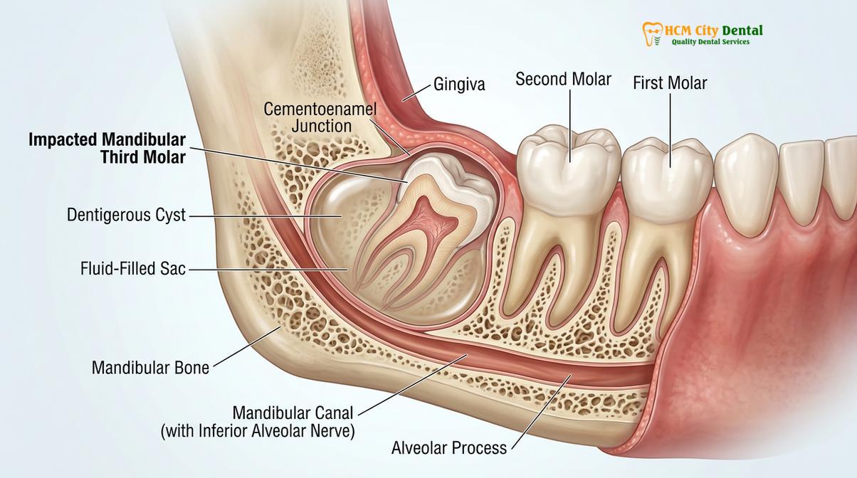
Histologically, the wall of a dentigerous cyst is composed of fibrous connective tissue, often containing a mild chronic inflammatory cell infiltrate, while the inner lumen is lined by non-keratinizing stratified squamous epithelium. According to Dr. Nguyen Van Cuong, a leading specialist in oral and maxillofacial surgery, distinguishing between a normal, slightly enlarged dental follicle and a true dentigerous cyst is a critical diagnostic step. A follicular space measuring less than 3 to 4 millimeters on a radiograph is generally considered normal hyperplastic tissue. However, when the pericoronal radiolucency exceeds 5 millimeters, a clinical diagnosis of a dentigerous cyst is highly suspected and warrants surgical intervention.
While these cysts are classified as benign (non-cancerous), their behavior is locally aggressive. They do not metastasize to other parts of the body, but their continuous, relentless expansion within the confined space of the mandible or maxilla can cause profound localized damage. The cyst acts as a space-occupying lesion, exerting outward pressure on all surrounding structures. Because the lower jaw (mandible) is composed of dense cortical bone on the outside and softer cancellous bone on the inside, the cyst typically expands along the path of least resistance—hollowing out the internal structure of the jawbone while leaving the outer contours seemingly intact until the late stages of the disease.
Why Cysts around Wisdom Teeth are Silently Destructive
These cysts expand slowly through hydrostatic pressure, causing painless but progressive resorption of the surrounding jawbone and adjacent tooth roots.
The most dangerous characteristic of a cyst around wisdom tooth structures is its silent progression. Unlike an acute dental abscess, which presents with rapid swelling, severe throbbing pain, and systemic signs of infection like fever, a dentigerous cyst grows insidiously. Patients can harbor a growing cyst for years, or even decades, without experiencing a single symptom. This asymptomatic nature is due to the slow rate of expansion, which allows the surrounding nerves and tissues to adapt to the gradual displacement without triggering acute pain signals.
The mechanism of wisdom tooth jaw bone loss associated with these cysts is deeply rooted in cellular biology. The expansion of the cyst is not merely a mechanical pushing of bone; it is an active biochemical process. The epithelial lining of the cyst and the surrounding fibrous capsule secrete various inflammatory mediators, including prostaglandins (specifically PGE2) and interleukins (IL-1 and IL-6)[2]. These biochemical signals stimulate osteoclasts—the specialized cells responsible for breaking down bone tissue. As the osteoclasts resorb the bone matrix, the cyst fluid volume increases, perpetuating a vicious cycle of expansion and bone destruction.
“The silent nature of odontogenic cysts is their most destructive feature. By the time a patient notices facial asymmetry or experiences pain, the cyst has often already compromised a significant volume of the mandibular bone, transforming a routine extraction into a complex reconstructive surgery.”
As the cyst expands anteriorly (forward), it encounters the roots of the adjacent second molar. The continuous pressure can cause root resorption, where the roots of the healthy second molar are slowly dissolved away. This can lead to tooth mobility, vitality loss, and eventually the need for root canal therapy or extraction of the second molar. Furthermore, the cyst can displace the impacted wisdom tooth itself, pushing it deep into the ramus of the mandible or dangerously close to the inferior border of the jaw.
In the upper jaw (maxilla), the destruction takes a different path. A dentigerous cyst associated with an upper wisdom tooth can expand upward and obliterate the maxillary sinus cavity. This can lead to chronic sinus issues, nasal obstruction, and an increased risk of oroantral communication (a hole between the mouth and the sinus) during surgical removal. The silent destruction underscores the critical importance of routine radiographic screening, as early detection is the only way to halt the bone loss before it causes irreversible anatomical damage.
Symptoms and How Cysts are Discovered on X-rays
Most cysts remain asymptomatic and are discovered incidentally on routine panoramic radiographs or 3D CBCT scans during standard dental check-ups.
Because early-stage dentigerous cysts rarely produce clinical symptoms, their discovery is almost always radiographic. When symptoms do occur, they usually indicate that the cyst has reached a massive size or has become secondarily infected. Clinical signs of an advanced or infected cyst may include a dull, aching pain in the posterior jaw, localized swelling of the gums behind the last molar, a bad taste or odor in the mouth (if the cyst has ruptured and is draining pus), and noticeable facial asymmetry or a hard, bony expansion on the side of the jaw.
In some cases, patients may experience a phenomenon known as “egg-shell crackling” (crepitus) upon palpation. This occurs when the cyst has expanded so much that it has thinned the outer cortical bone of the jaw to the thickness of an eggshell. Pressing on the swollen area produces a distinct crackling sensation as the thin bone flexes over the fluid-filled cavity. However, waiting for such clinical signs to appear is highly discouraged, which is why modern dentistry relies heavily on advanced imaging.
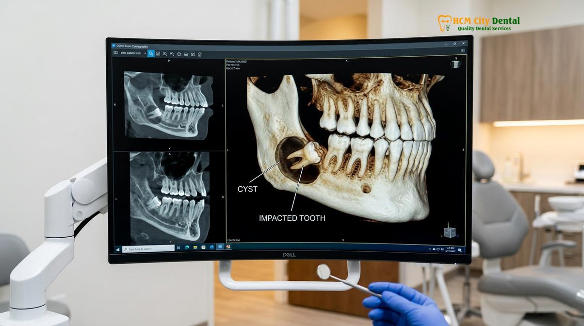
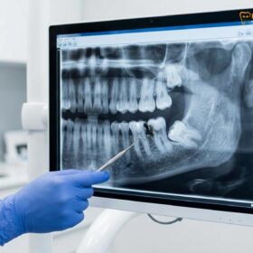
The initial screening tool is typically a panoramic radiograph (Panorex), which provides a broad, two-dimensional view of the entire upper and lower jaws. On a panoramic X-ray, a dentigerous cyst appears as a well-defined, unilocular (single-chambered) radiolucency (dark area) that symmetrically surrounds the crown of an unerupted tooth. A key diagnostic feature is that the cyst attaches to the tooth exactly at the cementoenamel junction (CEJ)—the anatomical line where the enamel of the crown meets the root surface.
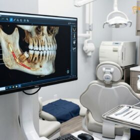
While a panoramic X-ray is excellent for initial detection, it lacks the three-dimensional detail required for precise surgical planning. For this, a Cone Beam Computed Tomography (CBCT) scan is the gold standard[3]. A CBCT scan provides high-resolution, 3D cross-sectional images of the jaw, allowing the oral surgeon to evaluate the exact volume of the cyst, its relationship to critical anatomical structures, and the degree of cortical bone thinning.
| Condition | Radiographic Appearance | Clinical Behavior | Treatment Approach |
|---|---|---|---|
| Hyperplastic Follicle | Radiolucency < 4mm around crown | Normal physiological state, asymptomatic | Routine extraction if impacted |
| Dentigerous Cyst | Radiolucency > 5mm, attaches at CEJ | Slow expansion, bone resorption | Enucleation and extraction |
| Odontogenic Keratocyst (OKC) | Multilocular, grows along bone length | Highly aggressive, high recurrence rate | Aggressive enucleation, peripheral ostectomy |
| Ameloblastoma | “Soap bubble” or “honeycomb” appearance | Benign tumor, severe local destruction | Wide surgical resection |
The CBCT scan is particularly crucial for differentiating a dentigerous cyst from more aggressive lesions, such as an Odontogenic Keratocyst (OKC) or an ameloblastoma. While a biopsy is ultimately required for a definitive histological diagnosis, the 3D imaging characteristics guide the surgeon in determining the appropriate level of surgical aggressiveness required during the initial procedure.
Risk of Jawbone Destruction and Pathological Fractures
Left untreated, an expanding cyst severely thins the mandibular bone, significantly increasing the risk of spontaneous jaw fractures and permanent nerve damage.
The structural integrity of the human mandible is heavily dependent on its dense cortical plates and the internal trabecular network. The angle of the mandible—the area where the horizontal body meets the vertical ramus, precisely where lower wisdom teeth are located—is already a natural biomechanical weak point. When a wisdom tooth cyst jaw lesion develops in this area, it hollows out the internal bone, replacing solid osseous tissue with a fluid-filled void.
As the cyst expands, it progressively thins the buccal (cheek side) and lingual (tongue side) cortical plates, as well as the inferior border of the mandible. This severe structural compromise puts the patient at a high risk for a pathological fracture. A pathological fracture occurs when a bone breaks not due to massive trauma, but because the bone itself has been weakened by disease. In patients with large dentigerous cysts, a fracture can occur during normal physiological activities, such as chewing hard food, yawning, or sustaining minor, otherwise harmless impacts to the face[4].
Clinical Warning: Patients diagnosed with large mandibular cysts should avoid contact sports, heavy lifting, and chewing excessively hard foods until the cyst is surgically removed and the jawbone has sufficiently regenerated. A pathological fracture complicates treatment significantly, often requiring rigid fixation with titanium plates and screws.
Beyond the risk of fracture, the expansion of the cyst poses a severe threat to the inferior alveolar nerve (IAN). The IAN is a major sensory nerve that runs through a bony canal within the mandible, providing sensation to the lower lip, chin, and teeth on that side of the face. As the cyst grows, it can compress the nerve against the dense cortical bone or even resorb the bony walls of the nerve canal, leaving the nerve exposed within the cystic cavity.
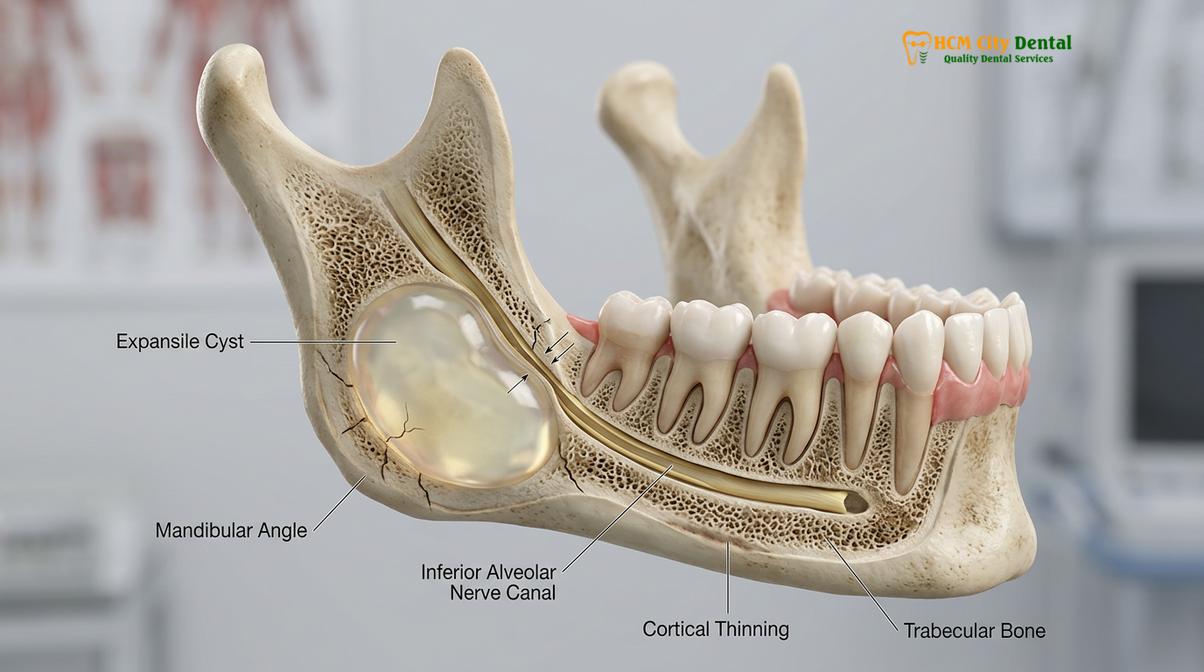
Compression of the IAN can lead to paresthesia—a condition characterized by numbness, tingling, or a “pins and needles” sensation in the lower lip and chin. If the nerve is severely stretched or damaged during the surgical removal of a massive cyst, this sensory deficit can become permanent. Therefore, early intervention is paramount. By removing the cyst while it is still small, the surgeon can preserve a safe margin of bone between the lesion and the nerve canal, drastically reducing the risk of neurological complications.
Surgical Enucleation and Cyst Removal Explained
The definitive treatment involves surgical extraction of the impacted tooth alongside complete enucleation of the cystic lining to prevent recurrence.
The management of a dentigerous cyst requires a precise and methodical surgical approach. The primary goal is twofold: to remove the source of the pathology (the impacted wisdom tooth) and to completely eradicate the cystic tissue to prevent recurrence. The standard procedure for achieving this is known as surgical enucleation. Enucleation involves shelling out the entire cystic sac intact, much like peeling the skin off a grape, without leaving any epithelial remnants behind.
The surgical workflow begins with the administration of profound local anesthesia, often supplemented with intravenous (IV) sedation to ensure patient comfort and cooperation. The surgeon designs a mucoperiosteal flap, carefully incising the gum tissue and reflecting it away from the bone to expose the surgical site. Because the cyst has often thinned or destroyed the overlying bone, accessing the cystic cavity may require minimal bone removal. However, if dense bone still covers the area, the surgeon will perform an ostectomy (bone removal) to create a window.
Modern oral surgery frequently utilizes piezoelectric surgery (piezosurgery) for this step. Piezosurgery uses ultrasonic micro-vibrations to cut bone with extreme precision while leaving soft tissues—such as the cystic lining and the inferior alveolar nerve—completely unharmed. Once access is gained, the impacted wisdom tooth is usually sectioned (cut into smaller pieces) using a surgical handpiece. Sectioning the tooth allows it to be removed through a smaller bony window, preserving as much of the patient’s natural jawbone as possible. For more information on standard extraction techniques, patients can review the protocols for surgical extraction of impacted molars.
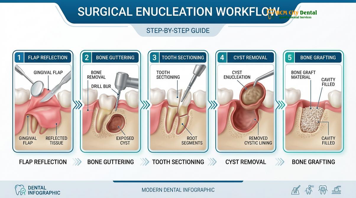
After the tooth is removed, the critical phase of cyst enucleation begins. Using specialized surgical curettes, the surgeon carefully separates the fibrous wall of the cyst from the surrounding bony cavity. It is imperative that the entire lining is removed; if fragments of the epithelium are left behind, the cyst can recur. Dr. Cuong emphasizes that in cases where the cyst is tightly adherent to the bone, peripheral ostectomy—using a bur to lightly shave the walls of the bony cavity—may be performed to ensure all microscopic cystic cells are eliminated.
In rare instances where a wisdom tooth cyst surgery involves a lesion so massive that immediate enucleation would risk a jaw fracture or severe nerve damage, a two-stage procedure called marsupialization may be employed. Marsupialization involves creating a surgical window into the cyst and suturing the cyst lining to the oral mucosa. This relieves the internal hydrostatic pressure, allowing the cyst to drain and gradually shrink over several months. As the cyst shrinks, new bone fills in from the periphery. Once the cyst has reduced to a safe size, a second surgery is performed to enucleate the remaining tissue and extract the tooth.
Recovery and Bone Grafting Solutions in Saigon
Following cyst removal, large bony defects often require bone grafting and PRF therapy to regenerate lost jawbone and ensure structural stability.
The removal of a large dentigerous cyst leaves behind a significant void, or “dead space,” within the jawbone. The body’s natural healing process will attempt to fill this space with a blood clot, which eventually organizes into granulation tissue and, over many months, ossifies into new bone. However, for large defects, relying solely on natural healing can lead to incomplete bone fill, leaving a permanent depression in the jaw or a persistent structural weakness.
To optimize healing and restore the jaw’s original strength, oral surgeons frequently employ Guided Bone Regeneration (GBR) techniques at the time of cyst removal. This involves packing the bony defect with a bone graft material. The graft acts as a scaffold (osteoconduction), providing a framework for the patient’s own bone cells to migrate into and deposit new bone matrix. The graft material may be an allograft (human donor bone), a xenograft (bovine or porcine origin), or an alloplast (synthetic material).
Clinical Case Example: A 28-year-old patient presented to HCMC Dental Clinic in Ho Chi Minh City with a large, asymptomatic dentigerous cyst discovered during a routine panoramic X-ray. The cyst had resorbed a significant portion of the mandibular angle. Following surgical enucleation and tooth extraction, the defect was grafted with a combination of particulate xenograft and PRF. A 6-month follow-up CBCT scan revealed excellent osseous integration and complete restoration of the mandibular bone density.
To further accelerate the healing process, advanced clinics utilize Platelet-Rich Fibrin (PRF) therapy[5]. PRF is created by drawing a small amount of the patient’s own blood just before surgery and spinning it in a centrifuge. This process isolates a concentrated matrix of platelets, white blood cells, and vital growth factors (such as PDGF and TGF-beta). The PRF membrane is mixed with the bone graft or placed directly into the surgical site. Because it is 100% autologous (from the patient’s own body), it carries no risk of rejection and significantly reduces post-operative pain, swelling, and the risk of infection.

“The integration of PRF technology in cystic defect management has revolutionized post-operative recovery. By delivering a concentrated dose of autologous growth factors directly to the surgical site, we observe faster soft tissue closure and a higher quality of dense, trabecular bone regeneration.”
The recovery timeline following wisdom tooth cyst surgery requires patience and strict adherence to post-operative instructions. Initial soft tissue healing over the surgical site typically takes 10 to 14 days. During this period, patients must maintain excellent oral hygiene using prescribed chlorhexidine mouthwashes and adhere to a soft diet to avoid disturbing the surgical flap. Complete bone regeneration within the cystic cavity is a slow physiological process, often taking 6 to 12 months. Regular follow-up appointments and radiographic monitoring at HCMC Dental Clinic are essential to verify that the bone is healing correctly and that there are no signs of cyst recurrence.
When to See a Doctor (Important Clinical Considerations)
Because dentigerous cysts are largely asymptomatic in their early stages, proactive screening is the most effective preventive measure. However, immediate clinical evaluation is required if you experience any of the following red flag symptoms:
- Unexplained Swelling: Any hard or soft swelling in the gums, cheek, or jawline, particularly behind the last visible molar.
- Sensory Changes: Numbness, tingling, or a loss of sensation in the lower lip, chin, or tongue, which may indicate nerve compression.
- Tooth Mobility: Sudden looseness or shifting of the second molars adjacent to the wisdom tooth area.
- Purulent Discharge: A bad taste in the mouth accompanied by pus draining from the gums, indicating a secondary infection of the cyst.
- Facial Asymmetry: A noticeable difference in the shape or contour of one side of the jaw compared to the other.
If you have impacted wisdom teeth that have not been evaluated in the past two years, scheduling a comprehensive examination with a 3D CBCT scan is highly recommended to rule out the presence of silent cystic lesions.
Frequently Asked Questions
How common is a cyst around a wisdom tooth?
A cyst around a wisdom tooth is relatively common, accounting for approximately 20% of all jaw cysts. They most frequently develop around impacted lower third molars in patients during their late teens to early thirties, often remaining undetected until routine radiographic screening.
Can a wisdom tooth cyst turn into cancer?
While extremely rare, the epithelial lining of a wisdom tooth cyst can undergo neoplastic transformation into oral cancer. Squamous cell carcinoma, mucoepidermoid carcinoma, or benign but aggressive tumors like ameloblastoma can develop from untreated cystic linings, making surgical removal and biopsy highly recommended.
Is surgery for a wisdom tooth cyst painful?
Surgery for a wisdom tooth cyst is not painful during the procedure due to advanced local anesthesia and optional sedation. Post-operative discomfort is typical but can be effectively managed with prescribed analgesics, cold compresses, and adherence to clinical aftercare protocols.
How long does the bone take to heal after wisdom tooth cyst removal?
Initial soft tissue healing takes about two weeks, but complete bone regeneration in the cystic cavity takes six to twelve months. The exact timeline depends on the cyst’s size, the patient’s age, and whether bone grafting or PRF therapy was utilized during the surgery.
What is the cost of wisdom tooth cyst surgery in Vietnam?
The cost of wisdom tooth cyst surgery in Vietnam varies based on the cyst’s size, surgical complexity, and the need for bone grafting. Currently, procedures at specialized centers like HCMC Dental Clinic offer highly competitive rates compared to Western countries, while maintaining international clinical standards.
References
- Journal of Oral and Maxillofacial Surgery. Pathogenesis and management of dentigerous cysts. (2021).
- International Journal of Oral and Maxillofacial Surgery. Bone regeneration following cyst enucleation. (2019).
- British Dental Journal. Radiographic diagnosis of odontogenic cysts using CBCT. (2020).
- Journal of the American Dental Association. Risk factors for pathological jaw fractures. (2022).
- Clinical Oral Investigations. Application of PRF in third molar extraction sockets. (2018).