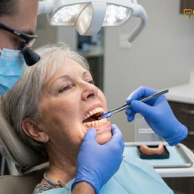
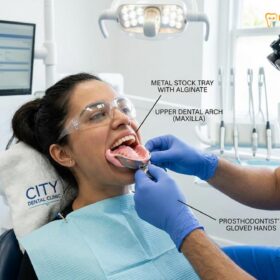
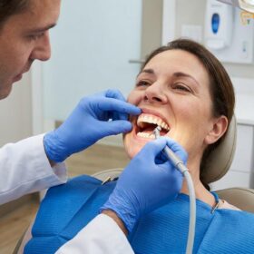
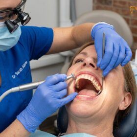
Transitioning to dentures requires a complex process of neuromuscular adaptation, mucosal conditioning, and psychological adjustment. The clinical timeline typically spans four to eight weeks as the oral cavity acclimates to the new prosthesis, necessitating dietary modifications, speech practice, and professional adjustments to ensure optimal retention, stability, and long-term comfort.
Clinical Summary:
The journey of adapting to a removable dental prosthesis involves distinct physiological and psychological phases. Initially, patients experience altered proprioception, increased salivary flow, and mild mucosal friction as the tissues bear new occlusal loads. Successful rehabilitation depends on the brain remapping sensory inputs and the perioral musculature learning to stabilize the appliance. Through a structured progression of an eating soft food diet, targeted phonetic exercises, and routine clinical evaluations, patients can achieve full functional restoration. Expert oversight ensures that pressure points are relieved promptly, preventing tissue hyperplasia and facilitating a seamless integration of the prosthesis into daily life.
Key Takeaways:
- Neuromuscular adaptation is required for the facial muscles to learn how to retain and stabilize the new prosthesis.
- Initial excessive salivation is a temporary parasympathetic response that resolves as the brain stops interpreting the appliance as food.
- Chewing must be relearned using a bilateral balanced technique to prevent the appliance from tipping or dislodging.
- Speech impediments, particularly with sibilant and fricative sounds, are common but correctable through consistent phonetic practice.
- Routine clinical adjustments are mandatory to relieve pressure points and prevent the formation of decubitus ulcers on the gums.
The Psychological Shift: Accepting Your Removable Prosthesis
Accepting a removable prosthesis involves overcoming the initial sensation of a foreign body and adapting to altered oral proprioception. Achieving psychological dental acceptance is a critical first step toward successful long-term rehabilitation.
The transition from natural dentition to a removable prosthetic device is as much a psychological journey as it is a physical one. Natural teeth are anchored into the jawbone by the periodontal ligament (PDL), a highly specialized connective tissue that provides intense sensory feedback to the brain regarding bite force, texture, and spatial awareness. When teeth are lost, this proprioceptive network is severed. The introduction of a complete or partial denture requires the central nervous system to rely on alternative sensory pathways, primarily the mucosal mechanoreceptors, to navigate the oral environment[1].
During the initial stages of wearing custom removable dentures, patients frequently experience a phenomenon known as somatosensory amplification. Because the acrylic base covers areas of the mouth that are normally exposed—such as the hard palate in the maxilla or the lingual vestibule in the mandible—the brain hyper-focuses on the appliance. It feels overwhelmingly large, intrusive, and unnatural. This is a standard neurological response to a sudden change in the oral architecture.
Dr. Nguyen Van Cuong, a leading specialist at HCMC Dental Clinic in Ho Chi Minh City, emphasizes that patient education is the cornerstone of overcoming this hurdle. When patients understand the biomechanical limitations and the required adaptation period of their new appliance, their anxiety significantly decreases. Achieving psychological dental acceptance means shifting the mindset from expecting the prosthesis to feel exactly like natural teeth to recognizing it as a highly functional medical device designed to restore aesthetics and mastication.
“The success of a removable prosthesis is dictated not only by the precision of the impression and the quality of the acrylic but by the patient’s willingness to embrace the adaptation process. The brain must literally rewire its sensory expectations, a process that requires patience, realistic expectations, and consistent wear.”
Grieving the loss of natural teeth is a recognized psychological phase in prosthodontics. Patients may feel self-conscious about their appearance, fear that the appliance will dislodge in public, or feel frustrated by the sudden inability to eat their favorite foods. Comprehensive comprehensive general dentistry protocols dictate that clinicians must provide empathetic counseling alongside mechanical adjustments. Support groups, detailed educational materials, and open communication with the dental team play a vital role in helping patients navigate this profound psychological shift.
Week 1: Excessive Saliva, Soreness, and the ‘Foreign Object’ Feel
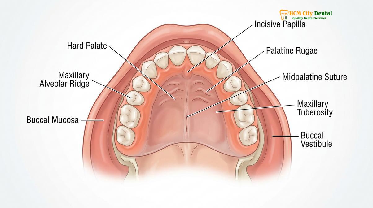
During the first week, the brain interprets the new denture as food, triggering excessive salivation and a bulky sensation. Mild mucosal soreness is expected as the tissues begin to bear occlusal loads.
The first seven days of wearing a new prosthesis are universally considered the most challenging phase of the transition. As soon as the appliance is seated in the oral cavity, the sensory receptors in the oral mucosa send signals to the brain indicating the presence of a large, foreign mass. Because the brain’s primary reference for an object of this size in the mouth is a bolus of food, it immediately activates the parasympathetic nervous system to stimulate the salivary glands[2].
This results in excessive salivation, a condition where the parotid, submandibular, and sublingual glands produce a high volume of saliva in an attempt to begin the digestive process and lubricate what it perceives as food. Patients often feel the need to swallow constantly, which can be exhausting and may occasionally trigger a mild gag reflex, particularly if the posterior border of the maxillary denture extends near the vibrating line of the soft palate. Fortunately, this salivary response is self-limiting. As the brain realizes that the object is not meant to be swallowed or digested, the salivary flow rates gradually return to baseline, usually within three to seven days.
Simultaneously, the patient will experience the classic “foreign object” sensation. The acrylic base of a maxillary denture covers the entire hard palate to achieve a suction seal, altering the tongue’s resting space. The mandibular denture, which rests on the much smaller surface area of the lower alveolar ridge, often feels loose because it lacks the palatal suction and is constantly subjected to the dynamic movements of the tongue and cheeks. This requires a significant new denture adjustment period as the oral environment accommodates the bulk of the material.
Mucosal soreness is another hallmark of the first week. The gingival tissues, which are naturally designed to deflect food rather than bear the constant, heavy load of mastication, must undergo a process of keratinization and toughening. As the rigid acrylic presses against the soft, non-keratinized mucosa, localized areas of ischemia (reduced blood flow) can occur, leading to tenderness and inflammation. It is imperative that patients do not abandon the prosthesis during this time; wearing it consistently is the only way the tissues will adapt and the dentist can identify exactly where adjustments are needed.
Weeks 2–3: Learning to Control Muscles and Chewing Techniques
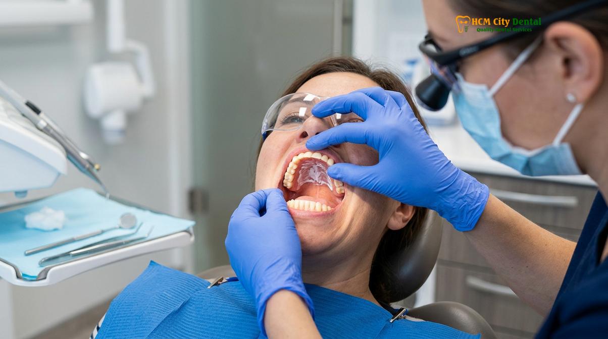
Weeks two and three focus on neuromuscular control, where the buccinator and orbicularis oris muscles learn to stabilize the prosthesis. Patients must adopt an eating soft food diet and bilateral chewing techniques.
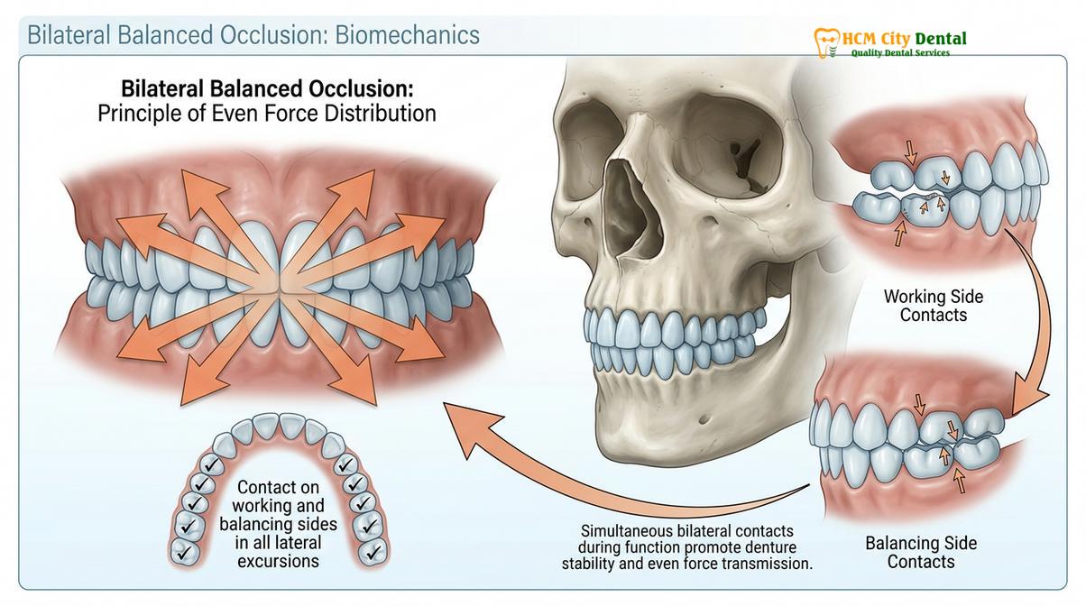
As the initial hypersalivation subsides and the acute soreness is managed through clinical adjustments, the focus of weeks two and three shifts entirely to biomechanics and neuromuscular adaptation. Unlike natural teeth, which are independently rooted in the jaw, a complete denture functions as a single, rigid unit. If pressure is applied unevenly to one side of the appliance, the opposite side will inevitably lift, breaking the suction seal and causing the denture to dislodge.
To counteract this lever effect, patients must master a technique known as bilateral balanced occlusion. This requires dividing food into smaller portions and chewing simultaneously on both the left and right sides of the mouth. This even distribution of masticatory force stabilizes the prosthesis against the alveolar ridges. Furthermore, patients must learn to avoid incising—biting directly into foods with the front teeth—as this applies a strong anterior force that will tip the posterior section of the denture downward.
During this phase, adhering to an eating soft food diet is non-negotiable. The underlying bone and mucosal tissues are still adapting to the compressive forces, and attempting to chew tough, fibrous, or sticky foods will cause severe tissue trauma and frustration. The diet should progress systematically to allow the muscles of mastication (the masseter, temporalis, and medial pterygoid) to rebuild their strength and coordination in harmony with the new appliance.
| Phase | Timeline | Recommended Dietary Choices | Clinical Rationale |
|---|---|---|---|
| Phase 1: Liquid & Puree | Days 1–5 | Broths, yogurt, smoothies, pureed soups, meal replacement shakes. | Minimizes occlusal load on inflamed mucosa; prevents dislodgement while swallowing reflexes adapt. |
| Phase 2: Soft Mechanical | Days 6–14 | Scrambled eggs, mashed potatoes, flaky fish, well-cooked pasta, soft cooked vegetables. | Introduces mild masticatory forces; allows practice of bilateral chewing without excessive tissue stress. |
| Phase 3: Firm Soft | Days 15–28 | Ground meats, soft breads (no crust), ripe fruits, soft cheeses. | Builds masseter muscle strength; tests the retention and stability of the prosthesis under moderate load. |
| Phase 4: Modified Normal | Week 5+ | Most normal foods, cut into small, bite-sized pieces. Avoid sticky candies and extremely hard nuts. | Achieves functional rehabilitation; requires ongoing mindfulness of bilateral chewing mechanics. |
Beyond chewing, the perioral musculature—specifically the buccinator muscles in the cheeks and the orbicularis oris muscle around the lips—must learn a new functional role. In a natural dentition, these muscles simply help keep food on the occlusal table. With a removable prosthesis, these muscles actively contract against the polished outer surfaces of the denture flanges to help hold the appliance in place. This subconscious muscle training is a critical milestone in the denture fabrication process and overall adaptation.
Week 4: Re-learning Pronunciation and Speaking with Confidence
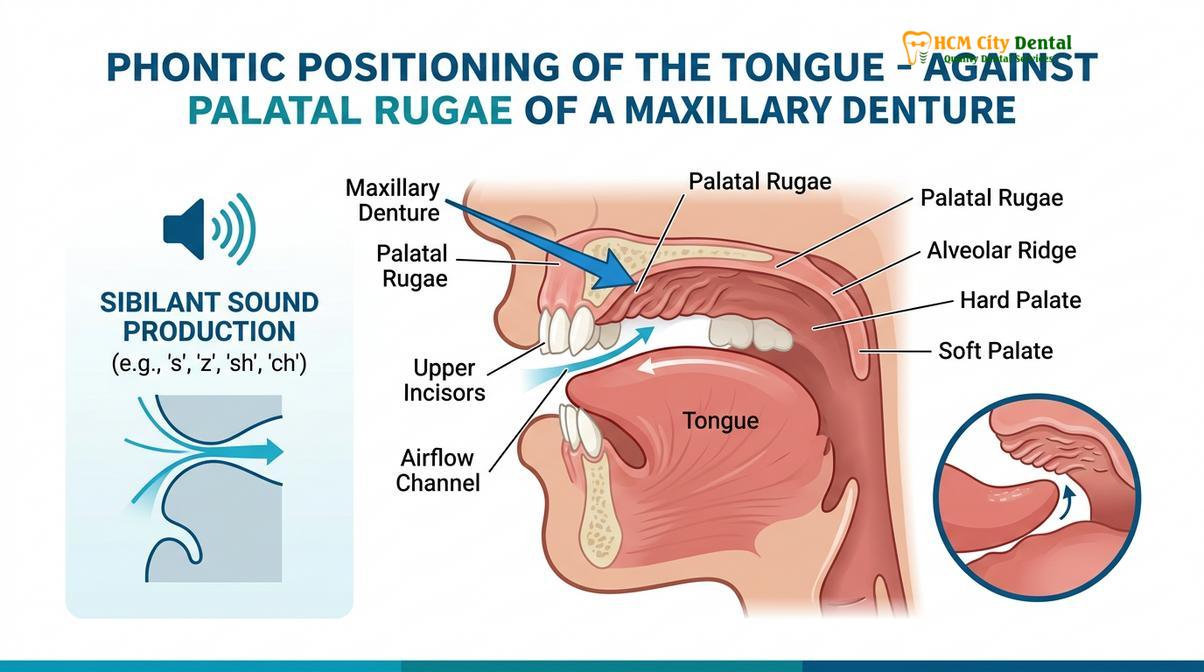
By week four, patients focus on speech pronunciation practice to correct phonetic challenges, particularly with fricative and sibilant sounds. Tongue adaptation to the palatal acrylic thickness is essential for clear articulation.
Speech is a highly complex motor function that requires precise coordination between the vocal cords, lips, cheeks, and tongue. The tongue, in particular, relies on exact spatial relationships with the teeth and the hard palate to articulate sounds clearly. When a patient receives a new maxillary denture, the acrylic base covers the palatal rugae—the ridges on the roof of the mouth that the tongue uses as a tactile guide for pronunciation. Additionally, the artificial teeth may be positioned slightly differently than the natural teeth to optimize the bite, altering the phonetic space[3].
As a result, patients frequently experience speech impediments during the first few weeks. The most commonly affected sounds are the sibilants (“S”, “Z”, “Sh”, “Ch”) and the fricatives (“F”, “V”). To pronounce an “S” sound correctly, the mandible must move forward and upward, creating a precise 1 to 1.5-millimeter gap between the upper and lower incisors, known as the closest speaking space. If the acrylic is too thick, or if the patient has not yet mastered mandibular control, the “S” may sound like a lisp or a whistle.
Similarly, the “F” and “V” sounds require the lower lip to lightly touch the incisal edges of the maxillary anterior teeth. If the patient is hesitant or lacks confidence in the stability of the appliance, they may not bring the lip up far enough, resulting in muffled or indistinct speech. Engaging in dedicated speech pronunciation practice is the most effective way to overcome these phonetic hurdles.
“The tongue is an incredibly adaptable muscle, but it requires repetitive phonetic training to map the new contours of a dental prosthesis. Reading aloud slowly and deliberately forces the neuromuscular system to recalibrate its spatial awareness, rapidly eliminating lisps and articulation errors.”
Clinicians recommend that patients spend at least fifteen to twenty minutes a day reading a book or newspaper aloud in front of a mirror. This visual and auditory feedback loop accelerates the brain’s ability to adjust the tongue’s trajectory. If a specific sound remains problematic after several weeks of practice, it may indicate that the acrylic base is too thick in a specific area or that the anterior teeth are set too far forward, requiring a minor clinical adjustment by the prosthodontist.
Preventing and Treating Initial Sore Spots: When to Request Adjustments
Sore spots occur due to uneven pressure distribution on the mucosal tissues and require professional occlusal or intaglio surface adjustments. Prompt sore spots relief prevents ulceration and tissue hyperplasia.
Despite the highest level of precision in impression-taking and fabrication, it is virtually impossible for a new denture to fit perfectly without requiring minor modifications once it is subjected to the dynamic forces of chewing and speaking. The oral mucosa is highly compressible, and the underlying alveolar bone has microscopic irregularities. When the rigid acrylic base moves slightly during function, it can create localized areas of high friction, leading to the development of sore spots or decubitus ulcers[4].
Sore spots typically manifest as red, inflamed patches on the gums or in the vestibular folds (where the gums meet the cheeks). If left untreated, these areas can ulcerate, becoming extremely painful and making it impossible to wear the appliance. Achieving effective sore spots relief is a collaborative effort between the patient and the dental professional. Patients must wear the prosthesis for at least 24 hours prior to their adjustment appointment, even if it is uncomfortable, so that the dentist can accurately identify the exact location of the friction.
Clinical Warning: Never attempt to adjust, file, or alter your dental prosthesis at home using sandpaper, rotary tools, or household adhesives. DIY adjustments permanently destroy the precise biofunctional fit of the appliance, alter the occlusal balance, and can cause severe, irreversible damage to your oral tissues. Always seek professional clinical modifications.
During an adjustment visit, the dentist will use a specialized material called Pressure Indicating Paste (PIP). This white, zinc-oxide-based paste is painted onto the intaglio (tissue-facing) surface of the denture. The appliance is then seated in the mouth, and the patient is instructed to bite down firmly. When the denture is removed, areas where the paste has been completely displaced indicate points of excessive pressure. The dentist will then use an acrylic bur to carefully relieve these specific spots by a fraction of a millimeter, instantly improving comfort without compromising the overall suction seal.
It is important to differentiate between a sore spot caused by an overextended acrylic flange (which rubs against the moving tissues of the cheek or frenum) and one caused by an occlusal prematurity. If the upper and lower teeth do not meet perfectly simultaneously, the denture will shift slightly with every bite, causing a sore spot on the opposite side of the arch. In these cases, the dentist must adjust the biting surfaces of the artificial teeth rather than the tissue-bearing acrylic.
Tips for a Smooth Transition: Exercises for Your Cheeks and Tongue
Specific myofunctional exercises strengthen the perioral musculature, enhancing the retention and stability of the removable prosthesis. Consistent practice accelerates the brain’s neuromuscular adaptation.
While the physical fit of the appliance is dictated by the dentist, the functional success of the prosthesis relies heavily on the patient’s muscular control. Myofunctional therapy—a series of targeted exercises designed to strengthen the tongue, lips, and cheeks—can drastically reduce the transition time and improve the stability of the appliance, particularly for the notoriously difficult lower denture[5].
The buccinator muscles, located in the cheeks, play a crucial role in keeping food positioned over the teeth during chewing. To strengthen these muscles, patients can practice the “cheek puff” exercise: with the dentures securely in place, close the lips tightly and puff the cheeks out with air, holding the pressure for five seconds before releasing. Repeating this ten times a day helps tone the musculature, allowing the cheeks to rest more firmly against the buccal flanges of the prosthesis, thereby enhancing the peripheral seal.
Tongue exercises are equally important. The tongue must learn to rest in a position that helps hold the lower denture down, rather than pushing it up and out of place. Patients should practice swallowing with their teeth gently closed together, focusing on pressing the tip of the tongue against the anterior palatal rugae (just behind the upper front teeth). This trains the tongue to stabilize the upper appliance while simultaneously keeping the floor of the mouth relaxed, preventing the dislodgement of the lower appliance.
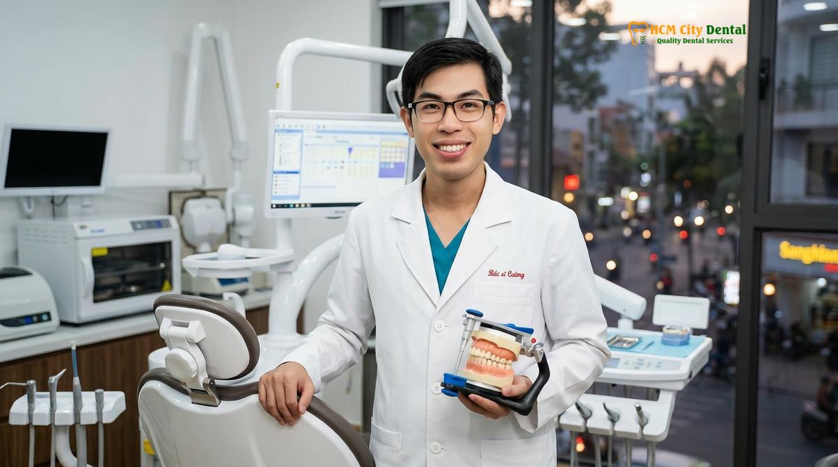
Clinical Case Example: A 65-year-old patient presented to HCMC Dental Clinic in Ho Chi Minh City struggling with the retention of a conventional mandibular denture. Dr. Nguyen Van Cuong implemented a structured myofunctional exercise regimen focusing on tongue positioning and buccinator strengthening. Within three weeks of consistent practice, the patient demonstrated a 60% improvement in prosthesis stability during mastication, significantly reducing the need for chemical denture adhesives and improving overall dietary intake.
Additionally, practicing exaggerated facial expressions—such as smiling widely, frowning, and puckering the lips—helps the orbicularis oris muscle adapt to the new vertical dimension established by the artificial teeth. These exercises ensure that the facial muscles remain toned, preventing the sunken facial appearance often associated with tooth loss and providing the necessary dynamic support for the general dental care prosthetics.
When to See a Doctor for Denture Complications
While a certain degree of discomfort and an adjustment period are entirely normal, specific clinical signs indicate the need for immediate professional intervention. Patients should not attempt to “tough out” severe complications, as this can lead to significant tissue damage or infection.
You should schedule an immediate evaluation with your dental provider if you experience any of the following:
- Severe or Bleeding Ulcers: Sore spots that bleed, become highly inflamed, or do not improve after a few days of wearing the appliance indicate a severe pressure point that requires mechanical relief.
- Signs of Oral Candidiasis (Thrush): White, cottage-cheese-like patches on the palate or gums, accompanied by a burning sensation, indicate a fungal infection. This often occurs if the appliance is not removed at night or if salivary flow is severely reduced.
- Numbness or Tingling (Paresthesia): A tingling sensation in the lower lip or chin can occur if the mandibular denture is compressing the mental nerve as it exits the jawbone. This requires immediate adjustment to prevent permanent nerve damage.
- Persistent Gag Reflex: If the gag reflex does not subside after the first week, the posterior border of the maxillary appliance may be overextended and requires shortening by the dentist.
- Inability to Chew Soft Foods: If the appliance constantly dislodges even when eating soft foods and practicing bilateral chewing, the occlusal balance or the peripheral seal may be fundamentally flawed, requiring a clinical reline or bite adjustment.
For patients experiencing ongoing difficulties, scheduling routine dental examinations is crucial. A thorough clinical assessment can determine whether a soft reline, a hard reline, or the integration of dental implants to create an overdenture is the most appropriate next step in your treatment plan.
Frequently Asked Questions
How long does the sore stage last with new dentures?
The initial sore stage typically lasts between two to four weeks as the oral mucosa adapts to the acrylic base. Regular clinical adjustments are necessary during this period to relieve pressure points and prevent tissue ulceration. As the tissues undergo mild keratinization and the muscles learn to stabilize the appliance, the soreness will progressively diminish.
Why do I have so much saliva after getting dentures?
Excessive salivation occurs because the brain initially interprets the new dental prosthesis as food, triggering the salivary glands. This parasympathetic response usually subsides within the first week as the nervous system acclimates to the appliance. Swallowing frequently and sucking on sugar-free mints can help manage the sensation during the first few days.
How can I speed up my speech adjustment with new dentures?
You can accelerate speech adjustment by reading aloud daily, speaking slowly, and practicing specific phonetic sounds like ‘S’, ‘F’, and ‘V’. Biting down gently before speaking also helps stabilize the prosthesis during articulation. The tongue requires repetitive motor training to map the new contours of the palatal acrylic, so consistent practice is essential.
Can I sleep with my new dentures in during the transition?
During the first 24 hours after extractions, your dentist may advise keeping immediate dentures in to act as a bandage. Afterward, you must remove them nightly to allow the mucosal tissues to rest and prevent fungal infections. Storing them in water or a designated cleaning solution overnight prevents the acrylic from warping and maintains optimal oral hygiene.
When can I return to eating a normal diet?
Most patients can transition from a soft diet to a relatively normal diet within four to eight weeks. However, extremely hard, sticky, or tough foods may always require careful chewing or should be avoided to prevent prosthesis damage. Mastering the bilateral balanced chewing technique is the key to expanding your dietary choices without dislodging the appliance.
References
- Journal of Prosthetic Dentistry. Neuromuscular adaptation and proprioceptive changes in complete denture wearers. (2021).
- International Journal of Oral and Maxillofacial Surgery. Salivary flow rates and mucosal keratinization during initial prosthesis placement. (2020).
- British Dental Journal. Phonetic adaptation and the closest speaking space in removable prosthodontics. (2019).
- Journal of the American Dental Association. Management of decubitus ulcers and sore spots in new denture patients. (2022).
- Clinical Oral Investigations. Myofunctional therapy and its impact on mandibular denture retention. (2018).