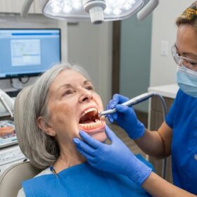
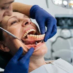
The Dr. Jiro Abe suction lower denture is an advanced prosthetic technique designed to achieve a stable, vacuum-like seal on the mandibular ridge without the need for surgical implants. By utilizing precise border molding and dynamic muscle activity, this protocol creates an atmospheric pressure seal, preventing the lower denture from floating or slipping during daily function.
Clinical Summary:
Suction-Effective Mandibular Complete Dentures (SEMCD), pioneered by Dr. Jiro Abe, revolutionize traditional prosthodontics by achieving true suction on the lower jaw. Unlike conventional dentures that rely on gravity and weak adhesion, the Abe protocol captures the dynamic movements of the oral musculature to form a continuous peripheral seal. This technique is particularly beneficial for patients with severe bone resorption who cannot undergo implant surgery. Clinical success depends on meticulous impression workflows, specifically capturing the sublingual fold and retromolar pad. At HCMC Dental Clinic, this non-invasive approach restores chewing efficiency and speaking confidence through advanced biomechanical engineering and precise anatomical adaptation.
Key Takeaways:
- SEMCD utilizes natural oral anatomy to create a vacuum seal without surgical implants.
- The technique relies heavily on capturing the sublingual fold and buccal mucosa during dynamic muscle movements.
- Dr. Jiro Abe’s protocol requires specialized slow-setting impression materials to record functional tissue states.
- It serves as a highly effective alternative for patients with severe mandibular ridge resorption.
- Proper border molding prevents the common “lifting and shifting” associated with traditional lower dentures.
The Lower Denture Dilemma: Why the Mandible is Hardest to Stabilize
The lower jaw lacks the broad surface area of the palate and is constantly disrupted by tongue and cheek movements, making traditional lower dentures highly prone to instability and dislodgement.
In the realm of removable prosthodontics, achieving stability in the maxillary (upper) arch is generally straightforward due to the broad, static surface area of the hard palate. This expansive anatomical structure allows for a natural suction effect, providing reliable retention for upper dentures. However, the mandibular (lower) arch presents a profoundly different biomechanical challenge. The mandible is shaped like a horseshoe, offering significantly less surface area for a prosthetic base to rest upon. Furthermore, the lower jaw is a highly dynamic environment, constantly influenced by the complex movements of the tongue, cheeks, and lips during speech, mastication, and swallowing [1].
One of the primary reasons traditional lower dentures fail to achieve stability is the progressive nature of alveolar bone resorption. Following the extraction of natural teeth, the jawbone undergoes continuous remodeling, often resulting in a flattened or concave ridge. Without the anchoring support of tooth roots, the bone diminishes, leaving a minimal foundation for a conventional denture to grip. This anatomical degradation forces the denture to rely almost entirely on gravity and weak salivary adhesion, which are easily overcome by the muscular forces of the oral cavity.
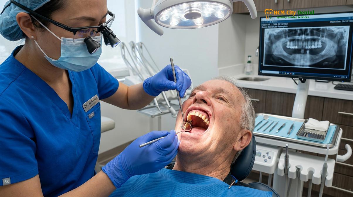
Dr. Nguyen Van Cuong frequently notes that patient frustration with lower dentures is one of the most common complaints encountered in clinical practice. When a traditional lower denture lacks a peripheral seal, air easily penetrates beneath the acrylic base. As the patient speaks or chews, the tongue and surrounding musculature inadvertently lift the prosthesis, causing it to float or shift painfully against the delicate gum tissue. This instability not only impairs chewing efficiency but also leads to chronic mucosal ulcerations and profound social embarrassment.
To address this dilemma, modern custom denture solutions must move beyond static anatomical impressions. The solution lies in understanding the functional boundaries of the oral cavity and engineering a prosthesis that harmonizes with, rather than fights against, the surrounding musculature. This paradigm shift forms the foundational philosophy of advanced mandibular prosthetic techniques.
The Science of SEMCD: Achieving a Vacuum Seal on the Gums
SEMCD achieves stability by creating a continuous peripheral border seal that traps saliva and air, generating an atmospheric pressure seal that locks the denture against the mandibular ridge.
The Suction-Effective Mandibular Complete Denture (SEMCD) represents a monumental leap in prosthetic engineering. Developed through decades of clinical research by Dr. Jiro Abe, this technique defies the long-held belief that true suction on the lower jaw is impossible. The science behind SEMCD is rooted in the principles of fluid dynamics and atmospheric pressure. To achieve a vacuum seal, the denture must establish a continuous, unbroken peripheral border that intimately contacts the yielding soft tissues of the oral cavity, effectively preventing the ingress of air [2].
When a perfectly adapted denture is seated, a thin layer of saliva is trapped between the acrylic base and the mucosal tissue. This salivary film acts as a fluid gasket. As forces attempt to dislodge the denture, the sealed borders prevent air from entering the space beneath the prosthesis. Consequently, the pressure beneath the denture drops below the ambient atmospheric pressure, creating a powerful suction effect that holds the denture firmly in place. This is the essence of the atmospheric pressure seal.
“The realization of mandibular suction is not a matter of chance, but the result of precise biomechanical engineering that transforms the dynamic oral musculature from a dislodging force into a retentive asset.”
Achieving this seal requires a profound understanding of the rheological properties of saliva and the viscoelastic nature of the oral mucosa. The denture flanges must be precisely contoured to fill the vestibular spaces without overextending into the active muscle attachments. If the flanges are too short, the seal is broken; if they are too long, the muscles will forcefully eject the denture during function. The Dr. Jiro Abe protocol meticulously maps these functional boundaries, ensuring that the prosthesis remains locked in place even during vigorous mastication.
Unlike conventional approaches that focus merely on the static shape of the bone, the SEMCD philosophy prioritizes the soft tissues. By utilizing the buccal mucosa, the retromolar pad, and the sublingual tissues to complete the seal, this technique offers a lifeline to patients who have suffered severe bone loss and are seeking advanced removable prosthetics without the need for invasive surgical interventions.
Border Molding with Muscle Activity: Capturing Gums in Motion
Dynamic border molding records the exact functional limits of the oral muscles during speech and swallowing, ensuring the denture borders harmonize with tissue movement rather than fighting it.
The cornerstone of the Dr. Jiro Abe protocol is the meticulous execution of border molding muscle activity. Traditional denture impressions are often static; they record the shape of the gums while the patient’s mouth is at rest. However, the oral cavity is rarely at rest. When a patient speaks, chews, or swallows, the muscles of the cheeks (buccinator), lips (orbicularis oris), and floor of the mouth (mylohyoid) contract and change shape. A denture designed from a static impression will inevitably clash with these moving tissues, leading to immediate dislodgement [3].
Dynamic border molding addresses this by capturing the gums in motion. During the impression phase, the clinician uses specialized, slow-setting elastomeric materials applied to the borders of a custom tray. While the material is still pliable, the patient is instructed to perform a series of exaggerated functional movements. These movements—such as puckering the lips, smiling widely, swallowing, and thrusting the tongue—actively shape the impression material. The resulting mold represents the absolute functional limits of the patient’s unique musculature.
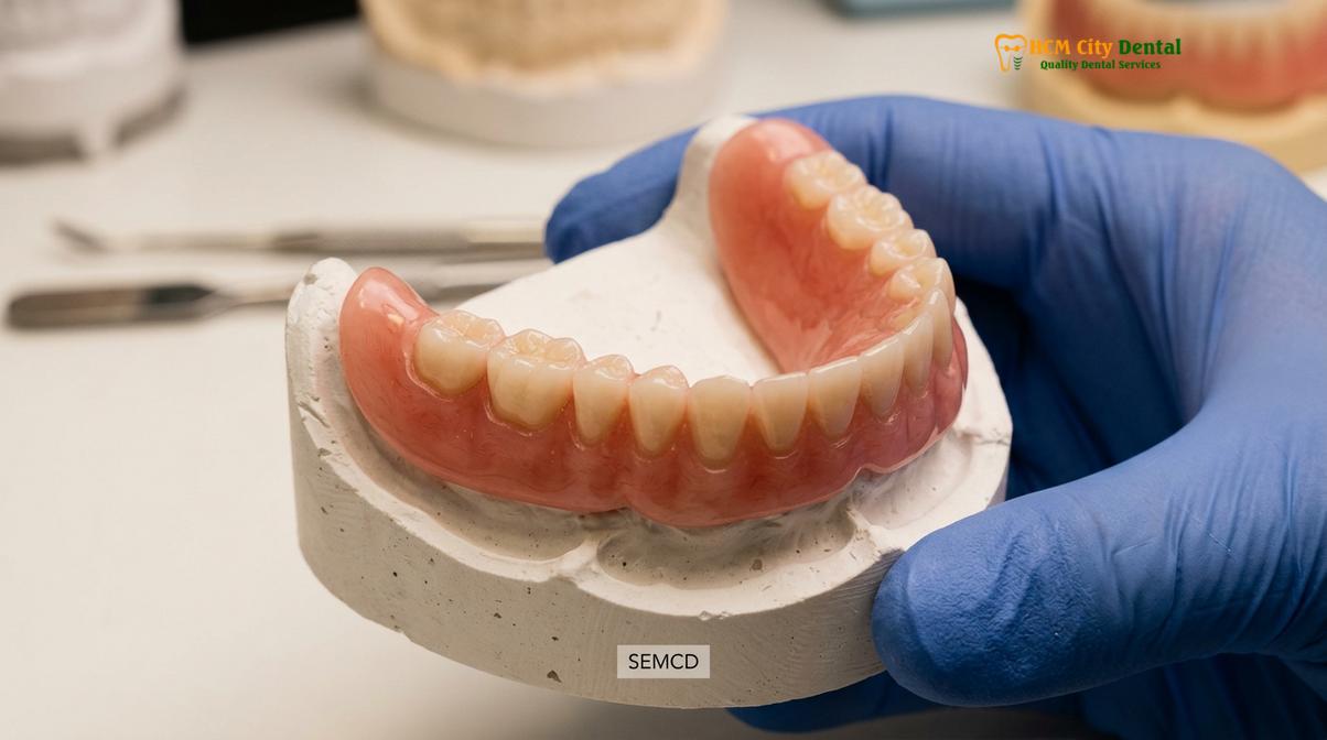
This dynamic capture ensures that the final denture flanges are perfectly contoured to accommodate muscle contraction. Instead of the muscles pushing the denture away, they drape over the polished surfaces of the prosthesis, actually aiding in its retention. This harmonious integration between rigid acrylic and dynamic soft tissue is what elevates the SEMCD technique above conventional methods.
| Feature | Traditional Static Impression | SEMCD Dynamic Border Molding |
|---|---|---|
| Tissue State | Recorded at rest (passive) | Recorded during active muscle function |
| Material Viscosity | Often fast-setting, high viscosity | Slow-setting, variable viscosity for tissue mapping |
| Peripheral Seal | Inconsistent, prone to air leakage | Continuous, engineered for atmospheric pressure seal |
| Patient Involvement | Minimal (mouth held open) | Active (swallowing, tongue movements, speaking) |
| Clinical Outcome | Relies on gravity and weak adhesion | Achieves true vacuum suction and high stability |
By prioritizing border molding muscle activity, clinicians can predictably achieve a level of stability previously thought impossible for lower dentures. This meticulous attention to functional anatomy is a hallmark of high-quality comprehensive general dentistry, ensuring that prosthetic solutions enhance the patient’s quality of life rather than adding to their daily struggles.
The Sublingual Fold Seal: The Secret to Locking Lower Dentures
The critical innovation of the Dr. Jiro Abe protocol is extending the denture base into the sublingual crescent, utilizing the soft tissues under the tongue to complete the posterior-lingual vacuum seal.
While capturing the buccal (cheek) and labial (lip) borders is essential, the true secret to locking a lower denture lies in the lingual (tongue) aspect. Historically, the lingual border of a lower denture was kept short to avoid interfering with tongue movement. However, this conservative approach leaves the posterior-lingual area open, allowing air to rush in and break the seal every time the patient swallows or speaks. The breakthrough of the Dr. Jiro Abe protocol is the strategic utilization of the sublingual fold seal [4].
The sublingual fold is a crescent-shaped area of soft tissue located in the floor of the mouth, beneath the anterior portion of the tongue. During the SEMCD impression process, the clinician carefully extends the impression material into this sublingual space. When the patient swallows or moves their tongue, the sublingual tissues elevate and drape over the extended lingual flange of the denture. This dynamic interaction creates a watertight and airtight seal along the most vulnerable border of the prosthesis.
Clinical Warning: Extending the denture base into the sublingual fold requires profound anatomical knowledge. Overextension or improper contouring in this delicate area can lead to severe mucosal ulceration, impingement of the lingual frenum, and restricted tongue mobility. This technique must only be performed by clinicians specifically trained in advanced biofunctional prosthetic workflows.
Achieving the sublingual fold seal requires a delicate balance. The flange must be long enough to maintain contact with the tissues during elevation, yet contoured precisely so as not to trigger the gag reflex or cause discomfort. The shape of the polished lingual surface is also critical; it must be slightly concave to allow the lateral borders of the tongue to rest comfortably upon it, further stabilizing the denture through passive weight distribution.
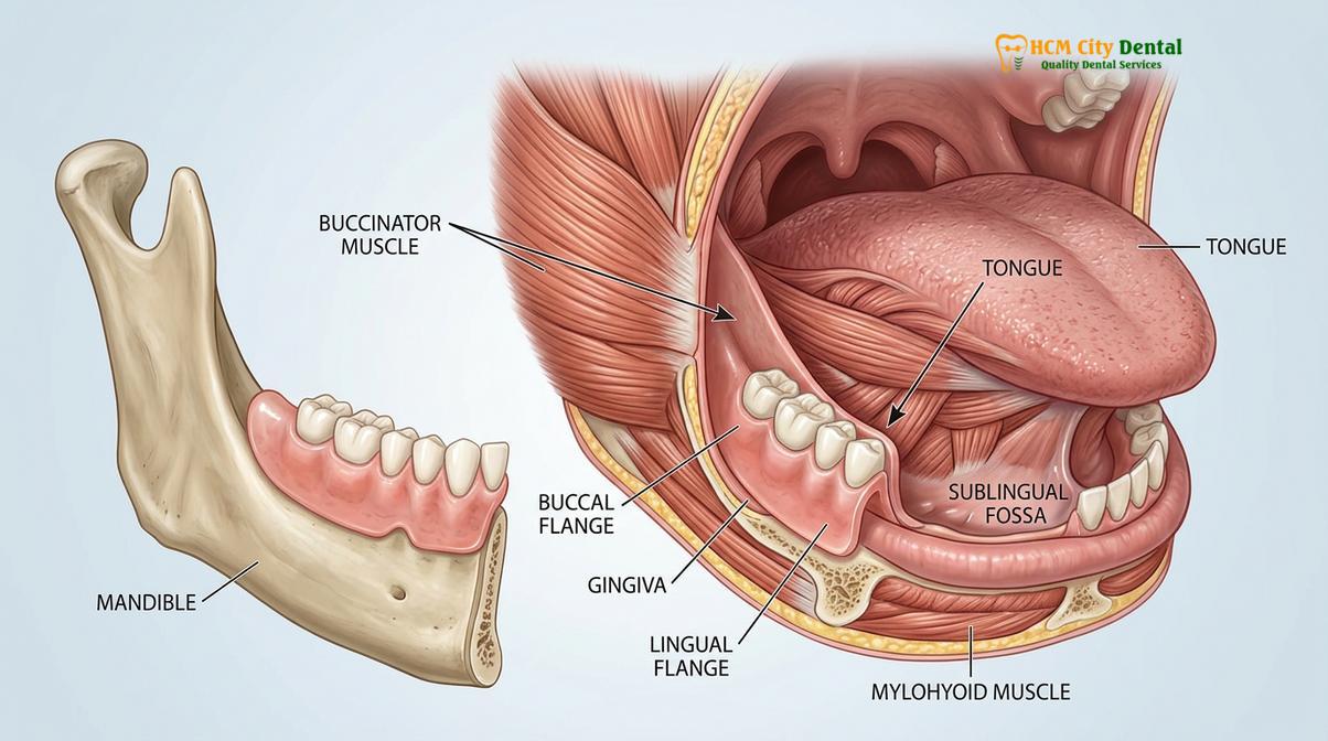
“The mastery of the sublingual crescent transforms the tongue from the denture’s greatest adversary into its most reliable stabilizing ally.”
Furthermore, the resting posture of the patient’s tongue plays a significant role in the success of this seal. Patients with a naturally retracted tongue posture may require specific tongue-training exercises to allow the sublingual tissues to properly engage the denture borders. Through comprehensive evaluation and precise execution, the sublingual fold seal effectively closes the final gap in the peripheral border, locking the lower denture firmly into place.
Abe Protocol Step-by-Step: Clinical Impression Workflow
The clinical workflow involves a rigorous multi-stage impression process using custom trays and specialized materials to capture the precise functional anatomy of the patient’s mandible.
The fabrication of a Suction-Effective Mandibular Complete Denture is not a standard procedure; it is a highly specialized clinical workflow that demands precision at every stage. The Dr. Jiro Abe protocol dictates a systematic approach to impression making, ensuring that no anatomical detail is overlooked. This rigorous process is what differentiates a premium suction denture from a conventional prosthetic [5].
Step 1: The Preliminary Functional Impression
The process begins with a preliminary impression using a specialized stock tray designed specifically for the SEMCD technique. Unlike standard trays, these trays feature extended lingual flanges to capture the retromolar pad and the sublingual crescent from the very first step. A high-quality, slow-setting alginate or preliminary silicone is used. During this phase, the clinician manually manipulates the patient’s cheeks and lips, and instructs the patient to perform specific tongue movements to establish the initial functional borders.
Step 2: Custom Tray Fabrication and Spacer Design
Based on the preliminary cast, a custom impression tray is fabricated in the dental laboratory. The design of this tray is critical. It must incorporate specific spacer thicknesses to allow room for the final impression materials while maintaining uniform pressure across the residual ridge. The tray is also equipped with a specialized handle that does not interfere with lip movement during the final impression.
Clinical Case Review: A 68-year-old patient presented to HCMC Dental Clinic in Ho Chi Minh City with a history of five failed conventional lower dentures due to severe mandibular resorption. By strictly adhering to the Dr. Jiro Abe protocol, Dr. Nguyen Van Cuong utilized a two-stage dynamic impression technique. The precise capture of the sublingual fold and buccal shelf resulted in a prosthesis that demonstrated immediate atmospheric pressure suction, restoring the patient’s ability to consume solid foods without adhesive.
Step 3: The Final Definitive Impression
This is the most crucial phase of the workflow. The clinician uses a combination of heavy-body and light-body elastomeric silicones. The heavy-body material is applied to the borders of the custom tray to perform the dynamic border molding. The patient is guided through a series of rigorous functional movements: swallowing forcefully, licking the upper and lower lips, and pushing the tongue against the tray handle. Once the borders are established, a light-body wash material is applied to capture the fine micro-anatomy of the mucosal tissues under light functional pressure.
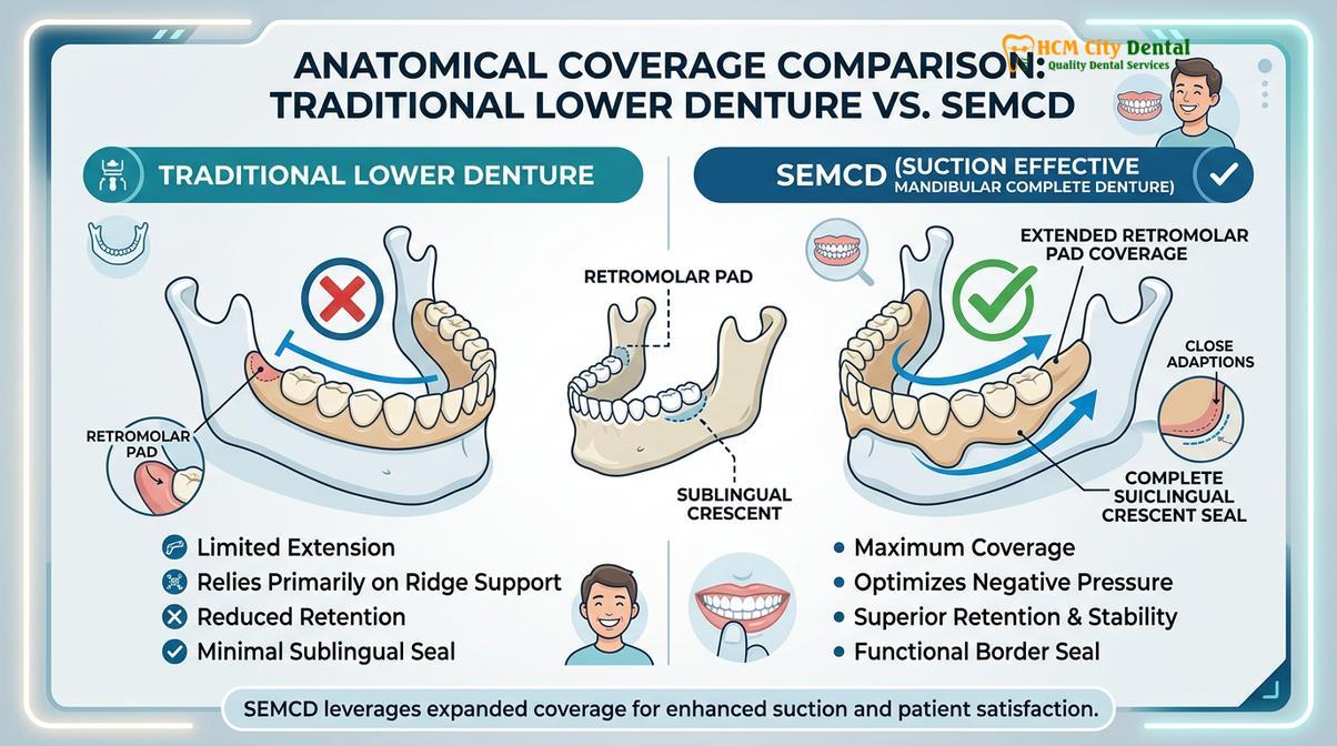
Step 4: Bite Registration and Biofunctional Try-In
Following the final impression, the precise vertical dimension of occlusion and centric relation are recorded. The trial dentures are then evaluated in the patient’s mouth. During this try-in, the clinician verifies the suction, stability, and phonetic function. Any necessary adjustments to the tooth arrangement or flange contours are made before the final acrylic processing.
This meticulous, step-by-step adherence to the protocol ensures that the final prosthesis is a true biofunctional device, seamlessly integrated into the patient’s stomatognathic system. Such dedication to clinical excellence is a core component of routine dental care and advanced rehabilitation.
Candidacy for SEMCD: Assessing Resorbed Lower Ridges
While highly effective, SEMCD candidacy depends on specific anatomical markers, including the shape of the residual ridge, the quality of the oral mucosa, and the patient’s tongue position.
While the Suction-Effective Mandibular Complete Denture offers a revolutionary solution for many, it is essential to understand that not every patient is an ideal candidate. The success of the atmospheric pressure seal relies heavily on specific anatomical and physiological factors that must be carefully evaluated during the initial consultation. A thorough clinical assessment is paramount to determine the prognosis of the treatment [6].
The primary factor in determining candidacy is the condition of the residual alveolar ridge. While the SEMCD technique is specifically designed to help patients with resorbed ridges, there must be a minimal amount of anatomical structure remaining to support the lateral forces of mastication. The presence of a well-defined retromolar pad and a relatively healthy buccal shelf are critical landmarks that contribute to the peripheral seal. Patients with completely flat or severely concave ridges may still achieve improved stability compared to conventional dentures, but absolute suction may be more challenging to attain.
Another crucial consideration is the quality and resilience of the oral mucosa. The tissues must be healthy, compressible, and free from chronic inflammation or hyperplastic tissue (such as epulis fissuratum). Patients suffering from severe xerostomia (dry mouth) present a significant challenge, as a sufficient volume of viscous saliva is required to maintain the fluid gasket that seals the denture borders. Without adequate saliva, the vacuum effect cannot be sustained.
Furthermore, the patient’s tongue posture and neuromuscular control are vital. A normal tongue position, where the lateral borders of the tongue rest gently against the lingual surfaces of the lower teeth, naturally aids in sealing the sublingual fold. Conversely, a retracted tongue posture—where the tongue pulls back into the throat during rest—can continuously break the lingual seal. In such cases, clinicians may need to incorporate tongue-training exercises into the treatment plan to improve the prognosis.
When to See a Doctor for Lower Denture Instability
If you are currently wearing a traditional lower denture and experiencing chronic instability, pain, or difficulty eating, it is crucial to seek professional clinical evaluation. Symptoms such as persistent sore spots, the need for excessive amounts of denture adhesive, or a denture that dislodges during normal speech indicate that the prosthesis is no longer harmonizing with your oral anatomy. Ignoring these signs can lead to accelerated bone loss, chronic mucosal infections, and significant nutritional deficiencies due to an inability to chew properly.
A specialized prosthodontist or a dentist trained in advanced biofunctional techniques can assess your specific anatomical condition. Through comprehensive diagnostics, they can determine if you are a candidate for the Dr. Jiro Abe protocol or if other full arch restorations might be more appropriate for your unique clinical situation. Early intervention is key to preserving your remaining jawbone and restoring your quality of life.
Frequently Asked Questions
Does the Jiro Abe suction denture require implants or surgery?
No, the Dr. Jiro Abe suction denture protocol does not require any surgical implants or invasive procedures. It relies entirely on advanced impression techniques that capture the dynamic movements of your oral muscles to create a natural atmospheric pressure seal along the gums. This makes it an excellent, non-invasive alternative for patients who are medically compromised or lack sufficient bone density for dental implants.
Why do most traditional lower dentures float or slip?
Traditional lower dentures often slip because the lower jaw lacks the broad surface area of the upper palate and is constantly disrupted by tongue and cheek movements. Without a precise peripheral seal, air enters beneath the acrylic base, breaking any weak adhesion and causing instability. Furthermore, traditional static impressions fail to account for the muscles in motion, leading to a prosthesis that fights against natural oral function.
How much does a suction-effective lower denture cost in Saigon?
The cost of a suction-effective lower denture in Saigon varies based on the specific materials used and the complexity of the patient’s mandibular ridge. According to the latest clinic fee schedule, these advanced prosthetics require specialized impression materials and extended clinical time, making them an investment in superior comfort and function. A personalized consultation is necessary to provide an accurate, individualized treatment estimate.
How long does it take to adapt to a suction lower denture?
Most patients adapt to their new suction lower dentures within a few weeks of consistent wear. Because the denture is designed to harmonize with your natural muscle movements, the neuromuscular adaptation period is often shorter and more comfortable compared to conventional floating dentures. However, minor adjustments by your dentist may still be required during the initial healing and settling phase.
Can an existing lower denture be relined to achieve suction?
Generally, an existing conventional denture cannot be simply relined to achieve true suction. The Suction-Effective Mandibular Complete Denture requires a specific base extension and border design that must be engineered from the very first preliminary impression to capture the sublingual fold accurately. Attempting to reline a poorly designed base will not predictably generate the necessary atmospheric pressure seal.
References
- Journal of Prosthodontic Research. Clinical outcomes of the Suction-Effective Mandibular Complete Denture. (2021).
- International Journal of Prosthodontics. Biomechanics of the sublingual fold seal in complete dentures. (2020).
- British Dental Journal. Dynamic border molding and its effect on mandibular denture retention. (2019).
- Journal of the American Dental Association. Non-surgical alternatives for severely resorbed mandibular ridges. (2022).
- Clinical Oral Investigations. Neuromuscular adaptation to biofunctional prosthetic systems. (2018).
- Journal of Oral Rehabilitation. Atmospheric pressure and surface tension in removable prosthodontics. (2023).