Diabetic dentures gum care requires meticulous attention because elevated blood sugar compromises gum microcirculation and reduces saliva flow. Successful management involves strict glycemic control, specialized low-pressure denture fittings, and rigorous daily hygiene to prevent infections, sore spots, and oral thrush while ensuring optimal prosthetic retention.
Clinical Summary:
Managing removable prosthetics in diabetic patients presents unique clinical challenges that require a deep understanding of both endocrinology and prosthodontics. Diabetes mellitus systematically alters the oral environment, leading to compromised tissue resilience, delayed healing, and a significantly higher susceptibility to opportunistic infections like oral candidiasis (thrush). The foundation of successful diabetic dentures gum care lies in mitigating mechanical trauma to the vulnerable oral mucosa. This is achieved through precise prosthetic engineering, including the use of resilient soft liners and mucostatic impression techniques that distribute masticatory forces evenly. Furthermore, managing diabetic xerostomia (dry mouth) is critical, as the lack of salivary lubrication destroys the peripheral seal necessary for denture retention and increases the risk of friction-induced ulcerations. By adhering to strict clinical protocols—ranging from preoperative blood sugar optimization to specialized daily antimicrobial cleaning routines—dental professionals can help diabetic patients achieve comfortable, functional, and infection-free prosthetic rehabilitation.
Key Takeaways:
- Elevated blood glucose impairs gum microcirculation, making the oral mucosa highly susceptible to mechanical trauma from dentures.
- Strict adherence to HbA1c control guidelines is mandatory before any pre-prosthetic surgical extractions to prevent severe socket infections.
- Diabetic xerostomia (dry mouth) eliminates the natural salivary seal, requiring specialized adhesives and low-pressure prosthetic designs.
- Daily antimicrobial cleaning protocols are essential to prevent Candida albicans (oral thrush), which thrives in high-glucose salivary environments.
- Successful rehabilitation requires seamless coordination between the prosthodontist and the patient’s endocrinologist to ensure systemic stability.
Diabetes and Gum Microcirculation: Why Healing takes Longer
Diabetes impairs the capillary networks within the gums, reducing oxygen and nutrient delivery to the tissues. This compromised microcirculation results in a fragile oral mucosa that heals slowly and is highly vulnerable to pressure from denture bases.
To understand the complexities of diabetic dentures gum care, one must first examine the profound impact of systemic hyperglycemia on the vascular architecture of the oral cavity. In patients with poorly controlled diabetes, the continuous presence of elevated blood glucose leads to the formation of Advanced Glycation End-products (AGEs). These harmful compounds accumulate in the vascular walls, causing a thickening of the capillary basement membranes. This structural alteration significantly restricts the flow of oxygen, immune cells, and essential nutrients to the gingival tissues, a condition known as compromised gum microcirculation.
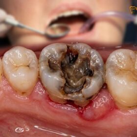
For a patient wearing custom removable prosthetics, this vascular impairment presents a critical challenge. The basal seat—the ridge of gum and bone that supports the denture—must withstand significant compressive forces during mastication (chewing). In a healthy individual, robust microcirculation allows the tissue to quickly recover from this mechanical stress. However, in a diabetic patient, the compromised gum microcirculation means the tissue is friable, less resilient, and prone to ischemic breakdown. Even minor pressure imbalances from a slightly ill-fitting denture can rapidly escalate into painful ulcerations or pressure sores.
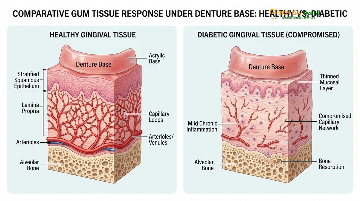
Furthermore, the impairment of diabetic dental healing is a major clinical concern. When a sore spot develops, the body’s natural inflammatory response is blunted. Leukocytes (white blood cells) exhibit reduced chemotaxis and phagocytosis in hyperglycemic environments, meaning they are less effective at migrating to the site of injury and clearing away necrotic tissue or invading bacteria[1]. Consequently, what might be a minor, self-resolving irritation in a non-diabetic patient can quickly become a chronic, non-healing ulcer in someone managing diabetes.
Prosthodontists must account for this delayed diabetic dental healing by scheduling more frequent postoperative adjustment visits. The goal is to identify and relieve areas of excessive pressure before the tissue breaks down. Patients are often advised to limit their wearing time during the initial adaptation phase, allowing the vulnerable mucosa adequate time to recover from the mechanical load of the prosthesis.
Pre-Extraction Checklist: Managing Blood Sugar and HbA1c Levels
Before extracting failing teeth to transition to dentures, diabetic patients must achieve stable glycemic control. Strict adherence to HbA1c guidelines minimizes the severe risk of postoperative socket infections and ensures proper ridge formation.
The transition from a failing natural dentition to complete or partial dentures often requires the surgical extraction of remaining teeth. For diabetic patients, this pre-prosthetic surgical phase carries elevated risks that must be meticulously managed. The primary concern is the prevention of a tooth extraction socket infection, a complication that can severely delay the fabrication and fitting of the final prosthesis.
Clinical protocols dictate that a comprehensive evaluation of the patient’s glycemic status is mandatory before any surgical intervention. The most reliable metric for this assessment is the HbA1c level, which reflects the average blood glucose concentration over the preceding two to three months. Adhering to established HbA1c control guidelines is not merely a recommendation; it is a critical safety measure to ensure predictable surgical outcomes and optimal diabetic dental healing.
When blood sugar levels are uncontrolled, the risk of a tooth extraction socket infection increases exponentially. High glucose levels in the blood and saliva provide an ideal breeding ground for pathogenic oral bacteria. Furthermore, the impaired immune response means the body cannot effectively wall off the extraction site, potentially leading to localized osteomyelitis (bone infection) or severe delayed healing of the alveolar ridge.
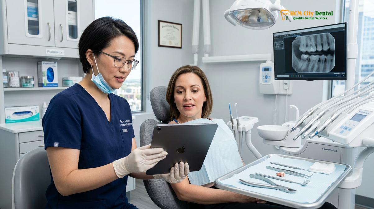
To mitigate these risks, dental professionals utilize strict preoperative parameters. The following table outlines the general clinical consensus regarding HbA1c levels and the corresponding surgical protocols for pre-prosthetic extractions:
| HbA1c Level (%) | Clinical Interpretation | Extraction & Prosthetic Protocol |
|---|---|---|
| Under 7.0% | Well-Controlled | Routine extractions can proceed safely. Standard healing protocols apply for immediate or delayed denture placement. |
| 7.0% – 8.0% | Moderately Controlled | Extractions proceed with caution. Prophylactic antibiotics may be prescribed to prevent tooth extraction socket infection. Frequent follow-ups required. |
| 8.0% – 9.0% | Poorly Controlled | Elective pre-prosthetic extractions are generally postponed. Consultation with an endocrinologist is required to stabilize levels before surgery. |
| Above 9.0% | Uncontrolled / High Risk | Strict contraindication for elective extractions. High risk of severe complications and impaired diabetic dental healing. Emergency care only. |
By strictly following these HbA1c control guidelines, the clinical team ensures that the alveolar bone heals smoothly and symmetrically, creating a stable and healthy foundation for the future denture[2]. Patients are often placed on a specialized antimicrobial rinse regimen immediately following the extractions to further suppress bacterial loads during the critical initial healing phase.
Xerostomia (Dry Mouth) in Diabetics: The Impact on Denture Retention
Diabetic xerostomia severely reduces salivary flow, eliminating the natural suction required to hold dentures in place. This lack of lubrication also increases mucosal friction, making specialized adhesives and hydration strategies essential.
One of the most pervasive and frustrating complications of diabetes in the oral cavity is xerostomia, commonly known as dry mouth. Saliva is a complex biological fluid that plays a multifaceted role in the success of removable prosthetics. It acts as a critical lubricant, a buffering agent against acidic byproducts, and, most importantly, the primary medium for creating the peripheral seal that holds a denture securely against the palate and alveolar ridges.
In diabetic patients, chronic hyperglycemia induces polyuria (excessive urination), leading to systemic dehydration that directly diminishes the output of the salivary glands. Additionally, many medications prescribed to manage diabetes and its associated comorbidities (such as antihypertensives or neuropathy drugs) list xerostomia dry mouth as a primary side effect. The resulting lack of saliva has a devastating impact on denture retention.
“Saliva provides the essential cohesive and adhesive forces required for prosthetic stability. In the diabetic patient presenting with severe xerostomia, the interfacial surface tension is lost, transforming a perfectly fitted denture into a loose, friction-inducing appliance.”
Without an adequate salivary film, the acrylic base of the denture rests directly against the dry, fragile oral mucosa. During speech and mastication, the micro-movements of the loose denture create severe mechanical friction. Because the tissue lacks the protective, gliding layer of saliva, this friction rapidly strips away the superficial epithelial cells, leading to painful abrasions and ulcerations. This is why managing xerostomia dry mouth is a cornerstone of diabetic dentures gum care.
To combat this, clinicians often recommend the strategic use of saliva substitutes and specialized hydration protocols. Patients are advised to take frequent sips of water and utilize oral moisturizing gels specifically formulated for dry mouth. Furthermore, the application of a high-quality denture adhesive becomes not just a convenience, but a clinical necessity to artificially recreate the lost peripheral seal. Patients must be educated on safe denture adhesive application to ensure they use the correct amount without altering the vertical dimension of occlusion or causing uneven pressure distribution[3].
Pressure Management: Specially Formulated Soft Gums Interfaces
To protect fragile diabetic gums, prosthodontists utilize low-pressure impression techniques and resilient soft liners. These specialized interfaces absorb masticatory shock and distribute forces evenly, preventing ischemic tissue breakdown.
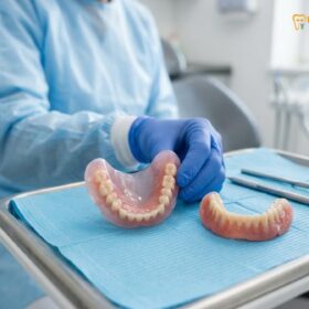
Given the compromised gum microcirculation and the heightened risk of mucosal ulceration, standard hard acrylic denture bases are often contraindicated for patients with advanced diabetes. The rigid nature of polymethyl methacrylate (PMMA) transmits the full force of the bite directly into the underlying bone and soft tissue. For a diabetic patient, this concentrated pressure can cause localized ischemia (lack of blood flow), leading to rapid tissue necrosis and accelerated alveolar bone resorption.
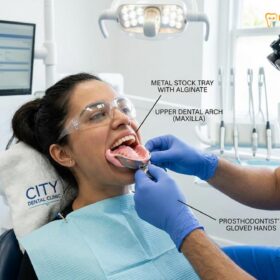
To address this biomechanical challenge, prosthodontists employ the concept of low pressure dentures fitting. This approach begins at the very first appointment during the impression phase. Instead of using mucocompressive techniques that displace the tissue, clinicians utilize mucostatic impression materials. These highly fluid materials capture the exact anatomical contours of the gums in their resting state, ensuring that the final denture base does not exert undue pressure on any specific anatomical landmark.
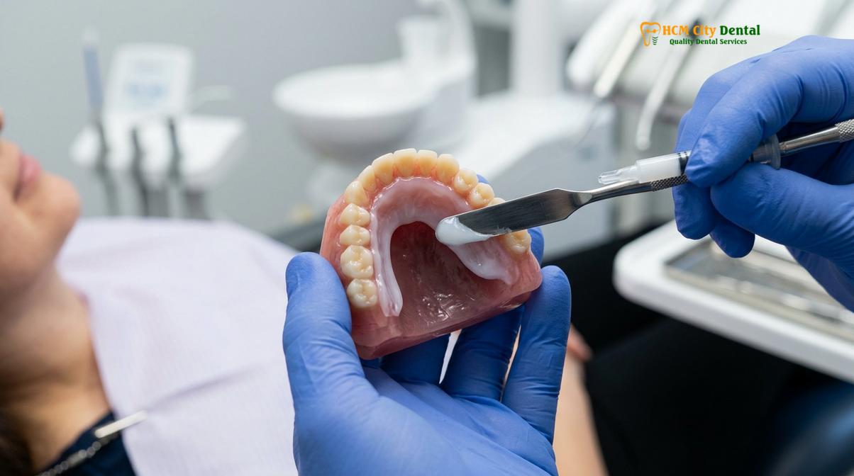
The most critical component of a low pressure dentures fitting, however, is the incorporation of a resilient soft liner. These specially formulated interfaces—typically made from medical-grade silicone or specialized plasticized acrylics—are permanently bonded to the tissue-bearing surface of the denture. The soft liner acts as a shock absorber, cushioning the fragile diabetic mucosa against the harsh impact of chewing forces.
When a patient bites down, the resilient liner compresses slightly, distributing the masticatory load over a much wider surface area. This even distribution prevents the formation of high-pressure focal points, thereby safeguarding the underlying gum microcirculation and preventing the development of sore spots[4].
It is important to note that these soft liners require meticulous maintenance. Because they are porous, they can harbor bacteria and fungi if not cleaned properly. Furthermore, the plasticizers in the material gradually leach out over time, causing the liner to harden. Therefore, diabetic patients utilizing soft-lined prosthetics must commit to regular clinical evaluations. When the liner begins to lose its resilience, undergoing clinical denture relining procedures is essential to restore the protective cushioning and maintain optimal tissue health.
Preventative Cleaning: Fighting Diabetic Oral Thrush (Candidiasis)
High salivary glucose levels in diabetics create an ideal breeding ground for Candida albicans. Rigorous daily cleaning protocols using specialized antimicrobial solutions are mandatory to prevent the development of painful oral thrush.
While mechanical trauma is a primary concern in diabetic dentures gum care, biological threats are equally formidable. Diabetic patients are significantly more susceptible to opportunistic fungal infections, the most common being oral candidiasis, widely known as oral thrush. The pathogen responsible, *Candida albicans*, is a naturally occurring yeast in the oral cavity. However, in a healthy mouth, it is kept in check by a robust immune system and a balanced oral microbiome.
In the diabetic mouth, the environment is drastically altered. Elevated blood sugar levels translate directly to increased glucose concentrations in the saliva and gingival crevicular fluid. This sugar-rich environment provides an abundant food source for *Candida albicans*, allowing it to proliferate rapidly. Compounded by xerostomia dry mouth—which reduces the mechanical flushing action of saliva—and a weakened local immune response, the yeast can easily colonize the porous surfaces of the denture acrylic and the underlying mucosal tissues.
Clinical Warning: Signs of Diabetic Oral Thrush
Diabetic denture wearers must be vigilant for symptoms of candidiasis, which include white, cottage-cheese-like plaques on the gums, tongue, or palate; severe redness and burning sensations under the denture base; and cracking at the corners of the mouth (angular cheilitis). If these symptoms appear, discontinue denture use immediately and seek professional antifungal treatment.
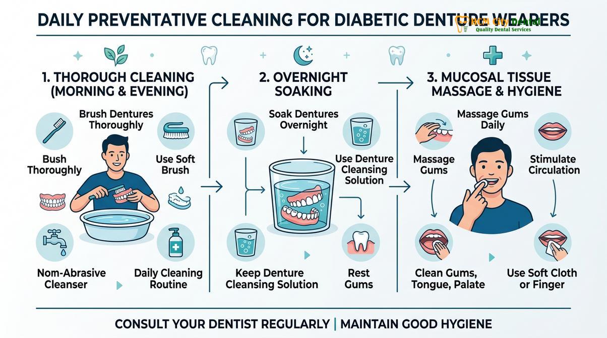
Preventing oral thrush requires an aggressive and consistent approach to prosthetic hygiene. Standard brushing with regular toothpaste is insufficient and can actually create micro-scratches in the acrylic where fungi can hide. Instead, diabetic patients must adhere to rigorous denture cleaning protocols that combine mechanical biofilm disruption with chemical disinfection.
The daily regimen should include brushing the appliance with a soft-bristled denture brush and a non-abrasive cleanser to remove gross debris. Crucially, the denture must then be soaked overnight in a specialized alkaline peroxide or chlorhexidine-based solution. This overnight soaking is vital, as it allows the antimicrobial agents sufficient time to penetrate the microscopic pores of the acrylic and eradicate fungal spores[5]. Furthermore, patients must never wear their dentures 24 hours a day; leaving the appliance out at night allows the oral tissues to breathe and recover from the day’s mechanical stress.
Coordination of Care: Syncing with Your Endocrinologist for Dental Work
Successful prosthetic rehabilitation for diabetics requires a multidisciplinary approach. Dentists and endocrinologists must collaborate to manage medication timing, stabilize blood sugar, and ensure systemic readiness for dental procedures.
The oral cavity is not an isolated system; it is deeply interconnected with the body’s overall systemic health. Therefore, providing exceptional diabetic dentures gum care requires a collaborative, multidisciplinary approach. A prosthodontist cannot successfully rehabilitate a diabetic patient’s smile without ensuring that the underlying metabolic condition is stable. This necessitates seamless coordination of care between the dental team and the patient’s managing endocrinologist or primary care physician.
Before initiating any complex treatment plan—especially those involving pre-prosthetic surgery or the management of a severe tooth extraction socket infection—the dental clinic must obtain a comprehensive medical clearance. This communication ensures that the dentist is fully aware of the patient’s current HbA1c control guidelines, their specific medication regimen (including insulin timing), and any associated comorbidities such as cardiovascular disease or diabetic neuropathy.
Clinical Case Review: Multidisciplinary Success
A 62-year-old patient with Type 2 Diabetes presented to HCMC Dental Clinic in Ho Chi Minh City experiencing severe, recurrent ulcerations under an old, ill-fitting lower denture. Initial diagnostics by Dr. Nguyen Van Cuong revealed an elevated HbA1c of 8.4% and profound xerostomia. Rather than immediately fabricating a new prosthesis, Dr. Cuong coordinated with the patient’s endocrinologist to adjust their glycemic management plan. After three months of systemic stabilization (HbA1c reduced to 7.1%), Dr. Cuong utilized a low pressure dentures fitting technique with a resilient silicone liner. The patient successfully adapted to the new prosthesis without any recurrence of mucosal breakdown, highlighting the necessity of integrated medical-dental care.
Coordination is also vital for appointment scheduling. Diabetic patients are generally advised to schedule their dental visits in the morning, shortly after breakfast and their morning medication. This timing helps prevent hypoglycemic episodes (dangerously low blood sugar) while in the dental chair. The dental team must be prepared with rapid-acting glucose sources and be trained to recognize the early signs of a diabetic emergency. By syncing clinical efforts, the medical and dental teams create a safe, predictable environment for the patient’s transition to a functional, comfortable smile, supported by a structured dietary adaptation plan that aligns with their nutritional requirements.
When to See a Doctor: Important Clinical Considerations
Diabetic patients must seek immediate professional evaluation if they experience persistent oral pain, non-healing sores, or signs of infection. Early intervention prevents minor irritations from escalating into severe systemic complications.
While meticulous daily care and adherence to clinical protocols significantly reduce the risk of complications, diabetic patients must remain vigilant regarding their oral health. The compromised nature of diabetic tissue means that minor issues can rapidly escalate into severe infections if left untreated. It is crucial to understand the distinction between normal adaptation discomfort and pathological warning signs that require immediate medical attention.
You should schedule an urgent evaluation with a dental professional if you experience any of the following symptoms:
- Non-Healing Ulcerations: Any sore spot or abrasion under the denture that does not show signs of improvement within 48 to 72 hours after removing the appliance.
- Signs of Acute Infection: Localized swelling, severe redness, throbbing pain, or the presence of pus (exudate) along the gumline or at previous extraction sites.
- Fungal Overgrowth: The appearance of white, easily scraped-off patches on the tongue, palate, or inner cheeks, often accompanied by a burning sensation or altered taste.
- Severe Prosthetic Instability: A sudden loss of denture retention that cannot be managed with standard adhesives, indicating potential rapid bone resorption or severe tissue dehydration.
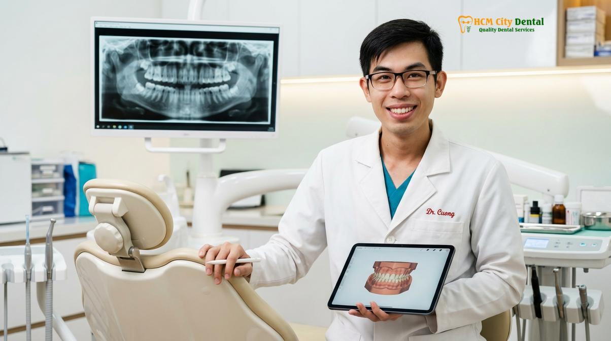
At HCMC Dental Clinic, our approach to comprehensive general dentistry emphasizes proactive monitoring for our diabetic patients. Dr. Nguyen Van Cuong strongly advises that diabetic denture wearers undergo professional clinical examinations at least every six months. These routine visits allow the clinical team to assess the integrity of the gum microcirculation, evaluate the fit of the prosthesis, and perform professional ultrasonic cleaning to eradicate hardened bacterial biofilms before they cause irreversible tissue damage.
Frequently Asked Questions
What HbA1c level is safe for teeth extractions before dentures?
An HbA1c level below 7.0% is generally considered safe for routine dental extractions, while levels between 7.0% and 8.0% require cautious clinical evaluation. Patients with levels above 8.0% face a significantly higher risk of delayed healing and postoperative infections, often necessitating a postponement of non-emergency surgical procedures until glycemic control improves. The dental team will coordinate with your physician to ensure your metabolic state is optimized before proceeding with pre-prosthetic surgery.
Why do diabetics get sore spots easier from dentures?
Diabetics develop sore spots more easily because elevated blood sugar compromises gum microcirculation and reduces salivary flow, leaving the oral mucosa thin, dry, and highly susceptible to mechanical friction. Without adequate lubrication and robust tissue resilience, even minor pressure imbalances from a denture base can quickly escalate into painful ulcerations. This necessitates the use of highly accurate impression techniques and often the incorporation of soft resilient liners to cushion the fragile tissues.
Can diabetic patients successfully get implant-supported overdentures?
Yes, diabetic patients can successfully receive implant-supported overdentures provided their blood sugar is well-controlled and they maintain rigorous oral hygiene. While the integration phase may require a slightly longer healing period, implant overdentures significantly reduce mucosal friction and bone resorption, making them an excellent long-term solution for patients managing diabetes. A thorough preoperative assessment of bone density and systemic health is required to determine candidacy.
How often should diabetics clean their dentures to prevent thrush?
Diabetic patients should clean their dentures thoroughly at least twice a day using a soft brush and non-abrasive cleanser, and soak them overnight in a specialized antifungal or antibacterial solution. Because high salivary glucose creates an ideal environment for Candida albicans, meticulous daily biofilm removal is mandatory to prevent oral thrush. Leaving the dentures out at night also gives the vulnerable oral tissues time to rest and recover from daily mechanical stress.
Can dry mouth from diabetes be cured for denture wearers?
While diabetic dry mouth cannot always be permanently cured, it can be highly effectively managed through strict glycemic control, the use of specialized saliva substitutes, and proper hydration. Denture wearers can also utilize specialized adhesives and low-pressure prosthetic designs to compensate for the lack of natural salivary retention and lubrication. Frequent sips of water and avoiding alcohol-based mouthwashes are also critical daily management strategies.
References
- Journal of Prosthodontic Research. Microvascular changes in diabetic oral mucosa and implications for removable prosthodontics. (2021).
- American Dental Association. Clinical guidelines for managing diabetic patients during oral surgery and prosthetic rehabilitation. (2022).
- International Journal of Oral Science. The impact of xerostomia on denture retention and mucosal health in diabetic populations. (2020).
- Journal of Clinical Periodontology. Biomechanical pressure distribution using resilient liners in compromised diabetic ridges. (2019).
- Oral Surgery, Oral Medicine, Oral Pathology. Fungal biofilm management and prevention of candidiasis in diabetic denture wearers. (2023).