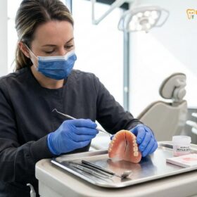
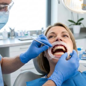
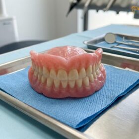
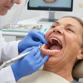
A comprehensive prosthodontic examination evaluates the residual alveolar ridge, oral mucosa, salivary flow, and muscle attachments to determine the optimal foundation for dental prosthetics. This precise diagnostic process aims to maximize retention, stability, and comfort for patients requiring removable or implant-supported restorations.
Clinical Summary:
A thorough prosthodontic evaluation is the cornerstone of successful dental rehabilitation. By systematically assessing hard and soft tissue anatomy, including bone volume, mucosa resilience, and frenum positions, clinicians can accurately predict prosthetic outcomes. Advanced radiographic imaging further aids in identifying anatomical limitations, allowing for tailored treatment planning that mitigates risks of denture instability, tissue trauma, and masticatory dysfunction. This meticulous approach ensures that the final prosthesis harmonizes with the patient’s unique oral biomechanics.
Key Takeaways:
- Comprehensive residual ridge assessment dictates the stability and load-bearing capacity of future dental prosthetics.
- Healthy, keratinized oral mucosa provides a superior foundation compared to hyperplastic or displaceable tissue.
- Adequate salivary flow is a critical requirement for creating the peripheral seal necessary for denture suction.
- High frenum attachments can actively dislodge prostheses and may require surgical modification prior to fabrication.
- Digital radiographic diagnostics, including CBCT, accurately map bone topography to guide customized treatment pathways.
The Residual Ridge Evaluation: Assessing Height, Width, and Shape
Evaluating the residual alveolar ridge’s dimensions and morphology is critical for determining the foundational support and stability available for any dental prosthesis.
The foundation of any successful removable prosthesis lies in the anatomical integrity of the underlying bone. Following tooth extraction, the alveolar bone undergoes a continuous, irreversible process of resorption. A meticulous residual ridge assessment is paramount to understanding the biomechanical limitations of the patient’s oral cavity. The maxilla typically resorbs in a superior and posterior direction, resulting in a progressively smaller upper arch. Conversely, the mandible resorbs inferiorly and anteriorly, creating a wider arch discrepancy that complicates the establishment of a harmonious occlusal relationship.
During the clinical examination, the prosthodontist evaluates the ridge based on its height, width, and cross-sectional shape. A broad, U-shaped ridge offers the most favorable prognosis, providing maximum surface area for stress distribution and excellent lateral stability against functional forces[1]. In contrast, a V-shaped or knife-edge ridge presents significant clinical challenges. The sharp bony crest concentrates occlusal loads onto a narrow band of overlying mucosa, frequently resulting in chronic pain and ulceration during mastication. In severe cases of atrophy, the ridge may become entirely flat, offering minimal resistance to lateral displacement.
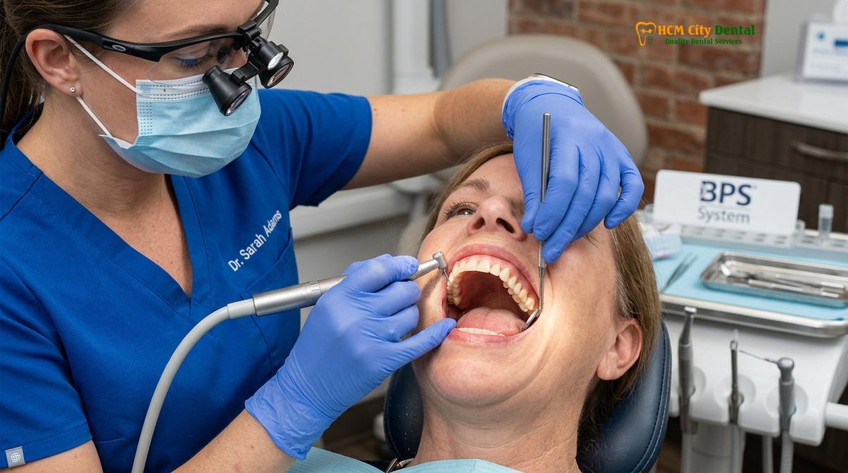
Beyond the primary ridge, secondary anatomical landmarks must be carefully palpated and evaluated. In the maxilla, the hard palate, maxillary tuberosities, and the hamular notches serve as critical support and retention areas. In the mandible, the buccal shelf—composed of dense cortical bone—acts as the primary stress-bearing region, while the retromolar pad provides a stable posterior boundary that resists resorption. Identifying these landmarks ensures that the future prosthesis can be designed to distribute forces optimally.
Understanding Mucosa Tissue Quality: Keratinized vs. Flabby Tissue
The resilience and health of the oral mucosa directly impact a patient’s ability to tolerate occlusal forces without experiencing pain, inflammation, or tissue ulceration.
The soft tissue overlying the residual bone acts as a biological cushion, absorbing and dissipating the immense pressures generated during chewing. The ideal oral mucosa health is characterized by a thick, firmly attached layer of keratinized stratified squamous epithelium. This resilient tissue can withstand the frictional and compressive forces of a rigid acrylic denture base without breaking down. When the mucosa is firmly bound to the underlying periosteum, it helps prevent the prosthesis from shifting, thereby enhancing overall stability.
However, many patients present with compromised soft tissue conditions. One of the most challenging clinical scenarios is the presence of a “flabby ridge.” This condition occurs when the underlying alveolar bone has resorbed rapidly, leaving behind a mass of mobile, hyperplastic fibrous tissue. Flabby tissue is highly displaceable; if a standard impression technique is used, the tissue will be compressed. Once the final denture is inserted, the tissue attempts to rebound to its resting state, causing the denture to unseat and lose retention. In some instances, alternative materials such as thermoplastic flexible partial dentures are evaluated for their clinical suitability to adapt to varying mucosal conditions[5].
“Prosthetic success relies heavily on the quality of the soft tissue foundation; placing a rigid denture base over highly displaceable mucosa without utilizing specialized mucostatic impression techniques often leads to chronic instability and severe patient discomfort.”
To manage displaceable tissue, clinicians must employ selective pressure or completely mucostatic impression techniques. These advanced methods capture the flabby tissue in its passive, undistorted state while applying functional pressure only to the healthy, load-bearing areas. Furthermore, the mucosa must be evaluated for signs of candidiasis, epulis fissuratum, or chronic inflammation, which generally need to be resolved through tissue conditioning before any final impressions are taken.
The Role of Saliva: Natural Lubrication and Denture Suction Retention
Saliva acts as a critical physical adhesive and biological lubricant, enabling the interfacial surface tension required for effective denture retention.
While bone and mucosa provide support and stability, retention—the resistance to vertical displacement—is heavily dependent on the physical properties of saliva. A healthy salivary flow rate is essential for creating the peripheral seal that locks a complete denture in place. The physics of denture retention rely on adhesion (the attraction between saliva and the acrylic base), cohesion (the internal attraction of saliva molecules), and interfacial surface tension. Together, these forces create a negative atmospheric pressure beneath the denture, commonly referred to as “suction.”
The quality of the saliva is just as important as the quantity. Thin, serous saliva forms an ideal, even film that maximizes surface tension and retention[2]. Conversely, thick, ropy, mucous saliva can accumulate unevenly, breaking the peripheral seal and causing the denture to lose its grip. Furthermore, saliva serves as a vital biological lubricant, reducing friction between the rigid prosthesis and the delicate oral mucosa during speech and mastication.
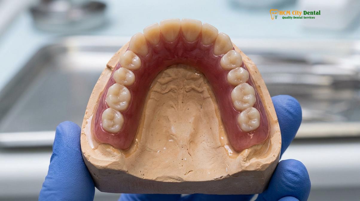
During the examination, the clinician will assess the pooling of saliva in the floor of the mouth and the moisture level of the buccal mucosa. Patients taking multiple medications, such as antihypertensives or antidepressants, frequently suffer from drug-induced xerostomia. For these individuals, the fabrication of customized removable dental prosthetics requires specialized approaches, including the use of artificial saliva substitutes, prolonged tissue conditioning, or the integration of dental implants to provide mechanical retention independent of salivary physics.
Evaluating Muscle and Frenum Attachments: Preventing Displacement
Identifying the location and tension of muscle and frenum attachments is essential to establish proper denture borders that resist functional dislodgment.
The oral cavity is a highly dynamic environment governed by complex muscular interactions. The borders of a dental prosthesis must rest in harmony with these moving structures; if a denture is overextended into active muscle tissue, the muscles will inevitably dislodge the appliance during normal functions such as speaking, swallowing, or smiling. Therefore, a meticulous evaluation of the surrounding musculature and frenum attachments is a critical phase of the prosthodontic examination.
Frenum attachments are fibrous bands of connective tissue that tether the lips and cheeks to the alveolar ridge. In a healthy, unresorbed ridge, these attachments are located far from the crest. However, as bone resorption progresses, the relative position of the frenum migrates closer to the crest of the ridge. If a high frenum attachment is not properly accommodated with a precise notch in the denture border, the tension from lip movement will break the peripheral seal and unseat the prosthesis.
| Anatomical Structure | Impact on Prosthesis Stability | Clinical Management Strategy |
|---|---|---|
| Labial/Buccal Frenum | Can break peripheral seal and dislodge denture during facial expressions. | Precise border molding to create relief notches; surgical frenectomy if severe. |
| Mentalis Muscle | Strong upward contraction can lift the anterior mandibular denture. | Careful determination of the labial vestibule depth; avoidance of overextension. |
| Mylohyoid Muscle | Elevates the floor of the mouth during swallowing, displacing lower dentures. | Dynamic functional impressions to capture the muscle in its contracted state. |
| Masseter Muscle | Can push against the distobuccal border of the lower denture, causing soreness. | Contouring the masseteric notch on the impression tray to allow muscle clearance. |
The dynamic nature of the floor of the mouth presents one of the greatest challenges in mandibular denture fabrication. The mylohyoid muscle, which forms the muscular floor of the mouth, elevates during swallowing. If the lingual flange of the lower denture is overextended, this muscular action will lift the denture entirely off the ridge. Capturing the precise functional depth and width of these anatomical spaces requires advanced border molding techniques, ensuring the final appliance integrates seamlessly with the patient’s neuromuscular system.
Radiographic Diagnostics: Panoramic and CBCT Ridges Measurement
Advanced imaging techniques provide a comprehensive three-dimensional view of the underlying osseous structures, revealing hidden pathologies and precise bone volumes.
While a visual and tactile clinical examination provides invaluable data regarding the soft tissues and surface topography, it cannot reveal the internal architecture of the jawbones. Radiographic diagnostics are an indispensable component of a modern prosthodontic evaluation. A digital panoramic x-ray serves as the standard baseline imaging modality, offering a broad, two-dimensional overview of the maxilla, mandible, temporomandibular joints (TMJ), and surrounding vital structures.
The panoramic radiograph allows the clinician to screen for retained root tips, impacted teeth, radiolucent cysts, or other asymptomatic intraosseous pathologies that must be addressed before prosthetic fabrication. Furthermore, it provides a general assessment of the vertical bone height relative to critical landmarks, such as the inferior alveolar nerve canal in the mandible and the maxillary sinus floor in the upper jaw, which is vital for planning premium prosthetic solutions[3]. However, because panoramic images are two-dimensional and subject to magnification distortion, they have limitations in complex cases.
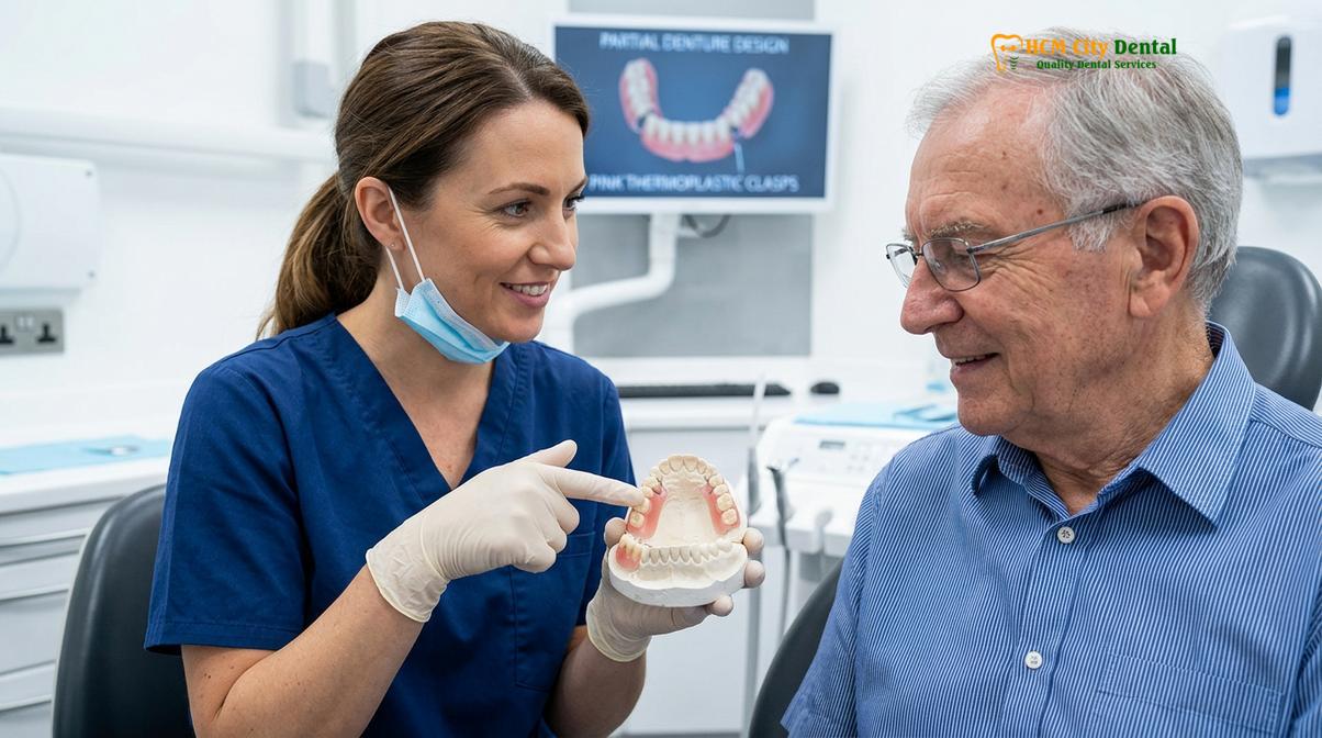
When anatomical limitations are severe, or when the patient is considering implant-supported solutions, a 3D Cone Beam Computed Tomography (CBCT) scan becomes highly recommended. CBCT imaging provides highly accurate, distortion-free, three-dimensional cross-sections of the alveolar ridge. It allows the prosthodontist to measure the exact buccolingual width of the bone, assess cortical bone density, and virtually plan the trajectory of dental implants. This level of diagnostic precision is routinely utilized to ensure predictable and safe surgical outcomes.
Creating the Customized Treatment Path: Managing Anatomic Limitations
Synthesizing clinical and radiographic findings allows the prosthodontist to design a personalized treatment plan that addresses specific anatomical challenges.
The culmination of the prosthodontic examination is the synthesis of all diagnostic data to formulate a tailored treatment strategy. No two mouths are identical, and a standardized approach to denture fabrication frequently leads to suboptimal results. During a detailed prosthodontist consultation, the clinician will discuss the specific anatomical limitations identified—such as severe bone resorption, high muscle attachments, or poor salivary flow—and present targeted solutions to overcome them.
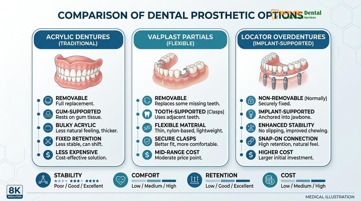
In many cases, pre-prosthetic surgery is required to optimize the foundation before impressions are taken. This may involve alveoloplasty to smooth sharp bony spicules, the removal of bulky bony exostoses (tori) that interfere with denture seating, or a vestibuloplasty to surgically deepen the vestibule and increase the surface area available for retention. If the anatomical limitations are too severe for a conventional removable appliance to function adequately, clinical studies often support transitioning to implant-supported overdentures for superior stability[4].
Clinical Case Review: A 65-year-old patient visited HCMC Dental Clinic in Ho Chi Minh City complaining of a completely unstable lower denture. Clinical evaluation revealed severe mandibular atrophy (a flat ridge) and a high floor of the mouth. The clinical team determined that conventional suction was highly unlikely due to the lack of vertical bone. The customized treatment path involved the placement of two symphyseal dental implants, transitioning the patient to an implant-retained overdenture, which immediately restored masticatory function and confidence.
By integrating advanced diagnostics with a deep understanding of oral biomechanics, modern dental practices can offer solutions ranging from premium biofunctional complete dentures to sophisticated implant-supported full-arch bridges. The goal is always to provide advanced removable prosthetics that not only restore aesthetics but also rehabilitate the patient’s ability to chew, speak, and live without discomfort.
When to See a Prosthodontist for an Evaluation
Timely clinical assessments are vital for identifying early signs of bone resorption and preventing irreversible damage to the oral structures beneath existing prosthetics.
Patients who currently wear dental prosthetics or those facing the prospect of complete tooth loss must be vigilant about their oral health. The jawbone and soft tissues change continuously over time, meaning a denture that fit perfectly several years ago may now be causing silent, irreversible damage to the underlying structures. According to guidelines aligned with the Vietnam Odonto-Stomatology Association (VOSA), regular prosthodontic assessments are crucial for aging populations to maintain optimal masticatory function and oral health.
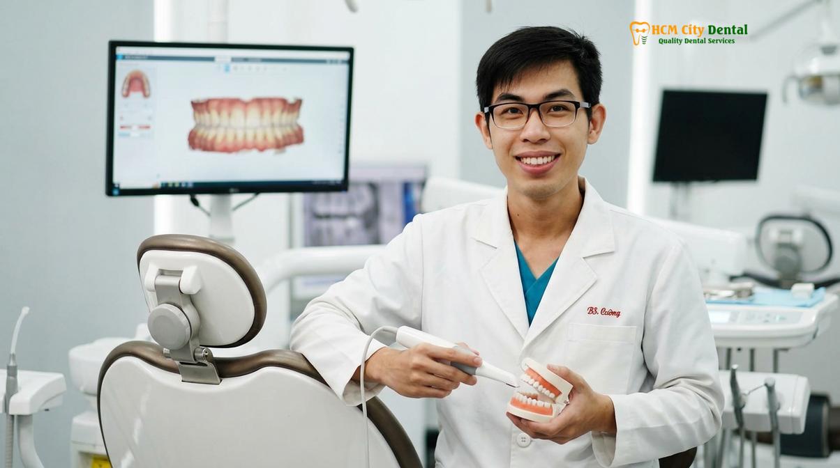
Dr. Nguyen Van Cuong, a leading specialist, emphasizes the importance of timely prosthodontic evaluations to prevent severe alveolar bone loss. He routinely advises patients that early intervention not only preserves the foundational jaw structure but also significantly improves the long-term success of any dental prosthesis. This proactive approach is a core component of comprehensive general dentistry care.
“Never ignore chronic sore spots or instability under a dental appliance. These are early warning signs of structural changes in the jawbone that require immediate professional reassessment to prevent further tissue degradation.”
Chronic sore spots, ulcerations, or bleeding gums beneath a denture are clear indicators that the appliance is no longer distributing forces evenly. Ignoring these signs can lead to accelerated bone loss and the development of hyperplastic flabby tissue. Additionally, if you notice a significant decrease in chewing efficiency, frequent dislodgment of the denture while speaking, or a collapsing facial profile (a sunken appearance around the lips and cheeks), your prosthetics require immediate reassessment.
References
- Journal of Prosthetic Dentistry. Biomechanical principles and tissue response in complete dentures. (2021).
- International Journal of Oral and Maxillofacial Surgery. Suction-effective lower dentures and border molding outcomes. (2020).
- British Dental Journal. Patient satisfaction and chewing efficacy with premium BPS dentures. (2019).
- Journal of the American Dental Association. Implant-supported overdentures vs. conventional removable prosthetics. (2022).
- Clinical Oral Investigations. Thermoplastic flexible partial dentures: clinical suitability and aesthetics. (2018).
If you are experiencing discomfort with your current dentures or are preparing for new dental prosthetics, a thorough clinical evaluation is the first step toward a stable, comfortable smile. Contact HCMC Dental Clinic in Ho Chi Minh City today to schedule your comprehensive prosthodontic assessment and explore personalized treatment options tailored to your unique oral anatomy.
For pricing, booking, and a free clinical assessment, visit our Dentures & Removable Prosthetics service page at HCMC Dental Clinic in Ho Chi Minh City.