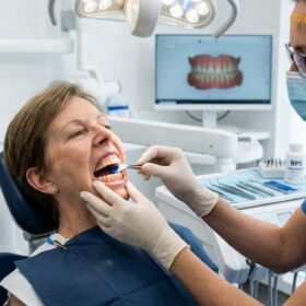
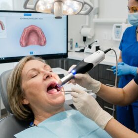
Bone loss after tooth loss occurs because the alveolar bone no longer receives the mechanical chewing stimulation required to maintain its density. Without a tooth root to signal cellular regeneration, the jawbone rapidly resorbs, leading to structural changes, facial collapse, and complications for future dental restorations.
Clinical Summary:
Following tooth extraction, the alveolar ridge undergoes immediate and progressive atrophy due to the cessation of biomechanical forces. This resorption is most aggressive during the first six to twelve months, resulting in significant horizontal and vertical bone volume reduction. While conventional removable prosthetics can restore chewing function, they do not halt this biological degradation and may even accelerate it through mucosal pressure. The only clinically proven methods to arrest alveolar bone resorption are socket preservation grafting at the time of extraction and the placement of endosteal dental implants, which mimic natural tooth roots to stimulate osteogenesis.
Key Takeaways:
- The alveolar bone requires constant mechanical stimulation from tooth roots to maintain its volume and density.
- Up to 25% of bone width can be lost within the first three months following an unpreserved tooth extraction.
- Traditional removable prosthetics resting on the gums do not prevent bone degradation and may increase resorption rates.
- Dental implants are the only restorative solution that provides internal stimulation to preserve jawbone architecture.
- Early intervention with socket grafting or immediate implant placement significantly improves long-term functional and aesthetic outcomes.
The Biology of the Alveolar Ridge: Why Bone Needs Teeth
The alveolar ridge exists solely to support teeth, relying on the mechanical forces of chewing to stimulate bone-building cells and maintain structural density.
To fully comprehend the phenomenon of bone loss after tooth loss, it is essential to understand the unique biological nature of the alveolar process. The jawbone is divided into two primary components: the basal bone, which forms the main structural body of the maxilla (upper jaw) and mandible (lower jaw), and the alveolar bone, which is the specialized ridge that houses the tooth sockets. Unlike the basal bone, which remains relatively stable throughout life, the alveolar bone is highly dynamic and completely dependent on the presence of teeth for its continued existence.
The relationship between a tooth and its surrounding bone is mediated by the periodontal ligament (PDL), a complex network of connective tissue fibers that suspends the tooth root within the socket. When you chew, bite, or swallow, mechanical forces are transmitted through the tooth, down the root, and into the PDL. The PDL acts as a shock absorber, translating these mechanical forces into tension and compression on the surrounding alveolar bone. This process, known as mechanotransduction, is the fundamental driver of bone health in the oral cavity.
According to Wolff’s Law of bone remodeling, bone tissue adapts to the loads under which it is placed. When mechanical stress is applied, it creates a microscopic piezoelectric effect—a tiny electrical charge that stimulates specific bone cells. Osteoblasts (cells responsible for building new bone) are activated to reinforce the bone structure, while osteoclasts (cells responsible for breaking down old bone) remove damaged or unnecessary tissue. In a healthy mouth with a full complement of teeth, this continuous cycle of osteoclasts activity and osteoblast regeneration maintains a dense, robust alveolar ridge.[1]
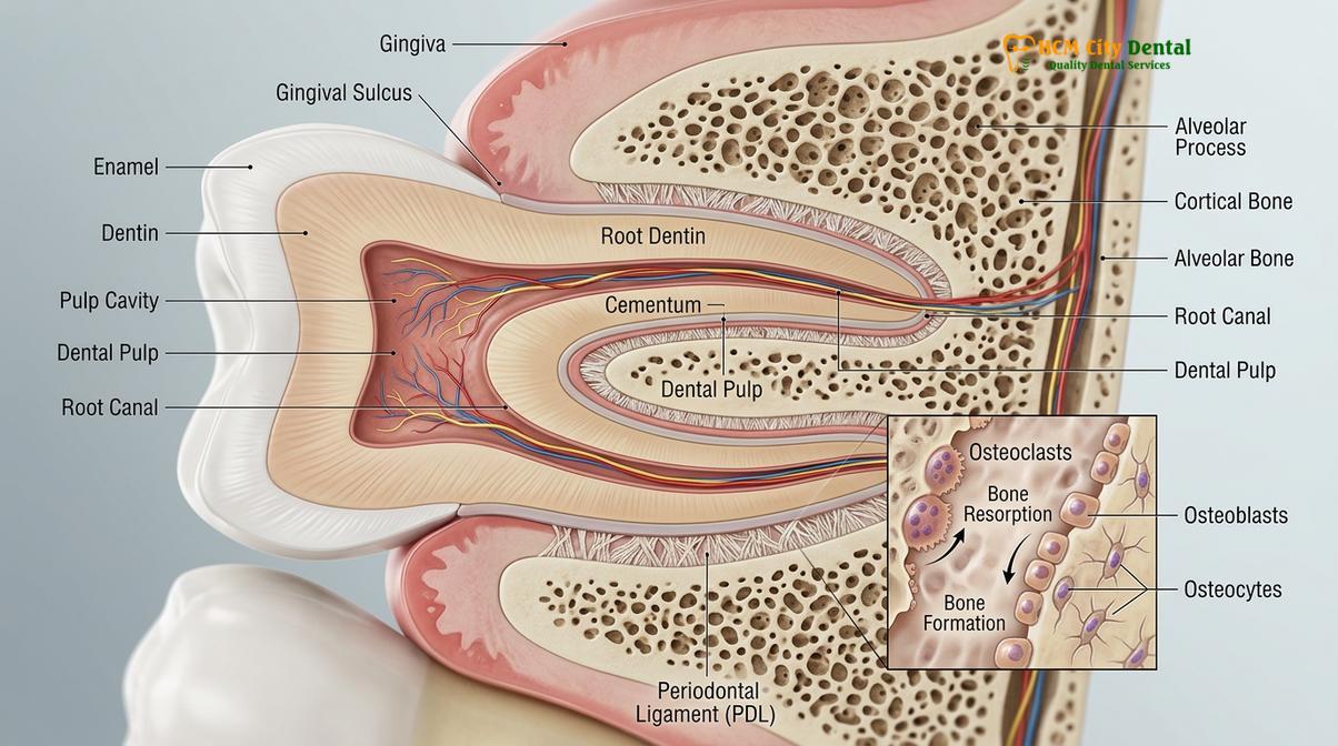
However, the moment a tooth is extracted, this delicate biological equilibrium is shattered. The severing of the periodontal ligament means that the alveolar bone in that specific area no longer receives any mechanical chewing stimulation. The body, which is highly efficient at conserving resources, interprets this lack of stimulation as a signal that the bone is no longer needed. Consequently, osteoblast activity ceases, while osteoclast activity continues unabated. The body begins to resorb the calcium and minerals from the redundant alveolar bone, redistributing them to other parts of the body. This process, clinically termed alveolar bone resorption, is an inevitable physiological consequence of tooth loss if preventative measures are not taken.
The cellular degradation of the jawbone is not merely a localized issue; it sets off a chain reaction that compromises the entire dental arch. As the bone shrinks, the adjacent teeth lose their lateral support, making them susceptible to shifting, tilting, and eventual mobility. Furthermore, the loss of bone volume drastically complicates future restorative efforts, as there may not be sufficient foundational tissue to support implants or even stable removable prosthetics.
Bone Loss Timeline: The Crucial First Year After Extraction
Jawbone deterioration begins immediately after extraction, with the most rapid and severe volume reduction occurring within the first six to twelve months.
The progression of bone loss after tooth loss is not a slow, linear process; rather, it is highly aggressive in the immediate aftermath of the extraction before tapering off into a chronic, lifelong condition. Understanding this timeline is critical for patients and clinicians when planning restorative treatments, as the window of opportunity for optimal intervention is relatively narrow.
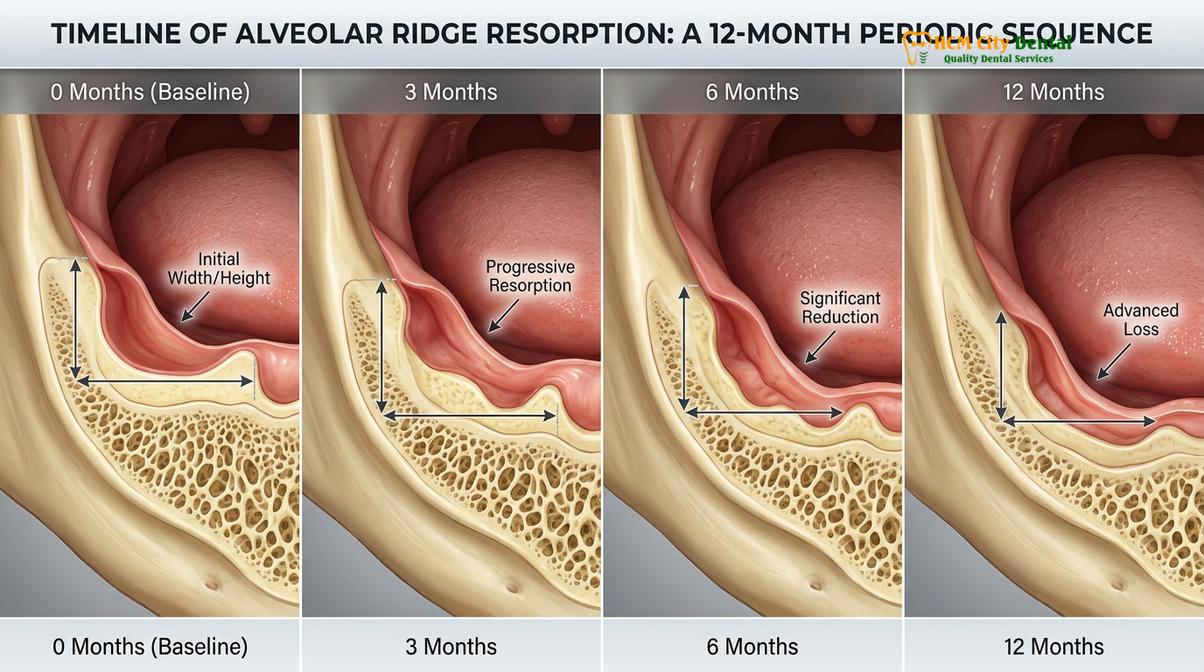
The acute phase of resorption begins within days of the extraction. Once the blood clot forms and the soft tissue begins to heal over the socket, the underlying bone remodeling process initiates. During the first three months, the alveolar ridge undergoes profound dimensional changes. Clinical studies indicate that the most significant loss occurs in the horizontal dimension (the width of the ridge). The buccal plate—the thin wall of bone on the cheek side of the tooth—is particularly vulnerable because it is naturally thinner and relies heavily on the blood supply from the periodontal ligament, which has been severed.[2]
By the six-month mark, the rate of resorption reaches its peak. Patients can experience up to a 25% reduction in bone width and a noticeable decrease in vertical height. The once broad and tall alveolar ridge begins to take on a knife-edge appearance, making it increasingly difficult to support any type of dental prosthesis. This rapid bone density loss is often imperceptible to the patient initially, as the gums heal and cover the defect, masking the structural collapse occurring beneath the surface.
| Time Post-Extraction | Biological Activity | Clinical Manifestation |
|---|---|---|
| 0 – 4 Weeks | Soft tissue healing; initial osteoclast activation. | Socket fills with granulation tissue; early crestal bone rounding. |
| 1 – 3 Months | Aggressive horizontal resorption; buccal plate degradation. | Noticeable narrowing of the ridge; up to 25% width reduction. |
| 3 – 6 Months | Continued horizontal loss; onset of significant vertical height reduction. | Ridge becomes knife-edged; adjacent teeth may begin to shift. |
| 6 – 12 Months | Peak of initial resorption phase; up to 50% total volume loss in severe cases. | Profound structural collapse; standard implants may require grafting. |
| 1 Year + | Chronic, continuous resorption at a rate of ~1% per year. | Long-term facial profile changes; severe prosthetic instability. |
As the timeline extends beyond the first year, the aggressive acute phase transitions into a chronic phase. While the rate of bone loss slows down to approximately 0.5% to 1% per year, it never truly stops. Over a decade, this continuous, insidious atrophy can result in the near-total obliteration of the alveolar ridge. In the mandible, severe resorption can expose the inferior alveolar nerve, causing chronic pain and numbness. In the maxilla, the bone loss often leads to the expansion of the maxillary sinus cavity (pneumatization), further reducing the available bone volume from the top down.
The anatomical differences between the upper and lower jaws also dictate the pattern of bone loss. The maxilla generally resorbs upward and inward, reducing the size of the upper dental arch. Conversely, the mandible tends to resorb downward and outward. This opposing pattern of deterioration creates a severe discrepancy in the bite relationship, making it exceptionally challenging for a dentist providing comprehensive general dentistry to align the jaws correctly without advanced surgical intervention.
Conventional Dentures and Bone Pressures: Accelerating Resorption?
Traditional removable dentures sit on the gum tissue, failing to provide internal bone stimulation and potentially accelerating bone loss through friction and pressure.
For generations, the standard treatment for extensive tooth loss has been the fabrication of removable complete or partial dentures. While these prosthetics successfully restore a degree of masticatory function and provide an immediate cosmetic improvement, they are fundamentally flawed from a biological perspective. Conventional dentures are tissue-borne, meaning they rest entirely on the oral mucosa (gums) and the underlying alveolar bone, rather than being anchored within the bone itself.
Because they sit on the surface, traditional dentures do not transmit the necessary mechanical forces into the bone to stimulate osteoblast activity. The bone remains biologically “unaware” that chewing is occurring, and therefore, the natural process of disuse atrophy continues uninterrupted. However, the situation is often worse than mere passive atrophy. The biomechanics of wearing a tissue-borne prosthesis can actively accelerate the rate of bone destruction.[3]
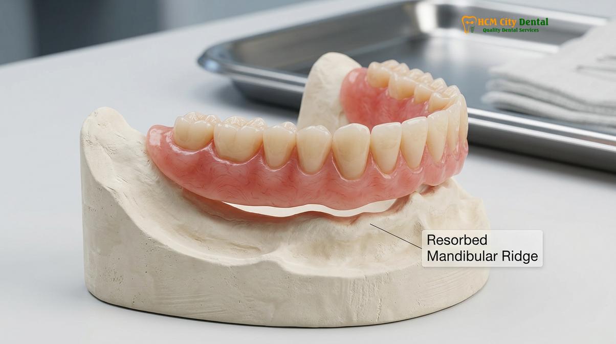
When a patient chews with a conventional denture, the rigid acrylic base compresses the soft gum tissue against the hard underlying bone. This compressive force can cause localized ischemia—a restriction of blood supply to the periosteum (the membrane covering the bone). The resulting lack of oxygen and nutrients, combined with the unnatural friction and micro-movements of a shifting denture, creates an inflammatory response. This chronic inflammation signals the body to increase osteoclast activity, leading to a faster breakdown of the bone tissue directly beneath the denture base.
This phenomenon creates a vicious cycle for the patient. As the bone shrinks away, the denture loses its foundational support and becomes loose. A loose denture moves more during speech and chewing, creating greater friction and uneven pressure spots, which in turn causes the bone to resorb even faster. Patients often find themselves needing frequent denture relines or entirely new prosthetics every few years just to maintain a basic level of function. Over decades of wear, the mandibular ridge can become so flat that it offers absolutely no retention for traditional removable prosthetics, leaving the patient functionally disabled.
Furthermore, the use of denture adhesives, while helpful for temporary retention, does nothing to mitigate the underlying biological problem. The mucoperiosteal burden—the stress placed on the gums and bone—remains high. It is a clinical reality that while conventional dentures replace the visible crowns of the teeth, they completely fail to replace the vital function of the tooth roots, making them a palliative rather than a curative solution for tooth loss.
Facial Collapse Syndrome: How Bone Loss Changes Your Facial Profile
Severe jawbone deterioration leads to a sunken facial appearance, decreased lower face height, and premature aging wrinkles around the mouth.
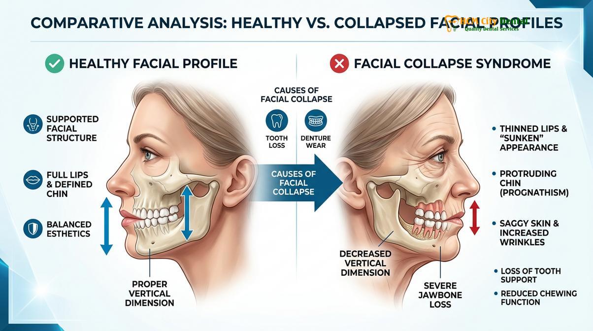
The consequences of alveolar bone resorption extend far beyond the confines of the oral cavity; they have a profound and devastating impact on the patient’s overall facial aesthetics. The jawbones provide the essential structural scaffolding for the lower third of the face, supporting the lips, cheeks, and surrounding musculature. When this scaffolding deteriorates, the soft tissues lose their foundation, resulting in a condition clinically referred to as facial collapse syndrome.
One of the most prominent signs of facial collapse is facial height reduction, specifically a decrease in the vertical dimension of occlusion (VDO). As the upper and lower jawbones shrink, the distance between the nose and the chin decreases. This loss of vertical height causes the lower jaw to rotate forward and upward, creating a pseudo-prognathic appearance—a severe underbite profile where the chin protrudes unnaturally. This structural shift drastically alters the natural proportions of the face, making the patient appear significantly older than their chronological age.
“The loss of vertical dimension in the lower third of the face not only compromises masticatory function but profoundly alters the patient’s aesthetic profile, often resulting in profound psychological impacts and a diminished quality of life.” – Journal of Prosthodontic Rehabilitation.
The soft tissue changes associated with bone loss are equally distressing. Without the underlying support of the teeth and alveolar ridge, the lips begin to cave inward, losing their fullness and definition. The vermilion border (the edge of the lips) thins out, and deep vertical wrinkles, often called “smoker’s lines,” form around the mouth even in non-smokers. The nasolabial folds—the lines running from the nose to the corners of the mouth—deepen significantly, and the corners of the mouth may turn downward, creating a permanent expression of sadness or fatigue.
Furthermore, the loss of bone in the posterior regions of the jaw causes the cheeks to hollow out, contributing to a gaunt, sunken appearance. The muscles of facial expression and mastication, lacking their proper attachment points and resting length, lose their tone and begin to sag. This comprehensive degradation of dental aesthetics can severely impact a patient’s self-esteem, leading to social withdrawal and depression.
Restoring a collapsed facial profile is one of the most challenging tasks in dentistry. While specialized prosthetics can artificially prop up the lips and cheeks, they cannot reverse the biological loss of bone. Preventing facial collapse requires proactive intervention at the time of tooth extraction, ensuring that the structural integrity of the jaw is maintained before the soft tissues have a chance to adapt to a diminished foundation.
Implant Stimulation: The Only Method to Signal Bone Preservation
Dental implants act as artificial tooth roots, transmitting chewing forces directly into the jawbone to stimulate cellular regeneration and halt resorption.
In the realm of modern restorative dentistry, endosteal dental implants represent a paradigm shift in the management of tooth loss. Unlike traditional bridges or dentures that merely sit above the gumline, dental implants are surgically placed directly into the alveolar bone. This fundamental difference in placement allows implants to perform the most critical function of a natural tooth root: biomechanical stimulation.
Dental implants are typically manufactured from biocompatible titanium or zirconia. When placed into the jaw, the bone tissue recognizes the implant surface and initiates a process called osseointegration. During osseointegration, osteoblasts grow directly onto the microscopic surface of the implant, fusing the titanium firmly to the living bone. This direct structural and functional connection is the key to halting bone loss.[4]
Once the implant is fully integrated and restored with a crown, bridge, or denture, it begins to transmit the forces of mastication deep into the surrounding bone. Just like a natural tooth, the implant creates the necessary mechanical stress that triggers the piezoelectric effect, signaling the body to maintain osteoblast activity. The bone is “tricked” into believing that a natural tooth is still present, effectively arresting the resorption process and preserving the crestal bone architecture.
For patients missing all their teeth, implant overdenture prevention strategies are highly effective. Instead of a traditional denture that rests on the gums, an overdenture snaps onto strategically placed implants. This hybrid approach provides the cosmetic replacement of a full arch of teeth while utilizing the implants to bear the occlusal load. The compressive forces that would normally destroy the bone are redirected through the implants, providing the necessary internal stimulation to keep the jawbone healthy and intact.
It is important to note that the success of implant therapy relies heavily on the quality and quantity of the existing bone. If a patient has waited years after tooth loss to seek treatment, significant resorption may have already occurred, necessitating complex bone grafting procedures before implants can be placed. Therefore, timely intervention is paramount to achieving the most predictable and conservative clinical outcomes.
Preventative Strategies: Bone Grafting at Extraction vs. Immediate Implants
Proactive treatments like socket preservation grafts or immediate implant placement at the time of extraction are critical to maintaining bone volume for future restorations.
The most effective way to manage bone loss after tooth loss is to prevent it from happening in the first place. Modern dental protocols dictate that a tooth should rarely be extracted without a concurrent plan for ridge preservation or immediate replacement. Clinicians have two primary strategies at their disposal to mitigate the inevitable biological collapse of the alveolar ridge: socket preservation grafting and immediate implant placement.
Socket preservation, also known as ridge preservation, is a proactive bone grafting procedure performed immediately after a tooth is extracted. The clinical workflow begins with an atraumatic extraction technique, where the dentist carefully removes the tooth while preserving the fragile buccal and lingual bone walls. Once the socket is thoroughly cleaned and debrided of any infected tissue, a bone grafting material is packed into the void. This material can be an allograft (human donor bone), xenograft (animal-derived bone, typically bovine), or a synthetic substitute.[5]
The graft acts as a biological scaffold, holding the space and preventing the surrounding bone and soft tissue from collapsing inward. A resorbable collagen membrane is often placed over the graft to prevent fast-growing gum tissue from invading the socket, allowing the slower-growing bone cells time to migrate into the graft matrix and regenerate new host bone. Over a period of four to six months, the graft material is gradually replaced by the patient’s own vital bone, resulting in a wide, dense ridge that is ideal for future implant placement.
“Implementing ridge preservation techniques immediately following tooth extraction significantly minimizes horizontal and vertical dimensional changes, providing a more predictable foundation for future implant therapy and optimal aesthetic outcomes.” – International Journal of Oral Implantology.
The second preventative strategy is immediate implant placement. In cases where there is no acute infection, sufficient bone beyond the apex of the root for primary stability, and a favorable gingival biotype, a dental implant can be placed into the socket during the same surgical visit as the extraction. This approach is highly advantageous because it immediately introduces a titanium root substitute into the bone, minimizing the time the bone is left without mechanical stimulation.
Immediate implants are often combined with a gap-filling bone graft to ensure that any space between the implant and the socket walls is completely filled, maximizing bone-to-implant contact. This streamlined protocol not only preserves the bone architecture but also significantly reduces the overall treatment time and the number of surgical interventions required for the patient.
Both socket preservation and immediate implants require meticulous surgical planning and execution. The choice between the two depends on a variety of clinical factors, including the location of the tooth, the presence of pathology, and the patient’s overall health. Consulting with a specialist for routine dental evaluations prior to any extraction is crucial to determining the most appropriate preventative strategy.
When to See a Doctor for Jaw Bone Loss
Jawbone loss is a silent condition that progresses without pain or obvious symptoms in its early stages. However, recognizing the clinical signs of alveolar atrophy is critical for seeking timely intervention before the damage becomes irreversible or requires extensive reconstructive surgery. If you have previously lost teeth or have been wearing traditional removable prosthetics, you should be vigilant for specific changes in your oral health and facial structure.
You should schedule a clinical consultation if you notice that your dentures, which once fit securely, have become loose, require excessive adhesive, or cause sore spots on your gums. This is a primary indicator that the underlying bone is shrinking. Additionally, if you observe shifting, tilting, or increased mobility in the teeth adjacent to an empty space, it suggests that the supporting bone architecture is failing. Changes in your facial profile, such as deepening wrinkles around the mouth, a sunken appearance to the cheeks, or a protruding chin, are late-stage signs of severe bone loss that warrant immediate professional evaluation.
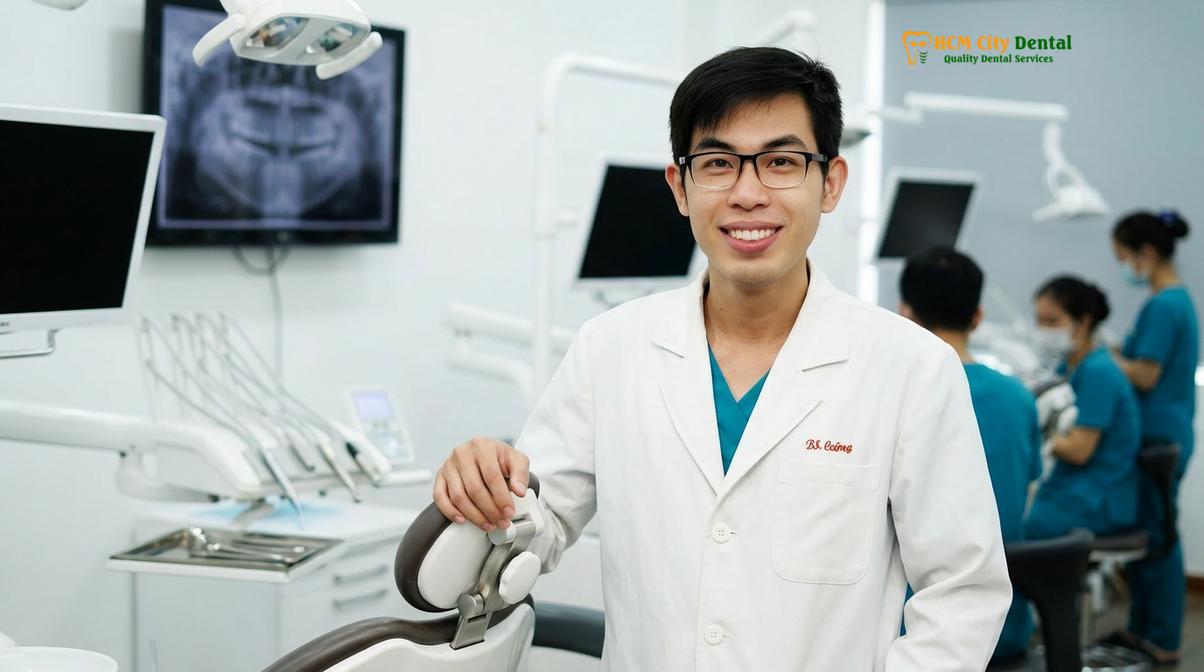
During a consultation, advanced diagnostic imaging, such as Cone Beam Computed Tomography (CBCT), is utilized to accurately measure the remaining bone volume in three dimensions. Dr. Nguyen Van Cuong and the clinical team at HCMC Dental Clinic in Ho Chi Minh City specialize in evaluating complex cases of bone atrophy. Based on a thorough diagnostic assessment, a personalized treatment plan can be developed, ranging from minimally invasive bone grafting to advanced implant-supported overdentures, ensuring the restoration of both function and aesthetics.
Frequently Asked Questions
How quickly does jaw bone shrink after teeth are pulled?
Jawbone shrinkage begins immediately after a tooth is extracted, with the most rapid loss occurring within the first six months. During this initial period, patients can lose up to 25% of the bone width and significant vertical height if the socket is left unpreserved. The rate of loss slows down after the first year but continues chronically for the rest of the patient’s life, eventually leading to severe structural collapse.
Can I get dental implants after wearing dentures for 10 years?
Yes, you can often receive dental implants after years of denture use, though preparatory procedures are usually required. Because prolonged denture wear leads to significant bone atrophy, most patients will need bone grafting or sinus lifts to rebuild the foundation before implant placement. Advanced imaging will determine the exact volume of bone remaining and dictate the necessary reconstructive steps.
Does a BPS denture stop bone loss better than standard dentures?
No, a BPS (Biofunctional Prosthetic System) denture does not stop bone loss, as it still rests on the gum tissue rather than integrating with the bone. While BPS dentures offer superior fit, better suction, and improved chewing efficiency compared to standard acrylic dentures, they cannot provide the internal mechanical stimulation required to halt the biological resorption of the alveolar ridge.
Is bone grafting painful when performed after a tooth extraction?
Bone grafting performed immediately after an extraction is generally not painful, as the area is already profoundly anesthetized. Post-operative discomfort is typically mild and easily managed with standard over-the-counter analgesics, similar to the recovery from a routine tooth extraction. The graft material is placed directly into the empty socket, requiring no additional surgical sites, making the procedure highly tolerable.
Can jaw bone loss from missing teeth cause headaches or jaw pain?
Yes, severe jaw bone loss can lead to bite misalignment and temporomandibular joint (TMJ) dysfunction, which frequently causes headaches and jaw pain. As the bone shrinks, the remaining teeth shift, altering the occlusion and placing unnatural stress on the facial muscles and joints. Restoring the lost vertical dimension with proper prosthetics or implants can alleviate these neuromuscular symptoms.
References
- Journal of Oral and Maxillofacial Surgery. Biomechanical principles of alveolar bone remodeling and resorption. (2021).
- International Journal of Oral Implantology. Dimensional changes of the alveolar ridge following tooth extraction. (2020).
- Journal of Prosthodontic Research. The impact of tissue-borne prosthetics on mandibular bone density. (2019).
- Clinical Oral Implants Research. Osseointegration and the preservation of crestal bone architecture. (2022).
- Journal of Periodontology. Efficacy of socket preservation techniques in preventing ridge collapse. (2018).