Splint therapy fitting in Saigon, District 1, involves a precise clinical protocol to design custom occlusal appliances that stabilize the temporomandibular joint (TMJ) and relax masticatory muscles. This specialized treatment requires accurate bite registration, advanced articulation, and meticulous micro-adjustments to ensure optimal neuromuscular harmony and long-term symptom relief.
Clinical Summary:
Temporomandibular disorders (TMD) encompass a complex group of musculoskeletal conditions affecting the jaw joint and surrounding musculature. Custom splint therapy remains the gold standard conservative intervention for managing these conditions. The process of splint therapy fitting in Saigon is a highly technical workflow that goes far beyond the provision of a simple mouthguard. It involves capturing high-precision intraoral data, utilizing facebow transfers to record the patient’s unique cranial anatomy, and mounting diagnostic casts on semi-adjustable articulators. The resulting appliance—typically a hard acrylic stabilization splint—is then meticulously calibrated during the delivery phase. Through precise clinical bite adjustment, the dentist ensures simultaneous, bilateral occlusal contacts that decompress the TMJ, reduce hyperactive muscle engrams, and provide a stable orthopedic platform for the mandible. This evidence-based approach significantly improves functional outcomes for patients suffering from chronic facial pain, bruxism, and articular disc displacements.
Key Takeaways:
- Custom splint therapy is a primary conservative treatment for decompressing the TMJ and reducing masticatory muscle hyperactivity.
- The fabrication process requires precise anatomical data, captured via digital intraoral scanners or high-fidelity elastomeric impressions.
- A facebow transfer is critical for accurately mounting diagnostic models and simulating the patient’s true mandibular hinge axis.
- Clinical delivery involves meticulous micro-reshaping of the hard acrylic to ensure simultaneous, frictionless occlusal contacts.
- Hard acrylic stabilization splints offer superior therapeutic benefits compared to soft, over-the-counter night guards.
- Successful therapy relies on continuous neuromuscular adaptation, often requiring follow-up visits for ongoing occlusal refinement.
| Splint Type | HCMC Dental Cost | Primary Clinical Goal |
|---|---|---|
| Michigan Stabilization Splint | from $100 USD (2,500,000 VND) | Bite balancing & muscle deprogramming |
| Anterior Repositioning Splint | from $150 USD (3,750,000 VND) | Disc displacement & joint decompression |
Understanding Splint Therapy for TMJ Disorders
Splint therapy utilizes custom-fabricated oral appliances to alter occlusal relationships, redistribute bite forces, and decompress the temporomandibular joint. This conservative approach is the first-line treatment for managing myalgia, arthralgia, and disc displacement.
Temporomandibular disorders (TMD) represent a heterogeneous group of conditions that affect the temporomandibular joint (TMJ), the muscles of mastication, and the associated contiguous tissue structures. Patients presenting with TMD often report a myriad of debilitating symptoms, including chronic pre-auricular pain, restricted mandibular range of motion, joint crepitus or clicking, and severe tension headaches. Due to the multifactorial etiology of these disorders—which can include occlusal discrepancies, parafunctional habits like bruxism, psychological stress, and macro-trauma—clinical guidelines universally recommend conservative, reversible therapies as the initial phase of management[1]. Among these conservative modalities, TMJ treatment protocols heavily emphasize the use of custom occlusal splints.
It is crucial to differentiate between a therapeutic stabilization splint and a standard protective device. While basic Nightguards are primarily designed to shield the dentition from the mechanical wear and tear associated with nocturnal grinding, a stabilization splint (often referred to as a Michigan splint or a flat-plane occlusal splint) serves a much more complex orthopedic function. The primary objective of a stabilization splint is to provide a temporary, ideal occlusal scheme that allows the mandible to seat itself in its most anatomically stable and orthopedically sound position—known as centric relation (CR)[2].
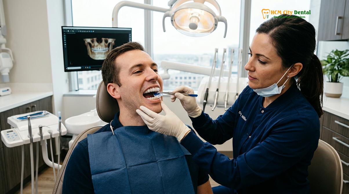
By introducing a hard, flat acrylic surface between the maxillary and mandibular teeth, the splint effectively disengages the patient’s habitual bite (maximum intercuspation). This disengagement interrupts the proprioceptive feedback loop from the periodontal ligaments to the central nervous system, which is often responsible for triggering hyperactive muscle spasms in the masseter and temporalis muscles. As Dr. Nguyen Van Cuong frequently emphasizes during clinical consultations, the success of splint therapy does not merely rely on placing a piece of plastic in the mouth; it depends entirely on the precise biometric calibration of that appliance to harmonize with the patient’s unique neuromuscular system.
Step-by-Step Custom Splint Fabrication Process
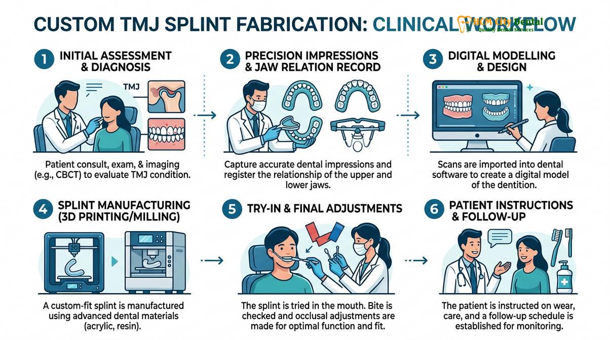
The fabrication of a therapeutic occlusal splint is a multi-stage clinical workflow that demands precise anatomical data capture. It transitions from initial diagnostics to laboratory manufacturing and final clinical calibration.
The creation of a highly effective stabilization splint is an intricate process that bridges the gap between clinical diagnostics and advanced dental laboratory technology. For patients undergoing splint therapy fitting in Saigon, the standard dental visit itinerary is carefully structured to ensure that every anatomical variable is accounted for. This systematic approach minimizes errors, reduces the need for excessive chairside adjustments, and maximizes the therapeutic efficacy of the final appliance.
The workflow generally encompasses three distinct phases. The first phase is dedicated to data acquisition, where the clinician captures the exact topography of the patient’s dentition and the spatial orientation of the maxilla relative to the cranial base. The second phase occurs in the dental laboratory, where skilled technicians utilize this data to mount the models, establish the correct vertical dimension of occlusion, and fabricate the acrylic appliance. The final phase is the clinical delivery, a meticulous process where the dentist seats the splint and performs dynamic occlusal adjustments to ensure perfect functional harmony.
Step 1: Taking High-Precision Impressions (Digital vs. Alginate)
Accurate impressions form the foundation of a well-fitting splint, utilizing either traditional elastomeric materials or advanced intraoral digital scanners to replicate the patient’s exact dental anatomy.
The foundation of any successful custom dental appliance is an exact replica of the patient’s oral anatomy. Any distortion, drag, or dimensional inaccuracy at this stage will inevitably translate into a poorly fitting splint, which can exacerbate TMJ symptoms rather than alleviate them. Historically, clinicians relied heavily on irreversible hydrocolloid materials (alginate) to capture these impressions. While alginate is cost-effective and relatively easy to use, it is highly susceptible to dimensional changes due to syneresis (loss of water) and imbibition (absorption of water). Therefore, when traditional methods are employed for complex TMJ cases, high-fidelity elastomeric materials such as polyvinyl siloxane (PVS) or polyether are strongly preferred due to their superior tear strength and exceptional dimensional stability over time.
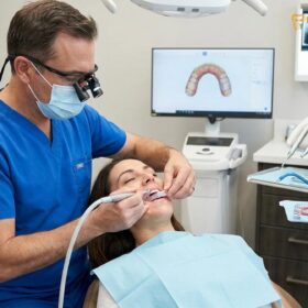
However, modern clinical practice has seen a massive paradigm shift toward digital dentistry. The use of advanced intraoral scanners (such as 3Shape TRIOS or Dentsply Sirona CEREC) has revolutionized the data acquisition phase. Digital scanning utilizes confocal microscopy or structured light to capture thousands of images per second, stitching them together to create a highly accurate, three-dimensional virtual model of the dental arches. This technology eliminates the discomfort associated with bulky impression trays and gooey materials, which is particularly beneficial for TMD patients who often suffer from restricted mouth opening (trismus) or severe gag reflexes.
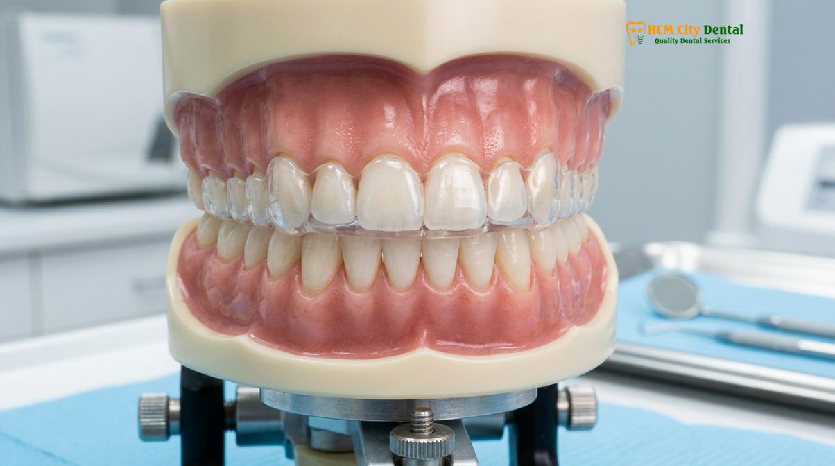
Furthermore, digital impressions offer unparalleled precision. The virtual models can be instantly magnified and evaluated on a computer screen, allowing the clinician to verify the capture of critical anatomical landmarks, such as the terminal molars and the palatal vault, before the patient even leaves the chair. Once the digital impression is approved, the stereolithography (STL) file is transmitted instantaneously to the dental laboratory. This seamless digital workflow not only expedites the fabrication process but also allows for the integration of the data with computerized occlusal analysis software, paving the way for highly sophisticated, digitally milled or 3D-printed splints.
Step 2: Facebow Transfer and Semi-Adjustable Articulation
A facebow transfer records the spatial relationship of the maxillary arch to the temporomandibular joints, allowing the dental laboratory to mount the models on a semi-adjustable articulator for accurate functional simulation.
Capturing the shape of the teeth is only the first part of the diagnostic puzzle. To design a splint that functions correctly during dynamic jaw movements—such as chewing, speaking, and grinding—the dental laboratory must understand how the patient’s upper jaw is positioned relative to their temporomandibular joints. This is where the facebow transfer becomes an indispensable clinical tool. A facebow is a caliper-like device used to record the spatial relationship of the maxillary arch to some anatomic reference point or points, and then transfer this relationship to an articulator.
The primary reference points captured by a facebow include the terminal hinge axis (the imaginary line through the two mandibular condyles around which the mandible rotates) and an anterior reference point (often the infraorbital notch or nasion). By securing the bite fork to the maxillary teeth and aligning the facebow with these cranial landmarks, the clinician captures a three-dimensional spatial record. This record is then sent to the laboratory along with the dental impressions.
“The omission of a facebow transfer in the fabrication of a stabilization splint forces the laboratory technician to mount the maxillary cast arbitrarily. This guesswork inevitably leads to occlusal interferences during excursive movements, requiring the clinician to spend excessive chair time grinding away the acrylic, which compromises the structural integrity and therapeutic design of the appliance.”
Once the laboratory receives the facebow record, the technician uses it to mount the maxillary cast onto a semi-adjustable articulator. An articulator is a mechanical instrument that represents the temporomandibular joints and jaws, to which maxillary and mandibular casts may be attached to simulate jaw movements. Unlike simple hinge articulators that only open and close, a semi-adjustable articulator can be programmed to mimic the patient’s specific condylar inclination and Bennett angle (the lateral shift of the working condyle). This custom articulation is critical. It allows the technician to design the splint with precise anterior guidance and canine disocclusion, ensuring that when the patient moves their jaw side-to-side or forward, the posterior teeth immediately separate, thereby protecting the TMJ from destructive lateral forces.
Step 3: Clinical Delivery and Micro-Reshaping of Acrylic
During the delivery appointment, the dentist seats the hard acrylic splint and performs meticulous clinical bite adjustments to ensure simultaneous, bilateral occlusal contacts and smooth excursive guidance.
The clinical delivery appointment is arguably the most critical phase of splint therapy. Even with the most accurate impressions and sophisticated laboratory articulation, the splint will require fine-tuning once it is placed in the patient’s mouth. The neuromuscular system is highly sensitive; a discrepancy as small as 10 to 20 microns (the thickness of a human hair) can be perceived by the proprioceptors in the periodontal ligaments, potentially triggering muscle spasms and defeating the purpose of the therapy.
The delivery process begins with evaluating the internal fit of the appliance. The dentist will carefully seat the splint on the maxillary arch, checking for adequate retention and stability. The splint should snap securely into place without placing excessive orthodontic pressure on any individual tooth. If the patient reports a feeling of tightness or if the splint rocks upon seating, the clinician will use pressure indicator paste (PIP) to identify internal binding points and relieve them using specialized acrylic burs.
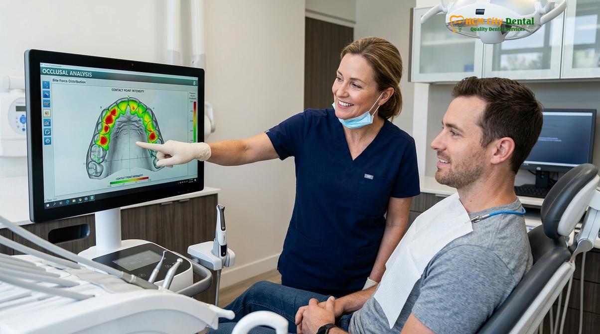
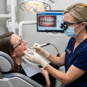
Once the internal fit is verified, the focus shifts to the occlusal surface. The goal of a stabilization splint is to provide mutually protected occlusion. This means that when the jaw closes into centric relation, all posterior mandibular teeth should contact the flat surface of the splint simultaneously and with equal intensity. To achieve this, the dentist performs a meticulous clinical bite adjustment. Using ultra-thin articulating foil (often 8 to 12 microns thick), the dentist asks the patient to tap their teeth together gently. The foil leaves colored marks on the splint, indicating the points of contact. The clinician then carefully grinds away the premature contacts (the heavy marks) until a uniform pattern of pinpoint contacts is achieved across the entire arch.
Clinical Warning: Inadequate occlusal adjustment during the delivery phase can lead to iatrogenic complications. If a splint introduces new occlusal interferences, it may exacerbate bruxism, increase joint loading, and cause severe masticatory muscle fatigue. Patients should never attempt to adjust or reshape their splints at home.
Following the static adjustment, the dentist must evaluate dynamic movements. The patient is instructed to slide their jaw forward (protrusion) and side-to-side (lateral excursion). During these movements, the splint must provide smooth guidance—typically via the mandibular canines gliding against a subtle ramp built into the anterior portion of the splint—while ensuring that all posterior teeth instantly disengage. This posterior disocclusion is vital for shutting down the elevator muscles (masseter and temporalis) during parafunctional grinding. In complex cases, advanced clinics may utilize computerized occlusal analysis systems, such as T-Scan, to digitally map the timing and force of the bite contacts, providing objective data to guide the micro-reshaping process[3]. For patients seeking comprehensive care, understanding the nuances of occlusal equilibration is essential for long-term joint health.
Material Science: Hard Acrylic vs. Dual Laminate Splints
The choice of splint material significantly impacts therapeutic outcomes, with hard heat-cured acrylic offering superior occlusal stability compared to softer, dual-laminate alternatives that may inadvertently stimulate bruxism.
The biomechanical properties of the material used to fabricate the splint play a profound role in its clinical efficacy. Patients often inquire about the differences between various types of appliances, particularly when comparing a hard vs soft night guard differences. For the management of true temporomandibular disorders, the overwhelming consensus in dental literature favors hard, rigid materials.
Stabilization splints are traditionally fabricated from heat-cured polymethyl methacrylate (PMMA) or, more recently, milled from highly cross-linked PMMA pucks using CAD/CAM technology. The rigidity of hard acrylic is essential for several reasons. First, it allows the dentist to perform precise, micron-level adjustments to the occlusal surface, creating the exact pinpoint contacts and smooth guidance ramps required for orthopedic stability. Second, a hard, flat surface allows the mandibular teeth to glide freely without resistance, which helps to deprogram the hyperactive jaw muscles.
| Material Type | Primary Indication | Occlusal Adjustability | Impact on Muscle Activity | Durability |
|---|---|---|---|---|
| Hard Acrylic (PMMA) | TMD, Severe Bruxism, Joint Decompression | Excellent (Micron-level precision) | Decreases hyperactivity (Deprogramming effect) | High (Resists heavy occlusal forces) |
| Dual Laminate (Hard out/Soft in) | Moderate Bruxism, Patient Comfort | Moderate (Surface can be adjusted) | Variable (May encourage slight chewing) | Medium (Soft liner degrades over time) |
| Soft Thermoplastic (EVA) | Athletic Mouthguards, Mild Clenching | Poor (Cannot be precisely calibrated) | Often increases hyperactivity (“Chewing gum” effect) | Low (Prone to rapid wear and perforation) |
Conversely, soft thermoplastic materials (like ethylene vinyl acetate, or EVA) are generally contraindicated for TMD patients. While they may feel initially comfortable, soft materials compress under biting force. This compressibility prevents the establishment of a stable occlusal endpoint. Furthermore, the resilient nature of soft guards often triggers a subconscious chewing reflex during sleep—frequently referred to as the “chewing gum effect.” This can actually increase the electromyographic activity of the masseter and temporalis muscles, exacerbating muscle pain and joint loading. For patients who struggle with severe muscle hypertrophy that does not fully respond to splint therapy alone, adjunctive treatments such as masseter Botox injections may be considered to chemically reduce muscle contractility[4].
Neuromuscular Adaptation and Long-Term Management
Successful splint therapy relies on the neuromuscular system adapting to the new occlusal scheme, leading to reduced muscle hyperactivity, decreased joint inflammation, and restored mandibular range of motion.
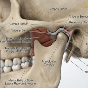
The delivery of the splint is not the conclusion of the treatment; rather, it marks the beginning of the neuromuscular adaptation phase. When a patient begins wearing a properly calibrated stabilization splint, the central nervous system receives new, harmonious proprioceptive input. The immediate disengagement of occlusal interferences allows the lateral pterygoid muscles—which are often in a state of chronic spasm in TMD patients—to finally relax. As these muscles release their tension, the mandibular condyles are permitted to seat themselves more superiorly and anteriorly within the glenoid fossa, effectively decompressing the highly innervated retrodiscal tissues.
This physiological shift often results in a rapid reduction of clinical symptoms. However, as the muscles relax and the joint decompresses, the patient’s jaw posture will inevitably change. This means that the initial bite contacts established on the splint during the delivery appointment will also shift. Therefore, follow-up appointments are an absolute necessity. During these visits, the dentist will re-evaluate the occlusion on the splint and perform further micro-reshaping to accommodate the jaw’s new, more relaxed position. This iterative process of adjustment and adaptation continues until the patient’s occlusion stabilizes and symptoms are fully resolved.
Clinical Case Review: A 34-year-old female patient presented to HCMC Dental Clinic in Ho Chi Minh City with chronic bilateral facial pain, limited mouth opening (28mm), and severe morning headaches. Clinical examination revealed masseter hypertrophy and a significant slide from centric relation to maximum intercuspation. The patient was fitted with a custom hard acrylic stabilization splint. Following the initial delivery and two subsequent adjustment visits over a four-week period, the patient’s jaw muscles relaxed significantly, allowing her maximum opening to increase to a healthy 45mm, with complete resolution of her morning headaches.
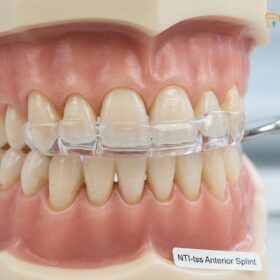
For some patients, particularly those with specific types of anterior disc displacement or severe clenching habits, alternative appliance designs may be explored. Understanding the occlusal splint vs night guard distinction is vital, and in certain specialized cases, anterior positioning devices (like the NTI-tss) may be utilized to specifically target incisal clenching forces, though these require extremely careful monitoring to prevent unwanted orthodontic tooth movement.
When to See a Doctor for TMJ Symptoms
Prompt clinical evaluation is essential when TMJ symptoms escalate from mild, intermittent discomfort to chronic pain, restricted jaw movement, or sudden changes in how the teeth fit together.
While occasional jaw clicking or mild muscle fatigue after chewing tough foods can be normal, certain signs and symptoms warrant immediate professional evaluation. Delaying treatment for progressive temporomandibular disorders can lead to irreversible degenerative changes within the joint capsule, such as osteoarthritis or permanent disc displacement, which may eventually necessitate invasive surgical interventions like arthrocentesis or open joint surgery[5].
You should seek a comprehensive clinical assessment if you experience any of the following red flag symptoms:
- Jaw Locking: Episodes where the jaw becomes stuck in either an open or closed position, requiring manual manipulation to release.
- Severe, Unrelenting Pain: Sharp or throbbing pain in the pre-auricular area (in front of the ear) that radiates to the temples, neck, or shoulders, and does not respond to over-the-counter analgesics.
- Sudden Occlusal Changes: A noticeable sensation that your upper and lower teeth no longer fit together correctly, or that your bite feels “off” or unstable.
- Significant Functional Limitation: Inability to open the mouth wide enough to comfortably insert two fingers vertically between the front teeth (typically less than 35mm of opening).
- Painful Crepitus: Grinding, crunching, or gravel-like sounds within the joint accompanied by pain during movement, which may indicate cartilage degradation.
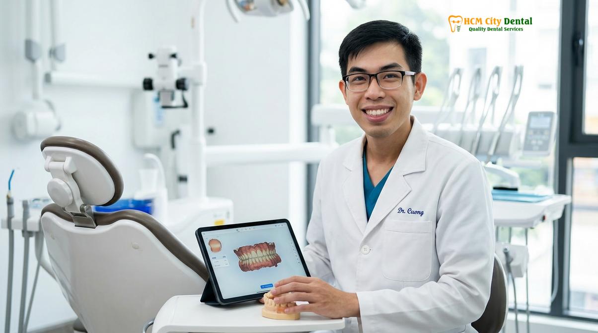
“Early intervention is the cornerstone of successful TMJ management. When patients ignore chronic joint sounds and muscular pain, the condition often progresses from a reversible muscular disorder to an irreversible structural joint derangement. A timely, accurate diagnosis and the implementation of a properly calibrated splint can halt this progression and restore normal function.” — Dr. Nguyen Van Cuong
If you are experiencing any of these symptoms, it is highly recommended to schedule a diagnostic consultation. A thorough clinical examination, often supplemented by advanced imaging such as Cone Beam Computed Tomography (CBCT), is necessary to determine the exact etiology of your pain and to formulate an appropriate, individualized treatment plan.
Frequently Asked Questions
How many dental visits are needed to get a TMJ splint?
A standard dental visit itinerary for a custom TMJ splint typically requires two to three clinical appointments. The first visit involves comprehensive diagnostics and high-precision impressions, while the second visit focuses on the clinical delivery, seating, and initial micro-reshaping of the acrylic appliance. A follow-up appointment is usually scheduled a few weeks later to evaluate neuromuscular adaptation and perform any necessary occlusal refinements as your jaw muscles relax into a new posture.
What is a facebow transfer?
A facebow transfer is a specialized clinical procedure used to record the exact spatial relationship between your upper jaw (maxilla) and your temporomandibular joints. This critical measurement allows the dental laboratory to mount your diagnostic models onto a semi-adjustable articulator accurately. By replicating your unique anatomical hinge axis, the technician can design a splint that functions in harmony with your natural jaw movements, minimizing the need for extensive chairside adjustments and ensuring optimal orthopedic support.
How does the dentist ensure the splint fits comfortably?
The dentist ensures a comfortable fit through meticulous clinical bite adjustment using ultra-thin articulating paper and specialized acrylic burs. During the delivery appointment, the internal surface of the splint is checked for pressure points using indicator paste. The biting surface is then carefully calibrated so that all teeth contact the splint simultaneously and evenly, preventing any single tooth from bearing excessive force and ensuring the jaw muscles can fully relax without triggering spasm-inducing interferences.
What is the difference between a stabilization splint and a standard night guard?
A stabilization splint is a rigid, precisely calibrated medical device designed to reposition the jaw and decompress the TMJ, whereas a standard night guard primarily protects teeth from grinding friction. While simple night guards are often made from softer materials and lack complex occlusal schemes, a stabilization splint features a flat, hard acrylic surface engineered to provide mutually protected occlusion, actively reducing muscle hyperactivity and joint inflammation by altering proprioceptive feedback.
How long will I need to wear my custom TMJ splint?
The duration of splint therapy varies based on your specific diagnosis, ranging from a few months for acute muscle spasms to indefinite nighttime wear for chronic joint degeneration. Initially, your dentist may recommend wearing the splint only at night or during periods of high stress. As your symptoms improve and your jaw muscles adapt to the new occlusal relationship, the wear time is often gradually reduced under clinical supervision, though many patients choose to continue nighttime wear for long-term protection.
References
- Journal of the American Dental Association. Occlusal splint therapy for temporomandibular disorders: a clinical review. (2020).
- Journal of Oral Rehabilitation. Efficacy of Michigan repositioning splints in TMD patients. (2019).
- Journal of Prosthetic Dentistry. Computerized occlusal analysis and bite mapping in TMJ therapy. (2021).
- Clinical Oral Investigations. Masseter Botox injections for chronic jaw pain and bruxism. (2018).
- International Journal of Oral and Maxillofacial Surgery. TMJ arthrocentesis and hyaluronic acid injections. (2022).
For personalized advice regarding TMJ diagnostics and custom splint therapy, please contact the clinical team at HCMC Dental Clinic to schedule a comprehensive evaluation.