The connection between sleep apnea and teeth grinding (bruxism) is deeply rooted in the body’s survival mechanisms. When the airway collapses during sleep, the brain triggers a micro-arousal and sympathetic response, causing the jaw muscles to clench and grind in an unconscious effort to reopen the obstructed airway.
Clinical Summary:
Sleep bruxism is increasingly recognized in modern dental medicine not merely as a standalone biomechanical issue, but as a secondary physiological response to obstructive sleep apnea (OSA). When upper airway resistance occurs, the central nervous system initiates a sympathetic micro-arousal to prevent asphyxiation. This arousal stimulates the masseter, temporalis, and lateral pterygoid muscles to contract forcefully, thrusting the mandible forward to restore airway patency. Consequently, treating the grinding without addressing the underlying respiratory obstruction—such as by prescribing a standard flat-plane night guard—can inadvertently worsen the apneic events by allowing the mandible to retrognathic (fall backward). Comprehensive management requires a dual-diagnostic approach, often utilizing mandibular advancement devices (MAD) to stabilize the airway while simultaneously protecting the dentition from catastrophic occlusal wear.
Key Takeaways:
- Teeth grinding during sleep often serves as an unconscious, life-saving reflex to reopen a collapsed airway.
- Standard flat-plane night guards can sometimes worsen sleep apnea by allowing the jaw and tongue to fall backward.
- Sympathetic nervous system arousals triggered by oxygen desaturation directly cause rhythmic masticatory muscle activity.
- Mandibular advancement devices (MAD) effectively treat both conditions by keeping the airway open and protecting teeth.
- Comprehensive polysomnography and detailed dental occlusal analysis are essential for an accurate dual diagnosis.
Patients comparing treatment options can read our comprehensive snoring guard comparison for clinical details.
Sleep Apnea and Bruxism: A Protective Airway Reflex
Sleep bruxism often acts as a physiological survival mechanism where the brain commands the jaw muscles to grind and thrust forward, attempting to mechanically reopen an obstructed airway during sleep apnea episodes.
For decades, the dental and medical communities viewed sleep bruxism primarily as a manifestation of psychological stress, anxiety, or localized occlusal discrepancies (a “bad bite”). However, contemporary sleep medicine and advanced polysomnographic research have fundamentally shifted this paradigm. Today, clinical evidence strongly suggests that a significant percentage of nocturnal teeth grinding is intricately linked to sleep-disordered breathing, specifically obstructive sleep apnea (OSA). In this context, bruxism is not merely a destructive habit; it is a highly coordinated, protective reflex orchestrated by the central nervous system to preserve life.
To understand this connection, one must examine the anatomy and physics of the human airway. During sleep, the muscles of the body, including those that support the soft palate, tongue, and pharynx, naturally relax. In individuals with anatomical predispositions—such as a narrow dental arch, a retrognathic (recessed) jaw, enlarged tonsils, or excess pharyngeal tissue—this relaxation leads to a narrowing or complete collapse of the airway. This phenomenon is clinically referred to as upper airway resistance. When the airway collapses, the flow of oxygen to the lungs is impeded, leading to a state of hypoxia (low oxygen) and hypercapnia (elevated carbon dioxide) in the bloodstream [1].
The brain, constantly monitoring blood gas levels, detects this dangerous drop in oxygen. To prevent asphyxiation, the brain’s reticular activating system triggers a sudden “micro-arousal.” This is not a full awakening where the patient becomes conscious, but rather a shift from deep, restorative sleep (such as REM or Stage 3 NREM) to a lighter sleep stage. Accompanying this micro-arousal is a massive surge in sympathetic nervous system activity—the body’s “fight or flight” response. Heart rate spikes, blood pressure increases, and motor pathways are activated.
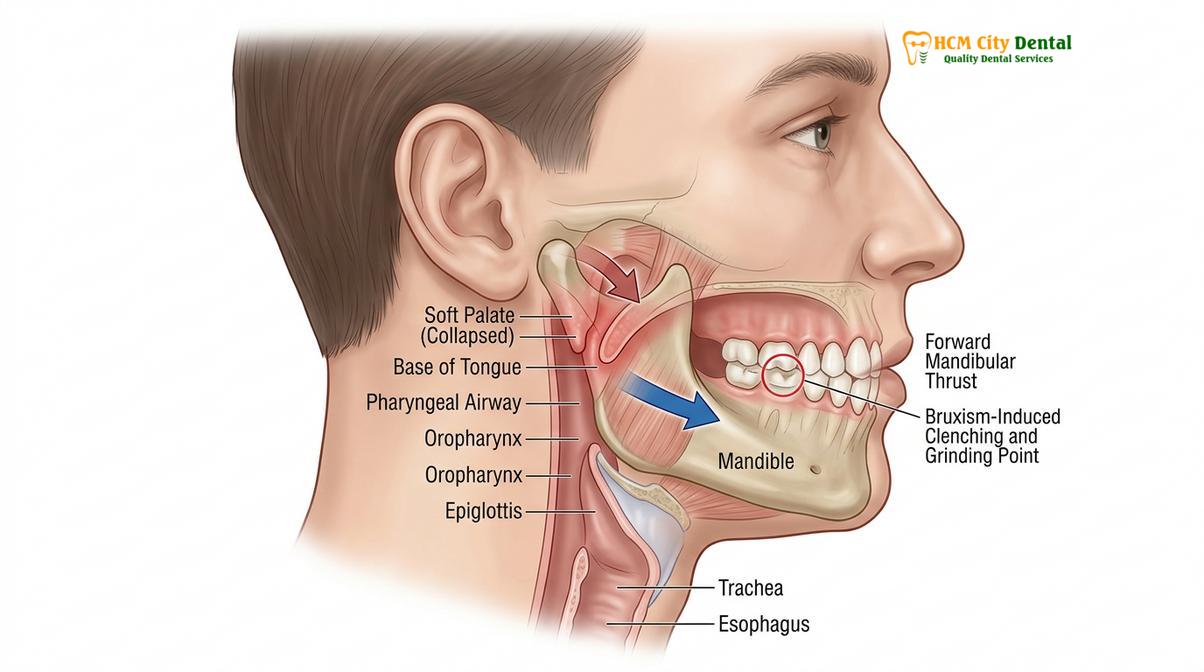
It is during this precise moment of sympathetic activation that the brain sends urgent signals to the muscles of mastication (chewing). The masseter and temporalis muscles contract forcefully, while the lateral pterygoid muscles pull the lower jaw forward. This forward thrusting and lateral grinding motion mechanically pulls the base of the tongue away from the posterior pharyngeal wall, effectively reopening the airway and allowing the patient to take a life-saving breath. Once breathing is restored, the patient settles back into deeper sleep, only for the cycle to repeat itself dozens or even hundreds of times per night.
Dr. Nguyen Van Cuong, a leading expert in complex restorative dentistry, frequently observes this phenomenon in clinical practice. “When we see a patient with severe, generalized tooth wear that does not align with their daytime habits, our first suspicion is no longer just stress,” notes Dr. Cuong. “We must immediately evaluate their airway. If we simply place a barrier between the teeth without addressing why the brain is commanding the jaw to move, we are doing the patient a profound disservice.” This underscores the critical importance of evaluating the airway before initiating comprehensive TMJ treatment protocols.
How Airway Collapse Triggers Sympathetic Arousal and Grinding
When the airway collapses, dropping oxygen levels trigger a sympathetic nervous system spike, causing a micro-arousal that activates the jaw muscles to clench and grind in order to restore breathing.
The physiological cascade that links a collapsed airway to the mechanical destruction of teeth is a fascinating study in human neurology and biomechanics. The sequence of events is highly predictable and has been extensively documented through concurrent polysomnography (sleep studies) and electromyography (EMG) of the jaw muscles. The specific pattern of muscle contraction observed during these events is termed Rhythmic Masticatory Muscle Activity (RMMA) [2].
The cycle begins with an apneic (complete cessation of breathing) or hypopneic (partial reduction in breathing) event. As the airway occludes, the patient’s respiratory effort increases. The diaphragm and intercostal muscles work harder against the closed airway, creating a negative intrathoracic pressure. This struggle continues for anywhere from 10 seconds to over a minute. During this time, the oxygen saturation in the blood (SpO2) steadily declines. The chemoreceptors in the carotid bodies and aortic arch detect this hypoxemia and send urgent distress signals to the brainstem.
The brainstem responds by initiating an autonomic and cortical arousal. This arousal is characterized by a sudden shift in electroencephalogram (EEG) frequencies, indicating that the brain is waking up just enough to rescue the body. Simultaneously, there is a surge in sympathetic tone. The heart rate, which may have been bradycardic (slow) during the apnea, suddenly becomes tachycardic (fast). Blood pressure spikes dramatically.
“The sequence of events in sleep apnea-induced bruxism is highly stereotyped: respiratory cessation leads to hypoxia, which triggers a cortical micro-arousal, followed immediately by sympathetic tachycardia, and culminating in forceful rhythmic masticatory muscle activity to restore airway patency.”
Following the autonomic arousal, the motor cortex activates the trigeminal nerve (Cranial Nerve V), which controls the muscles of mastication. The jaw is thrust forward and laterally in a grinding motion. This movement is not random; it is a highly specific biomechanical action designed to increase the anterior-posterior dimension of the pharyngeal airway. By moving the mandible forward, the genioglossus muscle (which attaches the tongue to the mandible) is pulled forward, lifting the tongue off the back of the throat.
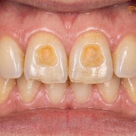
The sheer force generated during these nocturnal grinding episodes can be staggering. While normal chewing forces during the day rarely exceed 20 to 40 pounds per square inch, nocturnal bruxism forces can easily exceed 250 pounds per square inch. Because the patient is asleep, the normal protective proprioceptive reflexes that prevent us from biting too hard are suppressed. This leads to catastrophic damage to the dentition over time, including fractured cusps, abfraction lesions at the gumline, and severe attrition (flattening) of the occlusal surfaces.
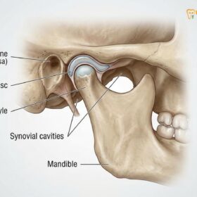
Furthermore, this constant, heavy workload on the masticatory muscles and the temporomandibular joint (TMJ) leads to profound muscular fatigue, lactic acid buildup, and joint inflammation. Patients frequently wake up with severe morning headaches, facial pain, and a jaw that feels locked or stiff. This is why addressing the root cause—the airway—is paramount. Simply treating the muscular pain with masseter Botox injections may provide temporary symptomatic relief, but it does not cure the underlying sleep-disordered breathing.
Diagnosing Sleep-Disordered Breathing in TMD Patients
Diagnosing the overlap between sleep apnea and temporomandibular disorders requires a combination of polysomnography to monitor respiratory events and clinical dental examinations to assess occlusal wear and jaw joint health.
The intersection of Temporomandibular Disorders (TMD) and Obstructive Sleep Apnea (OSA) presents a complex diagnostic challenge. Because the symptoms often overlap—morning headaches, chronic fatigue, neck pain, and jaw stiffness—patients are frequently misdiagnosed. A comprehensive, multidisciplinary approach is required to accurately identify the presence of both conditions and understand how they are interacting in the individual patient [3].
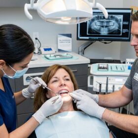
The diagnostic process begins with a thorough clinical dental examination. Dentists trained in dental sleep medicine are often the first healthcare providers to spot the subtle, early warning signs of a compromised airway. During the intraoral examination, the clinician looks for specific biomarkers of sleep-disordered breathing. One of the most prominent signs is a “scalloped tongue” (crenation). When the airway is narrow, the tongue is often forced forward against the lingual surfaces of the lower teeth, leaving distinct indentations along the lateral borders of the tongue.
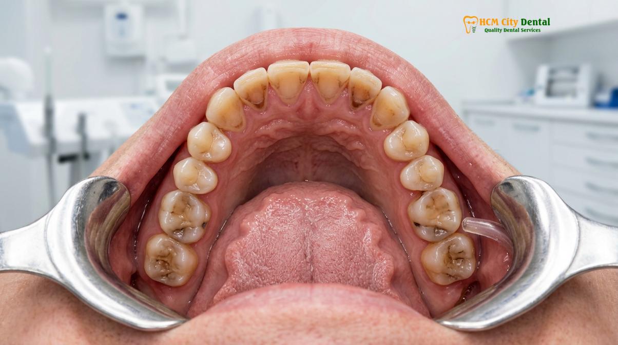
Another key indicator is the presence of a “linea alba,” a thick, white, hyperkeratotic line along the buccal mucosa (inside of the cheek) where the patient has been chronically clenching their teeth together. The teeth themselves tell a story of nocturnal trauma. The clinician will look for cupping or cratering on the chewing surfaces of the molars, where the hard enamel has been worn away, exposing the softer, yellow dentin underneath. The anterior teeth (incisors and canines) may appear shortened, chipped, or perfectly flat, having lost their natural anatomical curves.
Beyond the teeth, the anatomy of the airway itself is evaluated. The clinician will assess the Mallampati score, which grades the visibility of the soft palate and uvula when the mouth is open. A high Mallampati score (Class III or IV) indicates a crowded oropharynx and a higher risk of airway collapse. The size of the tonsils, the length of the soft palate, and the circumference of the neck are also carefully documented.
Clinical Case Study: The Hidden Airway Issue
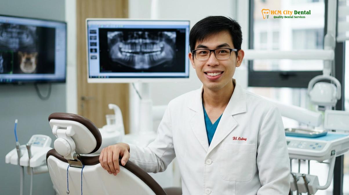
A 42-year-old male patient presented to HCMC Dental Clinic in Ho Chi Minh City complaining of severe, chronic jaw pain and multiple fractured dental restorations. Previous dentists had prescribed various soft night guards, which the patient reported chewing through within months. Upon comprehensive examination, Dr. Nguyen Van Cuong noted severe occlusal attrition, a heavily scalloped tongue, and a Class IV Mallampati airway. A referral for an at-home sleep study revealed an Apnea-Hypopnea Index (AHI) of 22, indicating moderate obstructive sleep apnea. The patient’s bruxism was a direct result of his airway collapsing. Treatment was shifted from standard night guards to a custom Mandibular Advancement Device (MAD), which successfully resolved both his apneic events and his nocturnal grinding.
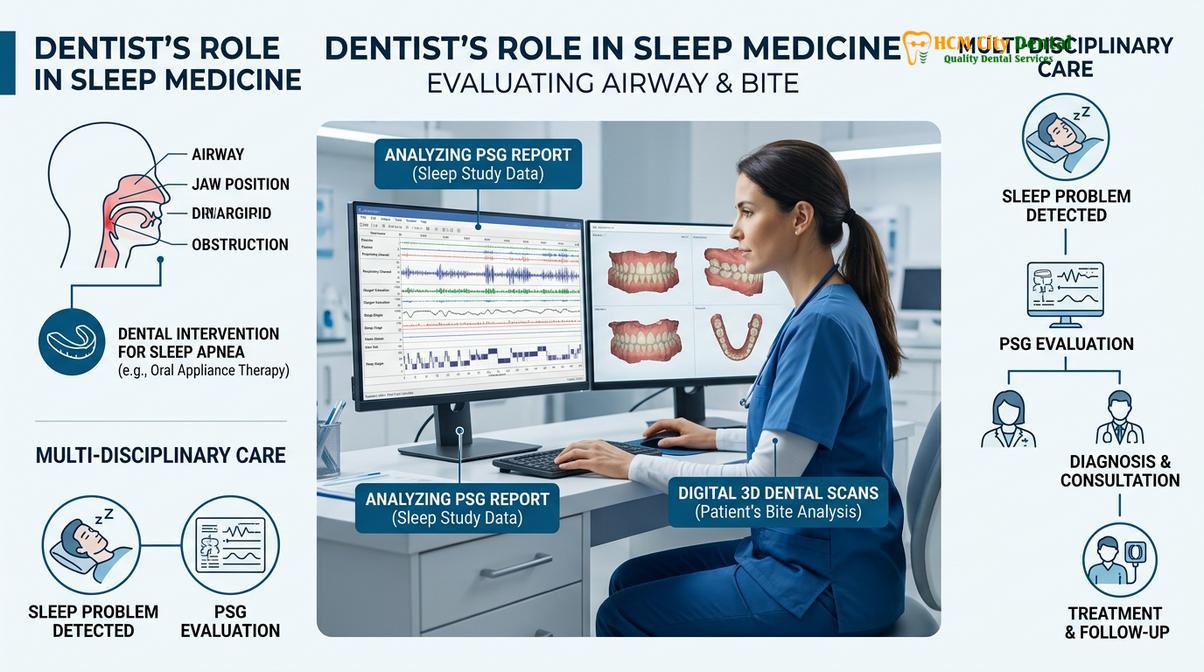
While dental signs provide strong circumstantial evidence, a definitive diagnosis of sleep apnea requires objective medical testing. This is typically achieved through Polysomnography (PSG), either conducted in a clinical sleep laboratory or via a high-quality Home Sleep Apnea Test (HSAT). The sleep study measures critical physiological parameters throughout the night, including airflow, respiratory effort, blood oxygen saturation (SpO2), heart rate, and brain wave activity (EEG).
The severity of sleep apnea is quantified using the Apnea-Hypopnea Index (AHI), which calculates the average number of breathing pauses (apneas) and partial obstructions (hypopneas) per hour of sleep. An AHI of 5-15 is considered mild, 15-30 is moderate, and greater than 30 is severe. In patients with concurrent bruxism, the sleep study will often reveal a direct temporal correlation: an apneic event occurs, oxygen drops, an EEG arousal is recorded, and immediately following, the EMG channels record intense masseter muscle activity (the Bruxism Episode Index, or BEI).
Mandibular Advancement Devices (MAD) vs. Stabilization Splints
While stabilization splints protect teeth from grinding, they can inadvertently worsen sleep apnea; conversely, mandibular advancement devices (MAD) pull the jaw forward, effectively treating both the airway obstruction and the resulting bruxism.
Once a dual diagnosis of sleep apnea and bruxism is established, the treatment paradigm must shift dramatically. The traditional dental response to teeth grinding has been the fabrication of a stabilization splint, commonly known as a night guard. However, in the presence of sleep-disordered breathing, the wrong type of appliance can be not only ineffective but potentially dangerous [4].
A standard stabilization splint (such as a Michigan splint) is typically a flat-plane, hard acrylic device worn over the upper or lower teeth. Its primary function is to provide a smooth surface for the opposing teeth to glide against, thereby protecting the enamel from wear and reducing strain on the TMJ. When considering choosing between a hard vs soft night guard, dentists generally prefer hard acrylic for heavy grinders, as soft materials can actually stimulate more chewing activity.
Clinical Warning: The Danger of Flat-Plane Splints in OSA Patients
Prescribing a standard flat-plane night guard to a patient with undiagnosed obstructive sleep apnea can exacerbate their respiratory distress. By increasing the vertical dimension of the bite without advancing the mandible, the splint can cause the lower jaw to rotate backward (retrognathia) during sleep. This backward rotation pushes the base of the tongue further into the pharyngeal space, narrowing the airway and potentially increasing the frequency and severity of apneic events.
To safely manage a patient with both conditions, a Mandibular Advancement Device (MAD) is often the treatment of choice. An MAD is a specialized oral appliance designed specifically for dental sleep medicine. Unlike a standard night guard, an MAD consists of two separate pieces—one for the upper arch and one for the lower arch—connected by a titration mechanism (such as hinges, straps, or telescopic rods). This mechanism allows the dentist to incrementally advance the lower jaw forward.
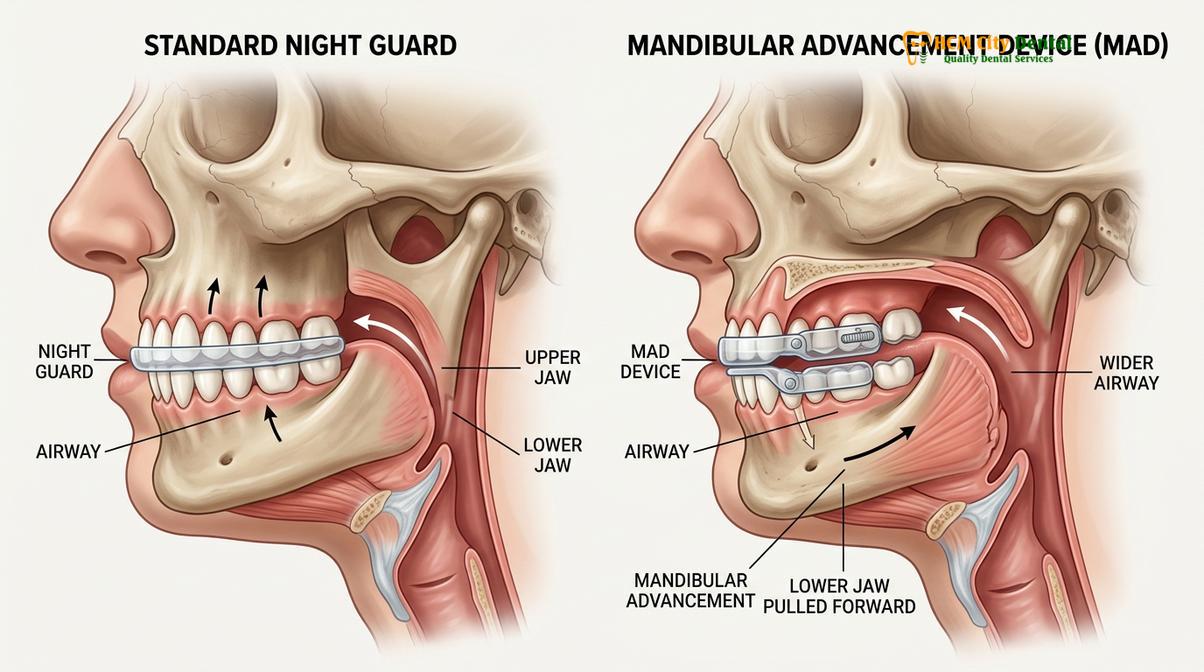
The biomechanical advantage of the MAD is twofold. First, by holding the mandible in a protrusive (forward) position, it physically pulls the tongue and soft tissues away from the back of the throat, maintaining a patent (open) airway throughout the night. This directly addresses the root cause of the sleep apnea. Second, because the upper and lower teeth are covered by the acrylic trays, the dentition is fully protected from the destructive forces of bruxism. Furthermore, because the airway is stabilized, the brain no longer needs to trigger the sympathetic micro-arousals that cause the grinding in the first place. Many patients experience a dramatic reduction or complete cessation of nocturnal bruxism once their airway is managed with an MAD.
The choice of appliance requires careful clinical judgment. For patients with severe TMJ dysfunction, advancing the jaw too quickly can exacerbate joint pain. In these cases, a highly customized approach is required, carefully balancing the need for airway patency with the orthopedic health of the jaw joints. This is where advanced diagnostics and precise bite adjustment procedures become critical components of the overall treatment plan.
| Feature | Stabilization Splint (Standard Night Guard) | Mandibular Advancement Device (MAD) |
|---|---|---|
| Primary Indication | Isolated bruxism, TMJ muscle pain | Obstructive Sleep Apnea, snoring, concurrent bruxism |
| Mechanism of Action | Provides a flat surface to protect teeth and relax muscles | Advances the lower jaw forward to open the pharyngeal airway |
| Effect on Airway | Neutral or potentially negative (can cause jaw to fall backward) | Highly positive (mechanically prevents airway collapse) |
| Design Structure | Single arch (usually upper or lower) | Dual arch (upper and lower connected by a mechanism) |
| Adjustability | Static (requires dental bur adjustment) | Dynamic (titratable to advance jaw incrementally) |
When to See a Doctor
Recognizing the signs of sleep-disordered breathing and concurrent bruxism early can prevent years of dental destruction and systemic health issues. Because these events occur during sleep, patients are often entirely unaware of the problem until significant damage has occurred or a sleep partner brings it to their attention. It is crucial to seek professional medical and dental evaluation if you experience any of the following clinical indicators.
First, if a bed partner reports that you snore loudly, gasp for air, or appear to stop breathing during the night, this is a primary red flag for obstructive sleep apnea and warrants immediate investigation. Second, if you consistently wake up with dull, aching pain in your jaw, temples, or neck, or if you experience frequent morning headaches, this suggests intense nocturnal muscle activity. Third, visual changes to your teeth—such as noticeable flattening, chipping, increased sensitivity to hot and cold, or receding gums—are clear signs of pathological occlusal forces.
Additionally, systemic symptoms of poor sleep quality should not be ignored. Excessive daytime sleepiness, chronic fatigue, difficulty concentrating, and mood disturbances are common consequences of the fragmented sleep architecture caused by repeated micro-arousals. If you have been diagnosed with hypertension (high blood pressure) that is difficult to control with medication, this can also be linked to the chronic sympathetic nervous system activation associated with sleep apnea.
Dr. Cuong emphasizes the importance of a proactive approach: “Patients often wait until a tooth fractures or their jaw locks before seeking help. By that point, the restorative work required is extensive. If you suspect you are grinding your teeth, do not assume it is just stress. A comprehensive airway and occlusal evaluation can identify the root cause and allow us to intervene conservatively.” If you are experiencing these symptoms, scheduling a consultation with a provider experienced in dental sleep medicine is the critical first step toward protecting your oral and systemic health.
Comprehensive Treatment Workflows at HCMC Dental Clinic
Effective management of concurrent sleep apnea and bruxism involves a multidisciplinary approach, combining custom oral appliance therapy, bite adjustments, and continuous airway monitoring to ensure optimal patient outcomes.
At HCMC Dental Clinic, the management of complex cases involving both sleep apnea and severe bruxism follows a rigorous, evidence-based clinical workflow. The goal is not merely to treat the symptoms, but to address the underlying physiological triggers while restoring the biomechanical harmony of the masticatory system. This comprehensive approach ensures long-term stability and significantly improves the patient’s quality of life [5].
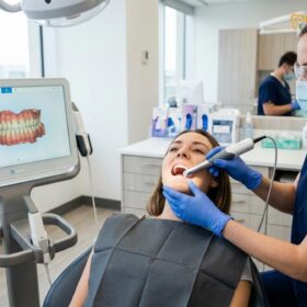
The workflow begins with an exhaustive diagnostic phase. Patients undergo high-resolution 3D Cone Beam Computed Tomography (CBCT) to visualize the temporomandibular joints and perform a volumetric analysis of the airway. Digital intraoral scanners are used to capture precise, micron-level impressions of the teeth, eliminating the need for uncomfortable traditional putty. If the patient has not yet had a formal sleep study, the clinic coordinates with local sleep physicians to facilitate polysomnography, ensuring an accurate baseline AHI and BEI are established.
“The integration of digital airway mapping with computerized occlusal analysis allows us to design appliances that not only advance the mandible to treat the apnea but also distribute occlusal forces perfectly to protect the TMJ.”
Once the diagnosis of obstructive sleep apnea HCMC is confirmed, the fabrication of a custom Mandibular Advancement Device (MAD) is initiated. Unlike over-the-counter “boil and bite” devices, which are bulky, uncomfortable, and often ineffective, custom MADs are milled from medical-grade polymers. At HCMC Dental, we construct custom appliances such as the Antisnoring Telescopic and Twinblock Snoring guards. They are designed to be low-profile, allowing for maximum tongue space and comfort. The devices feature precision titration mechanisms, allowing the dentist to advance the jaw in increments as small as 0.1 millimeters. This gradual advancement is crucial for allowing the TMJ ligaments and muscles to adapt to the new position without causing pain or spasm. For a detailed clinical discussion on this relationship, you can read our guide on the bruxism sleep apnea connection.
The delivery of the appliance is followed by a meticulous titration and follow-up protocol. The patient returns for periodic adjustments, during which the jaw is slowly advanced until the symptoms of snoring, apnea, and grinding are resolved. In some cases, particularly where the patient’s bite is fundamentally misaligned, adjunctive therapies may be required. This could involve anterior NTI devices for short-term muscle deprogramming, or comprehensive orthodontic intervention to permanently correct the arch dimensions and improve airway volume.
For patients whose teeth have already suffered significant damage from years of untreated bruxism, a full-mouth rehabilitation may be necessary once the airway is stabilized. This involves restoring the lost vertical dimension of the bite using crowns, veneers, or onlays, carefully engineered to function harmoniously with the TMJ. By addressing the airway first, the clinic ensures that these new restorations will not be destroyed by continued nocturnal grinding. For those seeking expert evaluation and custom protective nightguards or advanced MAD therapy, the multidisciplinary team at HCMC Dental Clinic in Ho Chi Minh City provides world-class, personalized care.
Frequently Asked Questions
Why does my body grind teeth when I stop breathing?
Your body grinds teeth during a sleep apnea episode as an unconscious survival reflex to reopen the collapsed airway. When oxygen levels drop, the brain triggers a micro-arousal, causing the jaw muscles to contract and thrust the lower jaw forward to restore airflow. This rhythmic masticatory muscle activity (RMMA) mechanically pulls the tongue away from the back of the throat, allowing you to take a breath and preventing asphyxiation.
Can a night guard worsen sleep apnea?
Yes, a standard flat-plane night guard can inadvertently worsen sleep apnea in some patients. By slightly opening the bite without advancing the lower jaw, a traditional guard can cause the mandible and tongue to fall backward, further obstructing the upper airway during sleep. This is why it is critical for patients who grind their teeth to be screened for sleep-disordered breathing before a standard stabilization splint is prescribed.
What are the symptoms of sleep apnea?
Common symptoms of sleep apnea include loud snoring, witnessed pauses in breathing during sleep, waking up gasping or choking, excessive daytime sleepiness, and morning headaches. Dental signs often include severe tooth wear, a scalloped tongue, and chronic jaw pain. Patients may also experience mood changes, difficulty concentrating, and unrefreshing sleep despite spending adequate time in bed.
How do I know if my jaw pain is from sleep apnea or just stress?
Determining the root cause of jaw pain requires a comprehensive clinical evaluation and often a sleep study. While stress-induced bruxism typically occurs without respiratory distress, sleep apnea-related jaw pain is usually accompanied by snoring, daytime fatigue, and specific oxygen desaturation patterns. A dentist trained in dental sleep medicine can evaluate your airway anatomy and tooth wear patterns to help differentiate between the two.
Is a CPAP machine required if I have both conditions?
A CPAP machine is not always required for mild to moderate cases of sleep apnea combined with teeth grinding. Many patients achieve excellent results using a custom mandibular advancement device (MAD), which simultaneously keeps the airway open and protects the teeth from grinding forces. However, for severe sleep apnea, CPAP remains the gold standard, and a dentist can fabricate a specialized night guard to be worn in conjunction with the CPAP mask to protect the teeth.
References
- Journal of Clinical Sleep Medicine. The Relationship between Sleep Bruxism and Obstructive Sleep Apnea Based on Polysomnographic Findings. (2019).
- Sleep Medicine Reviews. Rhythmic masticatory muscle activity during sleep: etiology and pathophysiology. (2020).
- Journal of the American Dental Association. The role of the dentist in the diagnosis and management of sleep-related breathing disorders. (2018).
- Journal of Prosthodontic Research. Effects of mandibular advancement devices on sleep bruxism and obstructive sleep apnea. (2021).
- International Journal of Prosthodontics. Occlusal splint therapy and its impact on upper airway dimensions. (2022).