TMJ physical therapy in Saigon combines targeted muscular rehabilitation, joint mobilization, and postural correction to alleviate temporomandibular joint pain. By addressing the biomechanical root causes of jaw dysfunction, specialized clinical protocols aim to restore normal range of motion, reduce masseter tension, and stabilize the articular disc using conservative, non-surgical methods.
Clinical Summary:
Temporomandibular Joint (TMJ) physical therapy represents a cornerstone of conservative management for craniomandibular disorders. This specialized rehabilitative approach focuses on restoring the complex arthrokinematics of the jaw through a combination of passive modalities and active neuromuscular re-education. Clinical protocols typically integrate intraoral myofascial release to address hypertonicity in the muscles of mastication, alongside targeted isometric exercises designed to stabilize the articular disc and strengthen the pterygoid complex. Furthermore, because the stomatognathic system is biomechanically linked to the cervical spine, comprehensive therapy mandates postural correction to relieve compensatory strain on the suprahyoid musculature. When synchronized with precise dental interventions—such as custom occlusal splint therapy—physical rehabilitation yields a high success rate in mitigating chronic facial pain, reducing joint crepitus, and preventing long-term degenerative joint disease. Expert clinical oversight ensures that therapeutic loads are carefully calibrated to the patient’s specific anatomical and pathological presentation.
Key Takeaways:
- Physical therapy for TMJ disorders prioritizes conservative, non-invasive rehabilitation of the masticatory muscles and joint capsule.
- Isometric exercises build essential muscular endurance without placing destructive sheer forces on the articular disc.
- Intraoral manual therapy effectively deactivates chronic trigger points within the masseter and medial pterygoid muscles.
- Correcting forward head posture is clinically vital for reducing secondary biomechanical stress on the temporomandibular joint.
- Combining active physical rehabilitation with passive occlusal splint therapy provides the most comprehensive long-term relief.
Supporting Splint Therapy with Active Physical Rehabilitation
Combining custom nightguards with active physical therapy ensures both passive joint decompression during sleep and dynamic muscular stabilization during waking hours.
The management of temporomandibular joint disorders (TMD) is rarely successful when relying on a single therapeutic modality. In modern clinical practice, the integration of passive dental appliances with active physical rehabilitation forms the gold standard of care. While advanced TMJ treatment protocols often begin with the fabrication of a custom orthotic device, the appliance alone only addresses part of the complex biomechanical equation. A hard acrylic stabilization splint provides a crucial mechanical barrier that prevents the destructive forces of nocturnal bruxism, effectively decompressing the highly innervated retrodiscal tissues and allowing localized capsular inflammation to subside[1]. However, a splint is fundamentally a passive intervention; it alters the proprioceptive feedback to the central nervous system while worn, but it does not actively retrain the dysfunctional muscle firing patterns that occur during daytime functions such as speaking, chewing, and swallowing.
This is where targeted physical therapy becomes indispensable. Active rehabilitation protocols are designed to complement the protective nature of custom-fitted nightguards by addressing the underlying neuromuscular imbalances. When a patient removes their splint in the morning, the masticatory muscles must be capable of maintaining the jaw in a physiologically stable, relaxed posture. Physical therapy facilitates this transition by improving the endurance of the elevator muscles and restoring symmetrical condylar translation. The synergistic effect of these two treatments is profound: the splint provides the necessary joint offloading required for tissue healing, while physical therapy builds the dynamic stability needed to prevent symptom relapse.
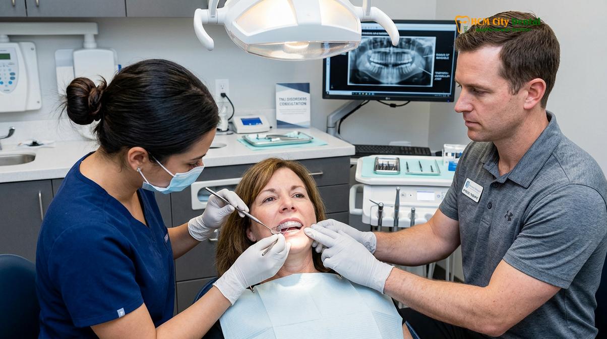
Furthermore, the adaptation period to a new occlusal appliance can sometimes induce transient muscle fatigue as the jaw acclimates to a new vertical dimension of occlusion. Physical therapists trained in craniomandibular rehabilitation can utilize specific modalities, such as thermal therapy, ultrasound, and gentle joint mobilization, to accelerate this adaptation process. By downregulating the hyperactive trigeminal nerve pathways, therapy ensures that the patient can comfortably tolerate the splint, thereby maximizing clinical compliance and therapeutic outcomes.
“An occlusal splint acts as a highly precise crutch for an injured temporomandibular joint. However, just as an orthopedic surgeon prescribes physical therapy after placing a cast on a fractured limb, a dental specialist must prescribe targeted jaw rehabilitation to restore functional muscular harmony once the joint is stabilized.”
Clinical evidence consistently demonstrates that patients who engage in a combined regimen of occlusal splint therapy and structured physical rehabilitation experience a faster reduction in visual analog scale (VAS) pain scores compared to those utilizing splint therapy alone. The active retraining of the neuromuscular system ensures that the therapeutic gains achieved during sleep are maintained and reinforced throughout the patient’s daily activities.
Isometric Jaw Exercises to Strengthen Pterygoid and Masseter Muscles
Isometric exercises build endurance in the lateral pterygoid and masseter muscles without straining the temporomandibular joint capsule, promoting stable jaw tracking.
Therapeutic exercise is a fundamental component of TMJ rehabilitation, but the specific type of exercise prescribed is critical to avoiding iatrogenic injury. The temporomandibular joint is a highly complex ginglymoarthrodial joint, meaning it functions through both hinge (rotation) and gliding (translation) movements. In patients with capsulitis, disc displacement, or hypermobility, traditional isotonic exercises—where the jaw moves through a range of motion against resistance—can exacerbate joint sheer forces and provoke painful clicking. Therefore, clinical protocols heavily emphasize isometric exercises. Isometric contractions involve muscle activation without any change in muscle length or joint angle. This allows the patient to build essential muscular endurance and neuromuscular control without placing mechanical stress on the vulnerable articular disc or the retrodiscal tissues[2].
The primary targets of these isometric protocols are the masseter, temporalis, and the intricate pterygoid muscles. The lateral pterygoid, in particular, plays a crucial role in controlling the movement of the articular disc during jaw opening. Dysfunction or spasm in the superior belly of the lateral pterygoid is a leading cause of disc displacement with reduction (the classic “clicking” jaw). By utilizing precise isometric holds, clinicians can help patients re-establish coordinated firing patterns between the lateral pterygoid and the elevator muscles, promoting smoother, more symmetrical condylar tracking.
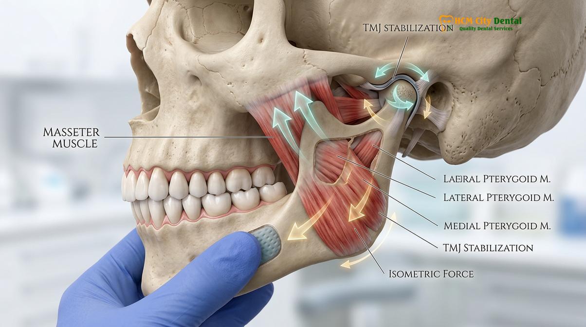
A standard isometric protocol often begins with resting posture awareness. The patient is instructed to place the tip of their tongue gently against the incisive papilla (the roof of the mouth just behind the upper front teeth), keep the teeth slightly apart, and breathe diaphragmatically. From this neutral position, gentle resistance is applied using the patient’s own fingers. It is important to note that these exercises are not designed to build massive hypertrophic strength, but rather to enhance proprioception and muscular endurance. The force applied should be minimal—typically no more than 10 to 20 percent of maximum voluntary contraction.
| Isometric Exercise | Clinical Execution | Target Musculature | Therapeutic Goal |
|---|---|---|---|
| Resisted Opening | Place thumb under the chin. Attempt to open the mouth slowly while applying gentle upward resistance. Hold for 5 seconds. | Suprahyoid muscles, Inferior Lateral Pterygoid | Improves controlled depression of the mandible without joint translation. |
| Resisted Closing | Place index fingers on the lower incisors. Attempt to close the mouth while applying gentle downward resistance. Hold for 5 seconds. | Masseter, Temporalis, Medial Pterygoid | Builds endurance in the elevator muscles, reducing fatigue-induced spasms. |
| Lateral Excursion | Place palm against the right side of the jaw. Attempt to move the jaw to the right against resistance. Repeat on the left. | Contralateral Lateral Pterygoid, Ipsilateral Masseter | Restores symmetrical lateral tracking and reduces unilateral deviations. |
| Mandibular Protrusion | Place fingers on the front of the chin. Attempt to push the jaw forward against gentle backward resistance. | Bilateral Lateral Pterygoids | Stabilizes the articular disc and improves anterior condylar glide control. |
In addition to active strengthening, controlled passive stretching techniques may be introduced in later stages of rehabilitation, provided there is no acute joint effusion. Passive stretching involves the clinician or the patient gently guiding the mandible to its maximum pain-free opening to restore elasticity to the joint capsule and the surrounding fascial networks. However, this must be performed with extreme caution, as overstretching a hypermobile joint can lead to subluxation. The integration of these exercises into a daily routine is paramount; neuroplastic changes in motor control require consistent, repetitive stimulus over several weeks to become permanent.
Manual Therapy: Intraoral Myofascial Release Techniques
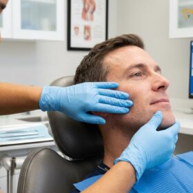
Intraoral myofascial release involves precise, gloved digital pressure applied directly to internal masticatory muscles to deactivate trigger points and relieve chronic tension.
While external massage and thermal therapies provide symptomatic relief for facial pain, they often fail to address the deep-seated muscular hypertonicity that drives chronic temporomandibular dysfunction. The most powerful muscles of the stomatognathic system—specifically the medial and lateral pterygoids, as well as the deep belly of the masseter—are largely inaccessible from the exterior of the face. To effectively treat these structures, specialized physical therapists and dental clinicians employ intraoral myofascial release techniques. This advanced form of manual therapy involves the practitioner utilizing gloved digits inside the patient’s oral cavity to palpate, identify, and deactivate myofascial trigger points that are responsible for referred pain patterns and restricted jaw mobility.
A trigger point is a hyperirritable nodule within a taut band of skeletal muscle. In the context of TMD, trigger points in the masseter frequently refer pain to the maxillary molars, the eyebrow, or deep into the ear, often leading to misdiagnoses of endodontic infections or otitis media. Intraoral masseter massage allows the clinician to grasp the muscle belly between the intraoral thumb and the extraoral index finger (a pincer grasp). By applying sustained, ischemic compression to the localized nodule, the clinician forces the contracted sarcomeres to release. The temporary restriction of blood flow is immediately followed by reactive hyperemia—a rush of oxygenated blood that flushes out accumulated metabolic waste products, such as lactic acid and substance P, thereby breaking the pain-spasm-pain cycle[3].
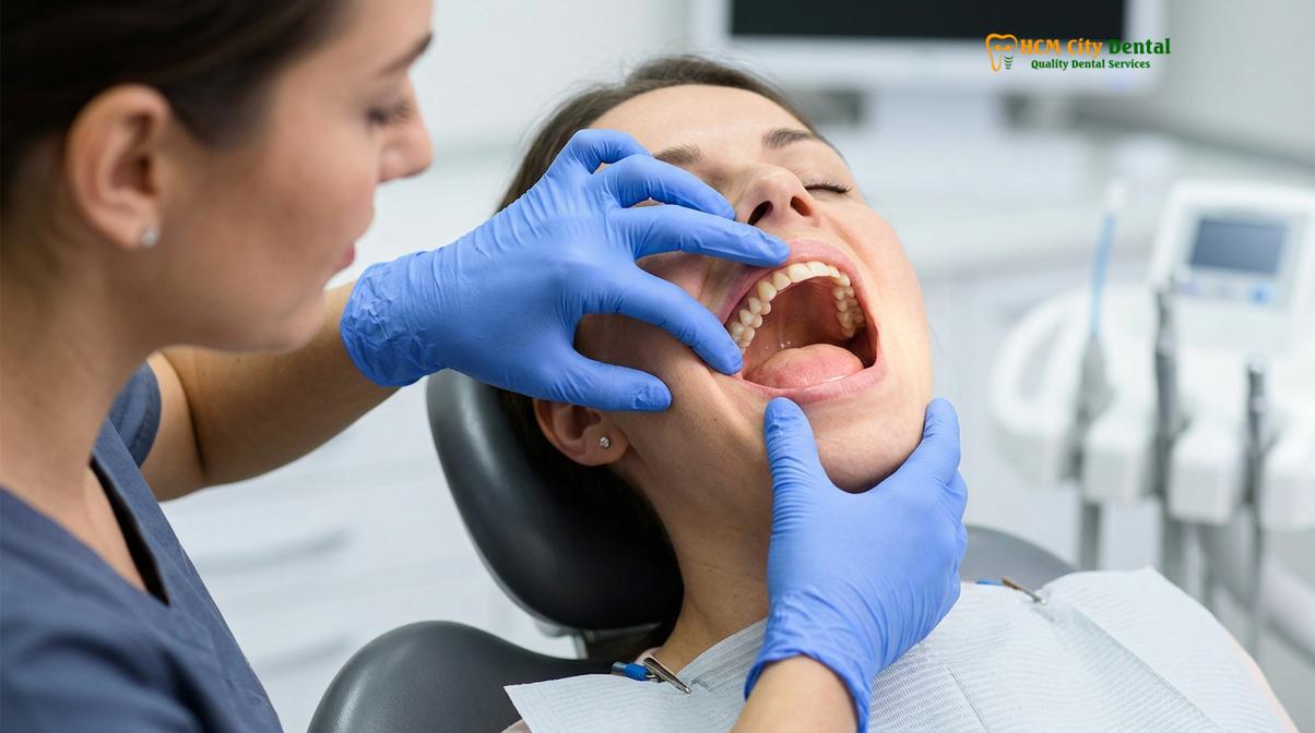
Clinical Contraindications: Intraoral myofascial release should be strictly avoided or modified in patients presenting with acute systemic infections, active oral ulcerations, severe joint effusion (swelling within the TMJ capsule), or unhealed mandibular fractures. Aggressive manual therapy in the presence of acute capsulitis can exacerbate inflammation and delay the healing process.
The medial pterygoid, which mirrors the masseter on the internal aspect of the mandibular ramus, is another frequent culprit in chronic jaw pain, particularly in patients who exhibit severe nocturnal clenching. Accessing the medial pterygoid requires the clinician to navigate the finger along the medial aspect of the mandible, posterior to the last molar. Release of this muscle often results in a dramatic and immediate improvement in the patient’s ability to open their mouth fully. Similarly, the tendon of the temporalis muscle, which inserts onto the coronoid process of the mandible, can be palpated and released intraorally by tracing the anterior border of the ascending ramus.
The physiological response to intraoral manual therapy extends beyond simple mechanical stretching. The sustained pressure stimulates the Golgi tendon organs and Ruffini corpuscles within the fascial network, triggering a parasympathetic nervous system response that promotes systemic relaxation and downregulates the hyperactive trigeminal nerve. While the procedure can induce transient discomfort—often described by patients as a deep, therapeutic ache—the subsequent restoration of normal muscle resting length is a critical step in temporomandibular joint therapy. Following a session of intraoral release, patients are typically instructed to perform gentle active range-of-motion exercises to maintain the newly acquired tissue extensibility.
Postural Re-Education: Cervical Spine Alignment and Jaw Recovery
Correcting forward head posture reduces compensatory strain on the suprahyoid muscles, directly decreasing compressive forces on the temporomandibular joint.
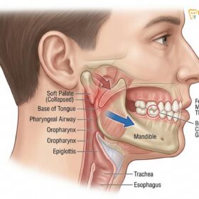
A comprehensive understanding of temporomandibular joint dysfunction is impossible without acknowledging the profound biomechanical relationship between the mandible, the cranium, and the cervical spine. The stomatognathic system does not function in isolation; it is intricately linked to the neck and shoulder girdle through a complex web of muscular, fascial, and neurological connections. Consequently, physical therapy for TMJ disorders must extend beyond the jaw itself to encompass postural re-education, specifically targeting the alignment of the cervical spine. Clinical studies consistently reveal a high prevalence of cervical spine disorders, particularly forward head posture (FHP), among patients suffering from chronic TMD.
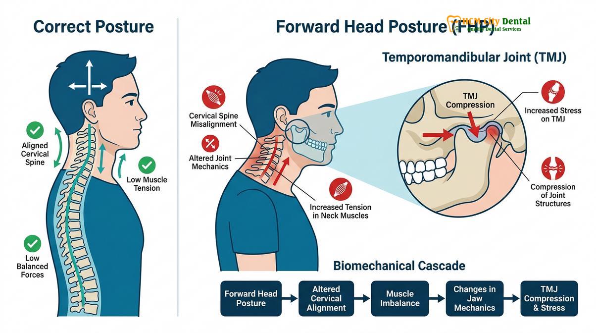
Forward head posture alters the resting trajectory of the mandible and significantly increases the mechanical load on the temporomandibular joints. When the head translates anteriorly relative to the cervical spine, the suprahyoid and infrahyoid muscles are placed under constant passive tension. This tension exerts a posterior and inferior pull on the mandible. To counteract this force and keep the mouth closed, the elevator muscles (masseter, temporalis, and medial pterygoid) must remain in a state of chronic hypertonicity. Over time, this constant muscular battle leads to fatigue, spasm, and increased compressive forces within the TMJ capsule, accelerating articular disc wear and tear[4].
Physical therapy addresses this biomechanical cascade through targeted cervical stabilization exercises. Deep neck flexor training is a primary focus, as these muscles are typically weak and inhibited in patients with FHP. Exercises such as cervical retractions (chin tucks) help restore the normal lordotic curve of the cervical spine and reposition the cranium directly over the shoulders. As cervical alignment improves, the passive tension on the hyoid musculature decreases, allowing the mandible to return to a more physiological resting posture. This reduction in baseline muscle tension is often accompanied by a spontaneous decrease in TMJ pain and an improvement in maximal incisal opening.
When to Consult a TMJ Specialist
Early clinical intervention is crucial for preventing progressive joint degeneration and chronic facial pain syndromes.
Patients experiencing persistent jaw pain, restricted mouth opening (trismus), or loud joint clicking accompanied by pain should seek professional evaluation promptly. According to the Vietnam Odonto-Stomatology Association (VOSA), early conservative intervention is highly recommended to prevent progressive articular disc degeneration and irreversible structural damage[6]. Delaying treatment can lead to chronic capsulitis or osteoarthritis of the temporomandibular joint, making non-invasive physical therapy less effective over time and potentially necessitating surgical intervention[5].
It is particularly important to consult a specialist if jaw pain begins to interfere with daily functions such as eating, speaking, or sleeping. Additionally, if you experience sudden changes in your bite (how your upper and lower teeth fit together) or if jaw pain is accompanied by frequent headaches, earaches, or tinnitus (ringing in the ears), a comprehensive diagnostic assessment is required. A qualified dental professional can accurately differentiate between muscular TMD, internal derangement of the joint, and other orofacial pain conditions, ensuring that the prescribed physical therapy protocol is both safe and effective.
Clinical Case Study: TMJ Rehabilitation in Saigon
A multidisciplinary approach combining custom occlusal appliances with targeted physical therapy yields optimal outcomes for complex TMJ disorders.
A clinical case highlights the efficacy of combined therapies for a patient visiting HCMC Dental Clinic in Ho Chi Minh City. The patient presented with chronic masseter hypertrophy, severe morning headaches, and limited jaw mobility due to long-standing nocturnal bruxism. Initial diagnostic imaging revealed mild anterior disc displacement with reduction, alongside significant hypertonicity in the lateral pterygoid muscles.
Dr. Nguyen Van Cuong, a leading specialist at the clinic, emphasizes the importance of a multidisciplinary approach for such complex cases. Dr. Cuong routinely integrates custom occlusal appliances with targeted physical therapy to ensure optimal joint stabilization. His clinical protocols focus on addressing the root biomechanical causes of jaw pain rather than merely masking the symptoms. By collaborating closely with physical rehabilitation principles, the treatment plan is tailored to the patient’s specific anatomical needs.
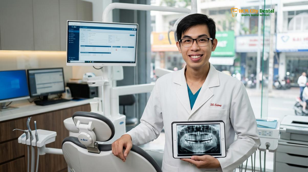
The patient was prescribed a custom nightguard to decompress the joint during sleep and protect the dentition from further occlusal wear. Concurrently, a structured physical therapy regimen was initiated, focusing on intraoral myofascial release to deactivate trigger points in the masseter and medial pterygoid. The patient was also instructed in a daily routine of isometric jaw exercises and cervical postural correction.
“The successful management of chronic temporomandibular dysfunction requires a commitment to both passive protection and active rehabilitation. By addressing the muscular, articular, and postural components simultaneously, we can restore harmonious function to the entire stomatognathic system.”
Following several weeks of consistent therapy and appliance wear, the patient reported a significant reduction in facial tension and a complete cessation of morning headaches. Clinical examination confirmed a restored, pain-free range of motion and symmetrical condylar tracking, demonstrating the profound benefits of integrating dental and physical rehabilitation modalities.
Frequently Asked Questions (FAQs)
Can physical therapy cure TMJ disorders?
Physical therapy aims to manage symptoms, restore normal range of motion, and prevent further deterioration rather than ‘curing’ structural joint degeneration. By addressing muscular imbalances, reducing capsular inflammation, and stabilizing the articular disc, targeted physical rehabilitation allows the temporomandibular joint to function with significantly reduced pain. Long-term success often depends on patient compliance with home exercise programs and concurrent dental interventions like occlusal splint therapy.
How do I massage my masseter muscle to stop jaw pain?
To massage the masseter muscle effectively, place your index and middle fingers on the lower corner of your jawline and apply firm, sustained pressure while slowly opening and closing your mouth. You can also use small, circular motions moving upward toward the cheekbone. For deeper relief, clinical intraoral massage involves a specialist placing a clean, gloved thumb inside the cheek and the index finger outside, gently squeezing the muscle belly to release localized trigger points.
What jaw exercises should I avoid if my joint clicks?
If your temporomandibular joint clicks, you should generally avoid wide-mouth opening exercises, forceful chewing of hard foods, and aggressive side-to-side jaw movements that can force the articular disc out of alignment. Isotonic exercises that require moving the jaw against heavy resistance can also exacerbate capsular ligament laxity. Instead, clinical protocols focus on gentle isometric holds where the jaw remains stationary while the muscles contract, promoting stability without stressing internal joint structures.
How long does it take for TMJ physical therapy to work?
Many patients begin to experience a noticeable reduction in muscular jaw pain and tension within two to four weeks of consistent, daily physical therapy. However, achieving long-term structural stability and correcting chronic postural imbalances typically requires three to six months of dedicated rehabilitation. The exact timeline varies significantly based on the severity of the temporomandibular disorder, the presence of underlying bruxism, and adherence to prescribed clinical protocols.
Is intraoral myofascial release painful?
Intraoral myofascial release can cause mild to moderate discomfort during the procedure, often described as a deep pressure sensation as chronic trigger points are deactivated. The clinician applies targeted ischemic compression to the internal masticatory muscles, which may temporarily refer pain to the teeth or ears. However, this transient discomfort is typically followed by a sense of muscular relaxation and improved jaw mobility once the pressure is released.
For detailed information on diagnostics and custom splint therapy options, visit our comprehensive TMJ & Jaw Pain Treatment Ho Chi Minh City guidelines.
References
- Journal of the American Dental Association. Occlusal splint therapy and physical therapy for temporomandibular disorders: a clinical review. (2020).
- Journal of Oral Rehabilitation. Efficacy of isometric jaw exercises in the management of chronic masticatory muscle pain. (2019).
- Clinical Oral Investigations. Intraoral myofascial release techniques for lateral pterygoid hypertonicity. (2018).
- Journal of Prosthetic Dentistry. The role of cervical posture correction in the comprehensive treatment of temporomandibular dysfunction. (2021).
- International Journal of Oral and Maxillofacial Surgery. Conservative management of TMJ disc displacement with reduction. (2022).
- Vietnam Odonto-Stomatology Association (VOSA). Guidelines on the conservative management of temporomandibular disorders. (2023).
If you are experiencing chronic jaw pain, restricted mobility, or persistent joint clicking, professional evaluation is essential. Contact the specialists at HCMC Dental Clinic in Ho Chi Minh City to schedule a comprehensive diagnostic assessment and explore personalized treatment options, including custom nightguards and targeted rehabilitation protocols designed to restore your oral health and comfort.