Diagnosing Temporomandibular Disorders (TMD) requires precise imaging to evaluate the jaw joint’s complex bony and soft tissue structures. In Vietnam, modern dental clinics utilize advanced 2D and 3D radiographic techniques to identify structural abnormalities, ensuring accurate, targeted treatment plans for chronic jaw pain and dysfunction.
Clinical Summary:
Temporomandibular disorder (TMD) diagnosis relies heavily on high-resolution radiographic imaging to differentiate between muscular pathologies and structural joint degeneration. While initial clinical screenings frequently utilize 2D panoramic radiographs to rule out gross dental anomalies, the definitive diagnosis of osseous changes—such as condylar flattening, subchondral sclerosis, or osteophyte formation—requires 3D Cone Beam Computed Tomography (CBCT). Soft tissue derangements, particularly articular disc displacement, may necessitate supplementary Magnetic Resonance Imaging (MRI). Combining meticulous clinical palpation with advanced imaging modalities ensures precise identification of joint space narrowing and biomechanical dysfunction. This evidence-based diagnostic workflow allows clinicians to formulate highly effective, conservative management strategies tailored to the specific anatomical needs of each patient.
Key Takeaways:
- 2D panoramic X-rays provide an excellent baseline screening for gross anatomical abnormalities but lack the 3D depth required for micro-structural joint analysis.
- CBCT is considered the clinical gold standard for visualizing bony architecture, offering sub-millimeter accuracy of the mandibular condyle and glenoid fossa.
- Radiographic signs of advanced TMD include condylar flattening, osteophyte (bone spur) formation, and subchondral sclerosis.
- Imaging must always be correlated with a comprehensive clinical examination, including muscle palpation and mandibular range-of-motion testing.
- Advanced imaging protocols in Ho Chi Minh City offer international-standard diagnostics at highly accessible rates for both local and international patients.
The Role of Dental Imaging in TMD Diagnosis
Dental imaging serves as the critical foundation for diagnosing structural temporomandibular joint disorders, allowing clinicians to visualize bone integrity, joint space, and anatomical alignment that cannot be assessed through physical examination alone.
The temporomandibular joint (TMJ) is one of the most complex anatomical structures in the human body. Classified as a ginglymoarthrodial joint, it uniquely combines both hinge (ginglymoid) and gliding (arthrodial) movements. This bilateral joint connects the mandible (lower jaw) to the temporal bone of the skull, facilitating essential daily functions such as mastication, phonation, and deglutition. Because the left and right joints are connected by a single bone—the mandible—they must function in perfect synchrony. When this harmonious movement is disrupted, patients often develop Temporomandibular Disorders (TMD).
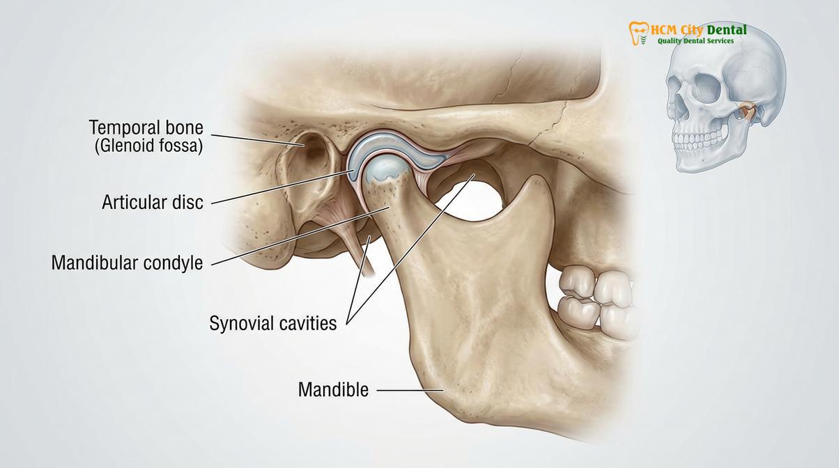
Clinically, TMD is broadly categorized into two main classifications: myogenous (originating from the masticatory muscles) and arthrogenous (originating from the joint structures themselves). While myogenous disorders, such as myofascial pain syndrome, are primarily diagnosed through clinical palpation and patient history, arthrogenous disorders necessitate sophisticated radiographic imaging. Without internal visualization, it is clinically impossible to accurately assess the integrity of the articular cartilage, the position of the intra-articular disc, or the morphological health of the subchondral bone[1].
The primary objective of TMJ imaging is to evaluate the anatomical relationship between the mandibular condyle and the glenoid fossa (mandibular fossa) at various stages of jaw opening and closing. Imaging helps clinicians identify structural anomalies, degenerative joint diseases (osteoarthritis), inflammatory conditions (rheumatoid arthritis), congenital malformations, and traumatic injuries. According to diagnostic frameworks supported by the Vietnam Odonto-Stomatology Association (VOSA), standardizing imaging protocols is essential for minimizing radiation exposure while maximizing diagnostic yield.
Orthopantomogram (OPG) X-Rays: What They Can and Cannot Reveal
An Orthopantomogram (OPG) provides a broad, two-dimensional overview of the entire maxillofacial region, making it an excellent initial screening tool for gross TMJ abnormalities, though it lacks the depth required for micro-structural analysis.
The Orthopantomogram, commonly referred to as a panoramic dental scan, is frequently the first line of radiographic investigation in general dental practice. This extraoral imaging technique captures the entire dentition, the maxilla, the mandible, and both temporomandibular joints in a single, continuous two-dimensional image. The fundamental mechanism of an OPG involves the X-ray tube and the image receptor rotating simultaneously around the patient’s head, utilizing a focal trough to keep the dental arches in sharp focus while blurring out adjacent structures.
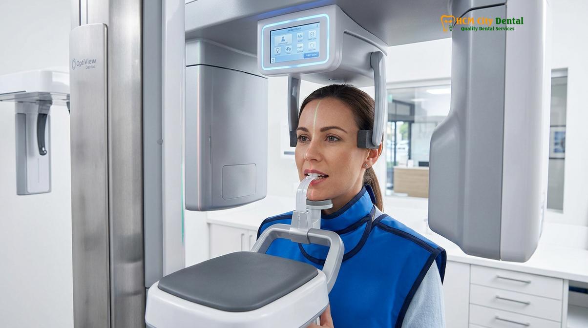
From a diagnostic perspective, the OPG is highly valuable for its broad field of view. It allows clinicians to rapidly assess the overall symmetry of the mandible, the vertical height of the condylar processes, and the general shape of the condylar heads. It is particularly effective at identifying gross pathologies that may refer pain to the TMJ region. For instance, an OPG can clearly reveal severely impacted wisdom teeth, large periapical abscesses, or mandibular fractures—all of which can produce symptoms that closely mimic primary temporomandibular joint dysfunction.
However, the clinical utility of the OPG in diagnosing specific, subtle TMJ pathologies is inherently limited by its two-dimensional nature. The temporomandibular joint is a complex, three-dimensional structure situated at an angle to the coronal and sagittal planes of the skull. When this 3D anatomy is compressed into a 2D image, significant superimposition of surrounding cranial bones occurs. The zygomatic arch, the base of the skull, and the cervical spine often overlap the TMJ space, obscuring fine osseous details[2].
Furthermore, panoramic radiographs are subject to magnification and distortion errors depending on the patient’s exact positioning within the focal trough. A slight tilt or rotation of the head can artificially alter the apparent size and shape of the condyles, potentially leading to false-positive diagnoses of condylar asymmetry. Because of these limitations, an OPG cannot reliably evaluate the precise width of the intra-articular joint space, nor can it detect early-stage erosive changes on the medial or lateral poles of the condyle.
Cone Beam Computed Tomography (CBCT): The Gold Standard for Jaw Joints
Cone Beam Computed Tomography (CBCT) delivers high-resolution, distortion-free three-dimensional images of the temporomandibular joint, making it the definitive imaging modality for detecting subtle osseous changes and joint space narrowing.
When clinical examinations and initial 2D screenings suggest the presence of structural joint pathology, Cone Beam Computed Tomography (CBCT) is universally recognized as the radiographic gold standard for evaluating the osseous components of the TMJ. Unlike traditional medical CT scanners that use a fan-shaped beam and multiple rotations to capture individual image slices, CBCT utilizes a divergent cone-shaped X-ray beam. This allows the machine to capture the entire maxillofacial volume in a single rotation, significantly reducing both the scanning time and the radiation dose administered to the patient.
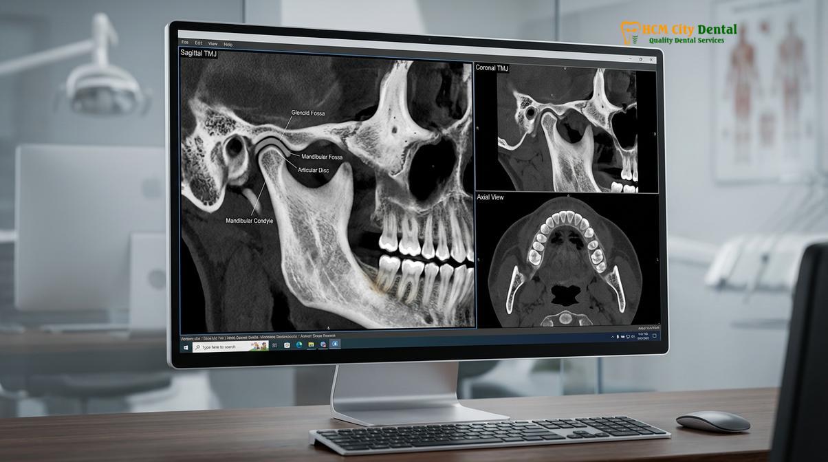
The technological superiority of CBCT lies in its ability to generate isotropic voxels—three-dimensional pixels that are perfectly cubical. This isotropic nature ensures that the reconstructed images are geometrically accurate and completely free from the magnification and distortion errors inherent in panoramic radiography. Clinicians can manipulate the 3D volume using specialized software to view the temporomandibular joint from any angle, generating precise sagittal, coronal, and axial cross-sections.
“The advent of Cone Beam Computed Tomography has revolutionized maxillofacial diagnostics. By eliminating anatomical superimposition, CBCT allows clinicians to visualize the intricate trabecular bone patterns and cortical outlines of the mandibular condyle with unprecedented clarity, fundamentally altering how we diagnose and stage degenerative joint diseases.”
In the context of TMD, CBCT is unparalleled in its ability to assess the true anatomical relationship between the condyle and the glenoid fossa. By evaluating the joint space in three dimensions, clinicians can accurately detect superior, posterior, or anterior displacement of the condyle within the fossa. A posteriorly displaced condyle, for example, often correlates clinically with anterior disc displacement and retrodiscal tissue compression, providing a critical piece of the diagnostic puzzle even without direct visualization of the soft tissue disc itself[3].
Clinical Case Review: HCMC Dental Clinic
A 42-year-old female patient presented to HCMC Dental Clinic in Ho Chi Minh City with chronic right-sided jaw pain and severe crepitus (grinding sounds) upon opening. Her initial 2D panoramic X-ray appeared largely unremarkable, showing only slight asymmetry. However, upon escalating the diagnostic protocol to a localized TMJ CBCT scan, the 3D coronal slices revealed significant erosive changes on the medial pole of the right condyle—an area completely obscured by superimposition on the 2D scan. This precise imaging allowed the clinical team to diagnose localized osteoarthritis and implement a targeted, conservative splint therapy protocol, avoiding unnecessary invasive procedures.
Comparing OPG and CBCT for Jaw Joint Analysis
Understanding the distinct capabilities of 2D and 3D imaging modalities helps patients appreciate why specific radiographic techniques are recommended for comprehensive temporomandibular joint evaluations.
To clarify the diagnostic differences between these two common imaging techniques, the following table outlines their respective clinical applications, strengths, and limitations when assessing the jaw joints.
| Diagnostic Feature | OPG (2D Panoramic X-Ray) | CBCT (3D Cone Beam Scan) |
|---|---|---|
| Dimensionality | Two-dimensional (2D) overview | Three-dimensional (3D) multi-planar |
| Superimposition | High (cranial bones overlap the TMJ) | None (cross-sectional slicing eliminates overlap) |
| Image Distortion | Subject to magnification and positioning errors | 1:1 ratio, geometrically accurate (isotropic voxels) |
| Primary Clinical Use | Initial screening, ruling out gross dental pathology | Detailed structural analysis, staging osteoarthritis |
| Joint Space Evaluation | Unreliable due to 2D compression | Highly accurate in sagittal, coronal, and axial planes |
Condylar Flattening and Subchondral Sclerosis: Clinical Signs
Advanced radiographic analysis often reveals specific degenerative markers such as condylar flattening, subchondral sclerosis, and osteophyte formation, which are critical indicators of osteoarthritis within the temporomandibular joint.
The temporomandibular joint is subjected to immense biomechanical forces during mastication and parafunctional habits such as bruxism (teeth grinding). Over time, if the adaptive capacity of the joint is exceeded, degenerative changes begin to manifest within the osseous structures. These changes, collectively referred to as osteoarthritis or degenerative joint disease (DJD), follow a specific pathophysiological progression that can be meticulously tracked using high-resolution CBCT imaging.
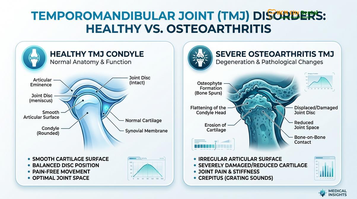
One of the earliest radiographic signs of bone condyle degeneration is the loss of the normal convex contour of the condylar head, a condition known as condylar flattening. In a healthy joint, the superior surface of the condyle is smoothly rounded, allowing it to glide effortlessly against the articular disc and the slope of the articular eminence. When abnormal loading occurs, the bone remodels in an attempt to distribute the excessive forces over a larger surface area, resulting in a flattened appearance. While mild flattening can sometimes be an adaptive, non-pathological response, progressive flattening is a hallmark of active joint degeneration.
As the degenerative process advances, the cartilage covering the condyle begins to break down, exposing the underlying subchondral bone to increased mechanical stress. In response to this stress, the bone undergoes a process called subchondral sclerosis. Radiographically, sclerosis appears as a thickening and increased radiopacity (whiteness) of the cortical bone layer just beneath the articular surface. This indicates that the bone is attempting to fortify itself against the abnormal biomechanical loads[4].
In more advanced stages of osteoarthritis, clinicians may observe the formation of osteophytes, commonly known as bone spurs. These are bony outgrowths that typically develop at the anterior margin of the condyle. Osteophytes represent a late-stage attempt by the body to stabilize a failing joint by increasing the articular surface area. Unfortunately, these bony projections often interfere with normal joint mechanics, leading to restricted mouth opening, severe crepitus, and chronic pain.
Integrating Imaging with Clinical Evaluation
Radiographic imaging is most effective when seamlessly integrated with a comprehensive clinical examination, ensuring that structural findings correlate directly with the patient’s functional symptoms.
While advanced imaging modalities like CBCT provide invaluable insights into the structural integrity of the temporomandibular joint, they must never be interpreted in isolation. A structural abnormality noted on an X-ray does not automatically equate to a clinical diagnosis of TMD. Many individuals possess radiographic signs of joint degeneration—such as mild condylar flattening or asymptomatic osteophytes—yet experience absolutely no pain or functional impairment. Conversely, a patient may suffer from severe myofascial pain with a completely normal CBCT scan.
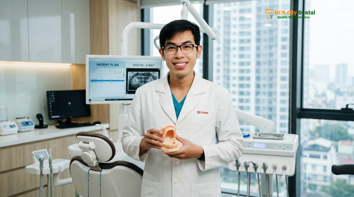
Dr. Nguyen Van Cuong, a leading expert in maxillofacial diagnostics, emphasizes that a thorough clinical evaluation must always precede advanced imaging. In his practice of General Dentistry at HCMC Dental Clinic, Dr. Cuong routinely integrates 3D CBCT scans to ensure precise, conservative management of temporomandibular disorders. By correlating the patient’s subjective pain reports with objective 3D data, the clinical team can develop highly targeted therapeutic interventions.
“Radiographic imaging should never replace a comprehensive physical examination; rather, it serves to confirm and precisely localize the structural pathologies suspected during clinical palpation.”
The integration of imaging into the Research Diagnostic Criteria for Temporomandibular Disorders (RDC/TMD) highlights the necessity of a dual-axis approach. Axis I involves the physical assessment of joint sounds, muscle tenderness, and range of motion, while Axis II evaluates the psychosocial factors contributing to chronic pain. Imaging serves to validate the Axis I findings, particularly when differentiating between disc displacement with reduction (clicking) and degenerative joint disease (crepitus)[5].
When to Consult a Specialist for Jaw Pain
Recognizing the early warning signs of temporomandibular joint dysfunction is crucial for seeking timely diagnostic imaging and preventing irreversible joint damage.
Patients should not wait for symptoms to become debilitating before seeking professional evaluation. Early intervention often allows for conservative management, whereas delayed treatment may necessitate complex rehabilitative procedures. You should consider scheduling a diagnostic consultation if you experience any of the following symptoms:
- Persistent Jaw Pain: A dull, aching pain in the jaw joint or surrounding facial muscles that does not resolve with rest or over-the-counter anti-inflammatory medications.
- Restricted Movement: Difficulty opening the mouth fully, or experiencing a “locked” jaw sensation where the mandible becomes temporarily stuck in an open or closed position.
- Joint Noises: Loud clicking, popping, or grinding (crepitus) sounds when chewing or speaking, especially if these sounds are accompanied by pain.
- Changes in Bite: A sudden feeling that your upper and lower teeth no longer fit together correctly, which may indicate a shift in the condylar position.
Important Clinical Note: If you experience sudden jaw locking, severe facial trauma, or rapid onset of swelling around the temporomandibular joint, seek immediate emergency dental care. Delaying evaluation can lead to permanent joint damage or exacerbate underlying structural issues.
Conclusion
Accurate diagnosis is the cornerstone of effective temporomandibular disorder management. While 2D panoramic X-rays offer a valuable initial screening tool, 3D CBCT imaging remains the definitive gold standard for evaluating the intricate osseous structures of the jaw joint. By identifying subtle degenerative changes such as condylar flattening and subchondral sclerosis early, clinicians can implement conservative therapies that preserve joint function and alleviate chronic pain. If you are experiencing persistent jaw discomfort, clicking, or restricted movement, comprehensive diagnostic imaging can provide the clarity needed to restore your oral health and quality of life. For expert evaluation and state-of-the-art 3D imaging, consider scheduling a consultation for General Dentistry at HCMC Dental Clinic in Ho Chi Minh City.
For detailed information on diagnostics and custom splint therapy options, visit our comprehensive TMJ & Jaw Pain Treatment Ho Chi Minh City guidelines.
References
- Journal of Oral Rehabilitation. Diagnostic accuracy of CBCT in temporomandibular joint osteoarthritis. (2021).
- Dentomaxillofacial Radiology. Panoramic radiography vs. CBCT in evaluating TMJ bony changes. (2019).
- Journal of the American Dental Association. Evidence-based clinical guidelines for TMD diagnostics. (2020).
- International Journal of Oral and Maxillofacial Surgery. Pathophysiology of subchondral sclerosis in the TMJ. (2018).
- Clinical Oral Investigations. The role of imaging in the Research Diagnostic Criteria for TMD. (2022).